

The DOI matches its Crossref record; 60 of the 60 references it cites were found in a registry or an index.
Targeting translocator protein (18 kDa) (TSPO) dampens pro-inflammatory microglia reactivity in the retina and protects from degeneration
Open accessAbstract
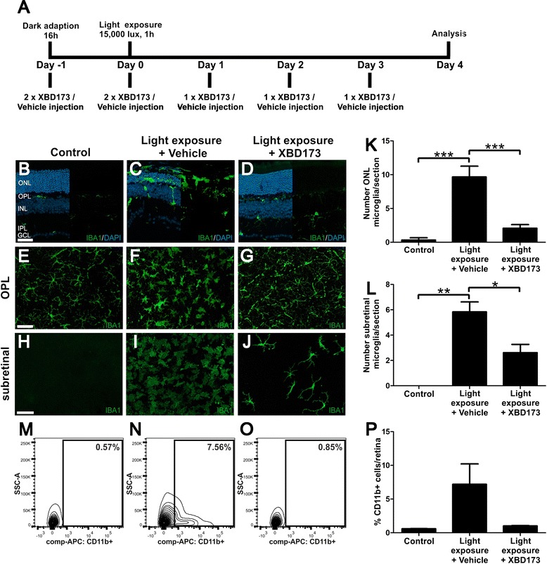
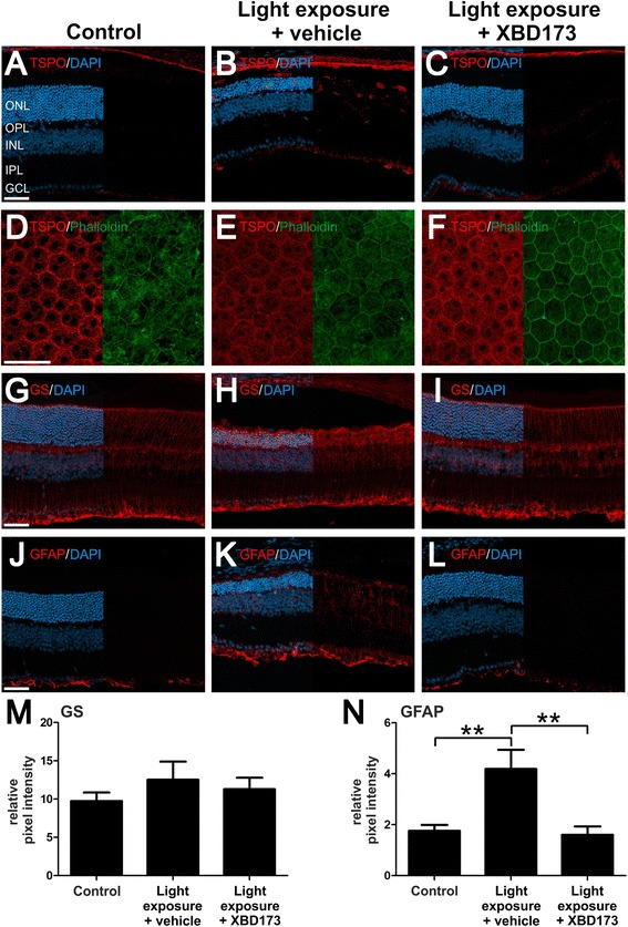
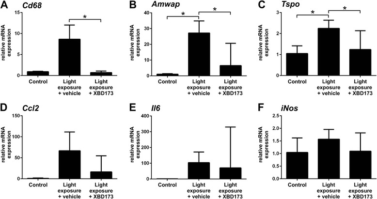
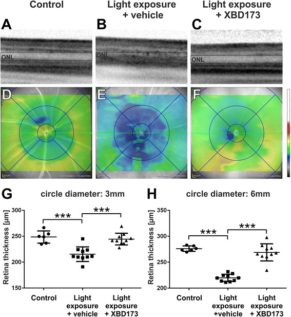
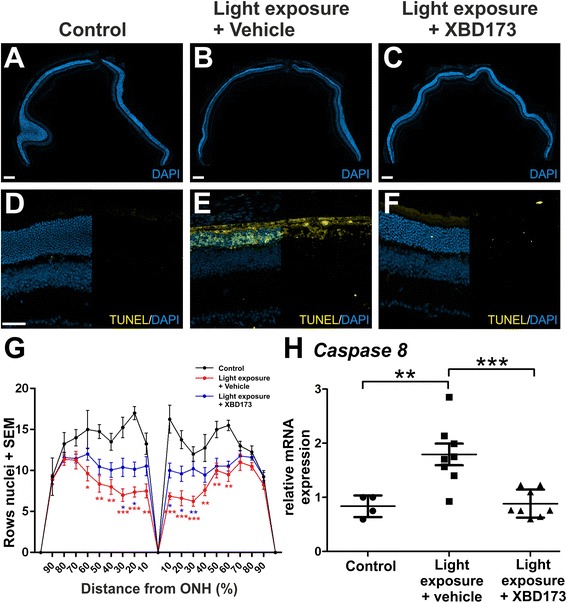
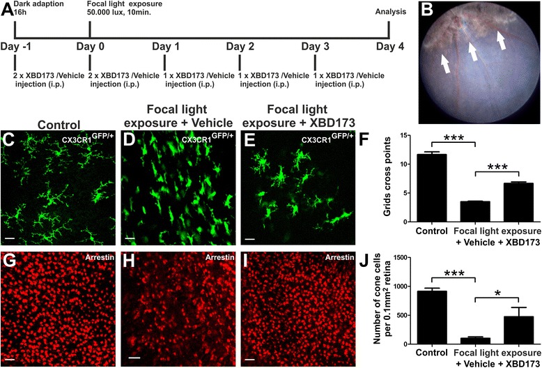
BACKGROUND: Reactive microglia are commonly seen in retinal degenerative diseases, and neurotoxic microglia responses can contribute to photoreceptor cell death. We and others have previously shown that translocator protein (18 kDa) (TSPO) is highly induced in retinal degenerations and that the selective TSPO ligand XBD173 (AC-5216, emapunil) exerts strong anti-inflammatory effects on microglia in vitro and ex vivo. Here, we investigated whether targeting TSPO with XBD173 has immuno-modulatory and neuroprotective functions in two mouse models of acute retinal degeneration using bright white light exposure. METHODS: BALB/cJ and Cx3cr1(GFP/+) mice received intraperitoneal injections of 10 mg/kg XBD173 or vehicle for five consecutive days, starting 1 day prior to exposure to either 15,000 lux white light for 1 h or 50,000 lux focal light for 10 min, respectively. The effects of XBD173 treatment on microglia and Müller cell reactivity were analyzed by immuno-stainings of retinal sections and flat mounts, fluorescence-activated cell sorting (FACS) analysis, and mRNA expression of microglia markers using quantitative real-time PCR (qRT-PCR). Optical coherence tomography (OCT), terminal deoxynucleotidyl transferase dUTP nick end labeling (TUNEL) stainings, and morphometric analyses were used to quantify the extent of retinal degeneration and photoreceptor apoptosis. RESULTS: Four days after the mice were challenged with bright white light, a large number of amoeboid-shaped alerted microglia appeared in the degenerating outer retina, which was nearly completely prevented by treatment with XBD173. This treatment also down-regulated the expression of TSPO protein in microglia but did not change the TSPO levels in the retinal pigment epithelium (RPE). RT-PCR analysis showed that the microglia/macrophage markers Cd68 and activated microglia/macrophage whey acidic protein (Amwap) as well as the pro-inflammatory genes Ccl2 and Il6 were reduced after XBD173 treatment. Light-induced degeneration of the outer retina was nearly fully blocked by XBD173 treatment. We further confirmed these findings in an independent mouse model of focal light damage. Retinas of animals receiving XBD173 therapy displayed significantly more ramified non-reactive microglia and more viable arrestin-positive cone photoreceptors than vehicle controls. CONCLUSIONS: Targeting TSPO with XBD173 effectively counter-regulates microgliosis and ameliorates light-induced retinal damage, highlighting a new pharmacological concept for the treatment of retinal degenerations.
Overview
The claims and statistics of this paper haven’t been checked yet.
Run the check — free, no account needed. It reads the open-access text and checks the sources it cites.
Integrity signals
Registry
Cites
Statistics
Figures
Verification
Claims the paper makes
The claims and statistics of this paper haven’t been checked yet.
Run the check — free, no account needed. It reads the open-access text and checks the sources it cites.
Claims other papers make about this one
Each is checked against this paper’s own text: does it say what the citing paper says it does?
In the citing paper
“Furthermore, increased TSPO expression may not be indicative of the inflammatory phenotype of microglia (Scholz et al, 2015).”
From Microglia Priming with Aging and Stress (2017) · cites this paper as Scholz et al, 2015
Self-check
Percentages, sums and reported differences re-computed from the paper’s own numbers.
The self-check needs the paper’s full text.
Citations
Cites
Resting microglial cells are highly dynamic surveillants of brain parenchyma in vivo
Nimmerjahn, A., Kirchhoff, F., & Helmchen, F. (2005) · Science · doi:10.1126/science.1110647 · Cited by 5,827
Kettenmann, H., Hanisch, U.-K., Noda, M., & Verkhratsky, A. (2011) · Physiological Reviews · doi:10.1152/physrev.00011.2010 · Cited by 3,547
Inflammation in neurodegenerative diseases
Amor, S., Puentes, F., Baker, D., & Van Der Valk, P. (2010) · Immunology · doi:10.1111/j.1365-2567.2009.03225.x · Cited by 1,355
Aloisi, F. (2001) · Glia · doi:10.1002/glia.1106 · Cited by 1,310
Wu, D. C., Jackson-Lewis, V., Vila, M., Tieu, K., Teismann, P., Vadseth, C., Choi, D.-K., Ischiropoulos, H., & Przedborski, S. (2002) · The Journal of Neuroscience · doi:10.1523/jneurosci.22-05-01763.2002 · Cited by 1,183
Rupprecht, R., Papadopoulos, V., Rammes, G., Baghai, T. C., Fan, J., Akula, N., Groyer, G., Adams, D., & Schumacher, M. (2010) · Nature Reviews Drug Discovery · doi:10.1038/nrd3295 · Cited by 930
Microglia derived from aging mice exhibit an altered inflammatory profile
Sierra, A., Gottfried‐Blackmore, A. C., McEwen, B. S., & Bulloch, K. (2007) · Glia · doi:10.1002/glia.20468 · Cited by 688
Para-Inflammation in the Aging Retina
Xu, H., Chen, M., & Forrester, J. V. (2009) · Progress in Retinal and Eye Research · doi:10.1016/j.preteyeres.2009.06.001 · Cited by 659 · cited 1×
“of microglia physiology contribute to degenerative pathologies of the retina and the entire CNS [3, 5–14].”
BackgroundSelf-citationWenzel, A., Grimm, C., Samardzija, M., & Remé, C. E. (2004) · Progress in Retinal and Eye Research · doi:10.1016/j.preteyeres.2004.08.002 · Cited by 655
Self-citationGupta, N., Brown, K. E., & Milam, A. H. (2003) · Experimental Eye Research · doi:10.1016/s0014-4835(02)00332-9 · Cited by 582
Cited by
Sorted by citations
Microglia Priming with Aging and Stress
Niraula, A., Sheridan, J. F., & Godbout, J. P. (2017) · Neuropsychopharmacology · doi:10.1038/npp.2016.185 · Cited by 398
Microglia in Retinal Degeneration
Rashid, K., Akhtar‐Schäfer, I., & Langmann, T. (2019) · Frontiers in Immunology · doi:10.3389/fimmu.2019.01975 · Cited by 365
Self-citationMicroglia and Inflammatory Responses in Diabetic Retinopathy
Kinuthia, U. M., Wolf, A., & Langmann, T. (2020) · Frontiers in Immunology · doi:10.3389/fimmu.2020.564077 · Cited by 332
Self-citationXu, G.-T., Zhang, J., & Tang, L. (2022) · Neural Regeneration Research · doi:10.4103/1673-5374.355743 · Cited by 293
Retinal microglia – A key player in healthy and diseased retina
Rathnasamy, G., Foulds, W. S., Ling, E., & Kaur, C. (2018) · Progress in Neurobiology · doi:10.1016/j.pneurobio.2018.05.006 · Cited by 250
The impact of high and low dose ionising radiation on the central nervous system
Betlazar, C., Middleton, R. J., Banati, R. B., & Liu, G.-J. (2016) · Redox Biology · doi:10.1016/j.redox.2016.08.002 · Cited by 196
Scholz, R., Sobotka, M., Caramoy, A., Stempfl, T., Moehle, C., & Langmann, T. (2015) · Journal of Neuroinflammation · doi:10.1186/s12974-015-0431-4 · Cited by 177
Self-citationTriazines: Syntheses and Inverse Electron-demand Diels–Alder Reactions
Zhang, F., Chen, Z., Tang, X., & Ma, J. (2021) · Chemical Reviews · doi:10.1021/acs.chemrev.1c00611 · Cited by 167
The Translocator Protein (TSPO) in Mitochondrial Bioenergetics and Immune Processes
Betlazar, C., Middleton, R. J., Banati, R. B., & Liu, G. J. (2020) · Cells · doi:10.3390/cells9020512 · Cited by 140
Retinal microglia: Functions and diseases
Fan, W., Huang, W., Chen, J., Li, N., Mao, L., & Hou, S. (2022) · Immunology · doi:10.1111/imm.13479 · Cited by 139
Modulation of three key innate immune pathways for the most common retinal degenerative diseases
Akhtar‐Schäfer, I., Wang, L., Krohne, T. U., Xu, H., & Langmann, T. (2018) · EMBO Molecular Medicine · doi:10.15252/emmm.201708259 · Cited by 129
Self-citationThe innate immune system in diabetic retinopathy
Pan, W. W., Lin, F., & Fort, P. E. (2021) · Progress in Retinal and Eye Research · doi:10.1016/j.preteyeres.2021.100940 · Cited by 117
Conedera, F. M., Pousa, A. M. Q., Mercader, N., Tschopp, M., & Enzmann, V. (2019) · Glia · doi:10.1002/glia.23601 · Cited by 94
Macrophage physiology in the eye
Chinnery, H. R., McMenamin, P. G., & Dando, S. J. (2017) · Pflügers Archiv - European Journal of Physiology · doi:10.1007/s00424-017-1947-5 · Cited by 87
Aires, I. D., Boia, R., Rodrigues‐Neves, A. C., Madeira, M. H., Marques, C., Ambrósio, A. F., & Santiago, A. R. (2019) · Glia · doi:10.1002/glia.23579 · Cited by 88
Pozzo, E. D., Tremolanti, C., Costa, B., Giacomelli, C., Milenkovic, V. M., Bader, S. M., Wetzel, C. H., Rupprecht, R., Taliani, S., Settimo, F. D., et al. (2019) · International Journal of Molecular Sciences · doi:10.3390/ijms20184467 · Cited by 89
The tspo-nox1 axis controls phagocyte-triggered pathological angiogenesis in the eye
Wolf, A., Herb, M., Schramm, M., & Langmann, T. (2020) · Nature Communications · doi:10.1038/s41467-020-16400-8 · Cited by 80
Self-citationPyroptosis in the Retinal Neurovascular Unit: New Insights Into Diabetic Retinopathy
Meng, C., Gu, C., He, S., Su, T., Lhamo, T., Draga, D., & Qiu, Q. (2021) · Frontiers in Immunology · doi:10.3389/fimmu.2021.763092 · Cited by 72
Roche, S. L., Ruiz‐Lopez, A. M., Moloney, J. N., Byrne, A. M., & Cotter, T. G. (2017) · Glia · doi:10.1002/glia.23243 · Cited by 73
Dimitrova‐Shumkovska, J., Krstanoski, L., & Veenman, L. (2020) · Cells · doi:10.3390/cells9040870 · Cited by 74
Madeira, M. H., Ortín-Martínez, A., Nadal-Nicolás, F. M., Ambrósio, A. F., Vidal‐Sanz, M., Agudo‐Barriuso, M., & Santiago, A. R. (2016) · Scientific Reports · doi:10.1038/srep27532 · Cited by 67
Koumine Attenuates Neuroglia Activation and Inflammatory Response to Neuropathic Pain
Jin, G., He, S.-D., Lin, S.-M., Hong, L.-M., Chen, W., Xu, Y., Yang, J., Li, S., & Yu, C. (2018) · Neural Plasticity · doi:10.1155/2018/9347696 · Cited by 65
Targeting Microglia to Treat Degenerative Eye Diseases
Wang, S. K., & Cepko, C. L. (2022) · Frontiers in Immunology · doi:10.3389/fimmu.2022.843558 · Cited by 66
Gong, J., Szegő, É. M., Leonov, A., Benito, E., Becker, S., Fischer, A., Zweckstetter, M., Outeiro, T. F., & Schneider, A. (2019) · Journal of Neuroscience · doi:10.1523/jneurosci.2070-18.2019 · Cited by 70
Control of Neuroinflammation through Radiation-Induced Microglial Changes
Boyd, A., Byrne, S., Middleton, R. J., Banati, R. B., & Liu, G. J. (2021) · Cells · doi:10.3390/cells10092381 · Cited by 65
Biswas, L., Zhou, X., Dhillon, B., Graham, A., & Shu, X. (2017) · Human Molecular Genetics · doi:10.1093/hmg/ddx319 · Cited by 57
Comprehensive analysis of mouse retinal mononuclear phagocytes
Lückoff, A., Scholz, R., Sennlaub, F., Xu, H., & Langmann, T. (2017) · Nature Protocols · doi:10.1038/nprot.2017.032 · Cited by 56
Self-citationThe Interaction Between Microglia and Macroglia in Glaucoma
Zhao, X., Sun, R., Luo, X., Wang, F., & Sun, X. (2021) · Frontiers in Neuroscience · doi:10.3389/fnins.2021.610788 · Cited by 58
Roche, S. L., Wyse‐Jackson, A. C., Gómez‐Vicente, V., Lax, P., Ruiz‐Lopez, A. M., Byrne, A. M., Cuenca, N., & Cotter, T. G. (2016) · PLoS ONE · doi:10.1371/journal.pone.0165197 · Cited by 55
Laquinimod protects the optic nerve and retina in an experimental autoimmune encephalomyelitis model
Wilmes, A. T., Reinehr, S., Kühn, S., Pedreiturria, X., Petrikowski, L., Faissner, S., Ayzenberg, I., Stute, G., Gold, R., Dick, H. B., et al. (2018) · Journal of Neuroinflammation · doi:10.1186/s12974-018-1208-3 · Cited by 54
Bian, M., Du, X., Cui, J., Wang, P., Wang, W., Zhu, W., Zhang, T., & Chen, Y. (2016) · Journal of Neuroinflammation · doi:10.1186/s12974-016-0516-8 · Cited by 54
Biswas, L., Farhan, F., Reilly, J., Bartholomew, C., & Shu, X. (2018) · International Journal of Molecular Sciences · doi:10.3390/ijms19123740 · Cited by 59
Microglia Activation and Immunomodulatory Therapies for Retinal Degenerations
Rashid, K., Wolf, A., & Langmann, T. (2018) · Frontiers in Cellular Neuroscience · doi:10.3389/fncel.2018.00176 · Cited by 51
Self-citationLitwińska, Z., Sobuś, A., Łuczkowska, K., Grabowicz, A., Mozolewska‐Piotrowska, K., Safranow, K., Kawa, M. P., Machaliński, B., & Machalińska, A. (2019) · Frontiers in Aging Neuroscience · doi:10.3389/fnagi.2019.00286 · Cited by 49
Mages, K., Graßmann, F., Jägle, H., Rupprecht, R., Weber, B. H. F., Hauck, S. M., & Grosche, A. (2019) · Journal of Neuroinflammation · doi:10.1186/s12974-019-1424-5 · Cited by 50
Fractalkine-CX3CR1 signaling is critical for progesterone-mediated neuroprotection in the retina
Roche, S. L., Wyse‐Jackson, A. C., Ruiz‐Lopez, A. M., Byrne, A. M., & Cotter, T. G. (2017) · Scientific Reports · doi:10.1038/srep43067 · Cited by 46
Regulation of Mitochondrial, Cellular, and Organismal Functions by TSPO
Gavish, M., & Veenman, L. (2017) · Advances in pharmacology · doi:10.1016/bs.apha.2017.09.004 · Cited by 49
Emerging therapies and their delivery for treating age‐related macular degeneration
Thomas, C. N., Sim, D. A., Lee, W. H., Alfahad, N., Dick, A. D., Denniston, A. K., & Hill, L. J. (2021) · British Journal of Pharmacology · doi:10.1111/bph.15459 · Cited by 46
Sun, W., An, X., Zhang, Y., Zhao, X., Sun, Y., Yang, C., Kang, X., Jiang, L., Ji, H., & Lian, F. (2023) · Frontiers in Endocrinology · doi:10.3389/fendo.2023.1270145 · Cited by 44
Effect of Inhibition of Colony-Stimulating Factor 1 Receptor on Choroidal Neovascularization in Mice
Schwarzer, P., Kokona, D., Ebneter, A., & Zinkernagel, M. S. (2019) · American Journal Of Pathology · doi:10.1016/j.ajpath.2019.10.011 · Cited by 40
Wang, W., Zhang, L., Zhang, X., Xue, R., Li, L., Zhao, W., Fu, Q., Mi, W., & Li, Y. (2016) · Frontiers in Pharmacology · doi:10.3389/fphar.2016.00384 · Cited by 40
Gene-agnostic therapeutic approaches for inherited retinal degenerations
John, M. C., Quinn, J., Hu, M. L., Cehajic‐Kapetanovic, J., & Xue, K. (2023) · Frontiers in Molecular Neuroscience · doi:10.3389/fnmol.2022.1068185 · Cited by 39
Leva, G., Klein, C., Benyounes, J., Hallé, F., Bihel, F. J. ‐., Collongues, N., Seze, J. D., Mensah‐Nyagan, A. G., & Patte‐Mensah, C. (2017) · Biochimica et Biophysica Acta (BBA) - Molecular Basis of Disease · doi:10.1016/j.bbadis.2017.09.007 · Cited by 42
Zhao, F., Gao, X., Ge, X., Cui, J., & Liu, X. (2021) · Bioengineered · doi:10.1080/21655979.2021.1996512 · Cited by 37
Neuroprotective effect of mitochondrial translocator protein ligand in a mouse model of tauopathy
Fairley, L. H., Sahara, N., Aoki, I., Ji, B., Suhara, T., Higuchi, M., & Barron, A. M. (2021) · Journal of Neuroinflammation · doi:10.1186/s12974-021-02122-1 · Cited by 37
Petrash, C. C., Palestine, A. G., & Canto-Soler, M. V. (2021) · Frontiers in Immunology · doi:10.3389/fimmu.2021.621007 · Cited by 36
Immune response in retinal degenerative diseases – Time to rethink?
Xu, H., & Chen, M. (2022) · Progress in Neurobiology · doi:10.1016/j.pneurobio.2022.102350 · Cited by 33
Self-citationZhao, N., Hao, X.-N., Huang, J.-M., Song, Z., & Tao, Y. (2024) · Aging and Disease · doi:10.14336/ad.2023.0823-3 · Cited by 32
Translocator protein in the rise and fall of central nervous system neurons
Cheung, G., Lin, Y. C., & Papadopoulos, V. (2023) · Frontiers in Cellular Neuroscience · doi:10.3389/fncel.2023.1210205 · Cited by 33
Song, D., Sulewski, M. E., Wang, C., Song, J., Bhuyan, R., Sterling, J. K., Clark, E., Song, W.-C., & Dunaief, J. L. (2017) · Europe PMC (PubMed Central) · Cited by 30
Oxysterols and retinal degeneration
Zhang, X., Alhasani, R. H., Zhou, X., Reilly, J., Zeng, Z., Strang, N. C., & Shu, X. (2021) · British Journal of Pharmacology · doi:10.1111/bph.15391 · Cited by 27
Müller cells trophism and pathology as the next therapeutic targets for retinal diseases
Arrigo, A., Cremona, O., Aragona, E., Casoni, F., Consalez, G., Dogru, R. M., Hauck, S. M., Antropoli, A., Bianco, L., Parodi, M. B., et al. (2025) · Progress in Retinal and Eye Research · doi:10.1016/j.preteyeres.2025.101357 · Cited by 27
Bian, M., Zhang, Y., Du, X., Xu, J., Cui, J., Gu, J., Zhu, W., Zhang, T., & Chen, Y. (2017) · Brain Research · doi:10.1016/j.brainres.2017.03.019 · Cited by 25
Wen, L., Hu, Y., Meng, T., Tan, Y., Zhao, M., Dai, S., Yuan, H., & Hu, F. (2018) · Colloids and Surfaces B Biointerfaces · doi:10.1016/j.colsurfb.2018.12.016 · Cited by 25
Stravalaci, M., Ferrara, M., Pathak, V., Davi, F., Bottazzi, B., Mantovani, A., Medina, R. J., Romano, M. R., & Inforzato, A. (2022) · Frontiers in Pharmacology · doi:10.3389/fphar.2021.811344 · Cited by 24
Deletion of TSPO Causes Dysregulation of Cholesterol Metabolism in Mouse Retina
Farhan, F., Almarhoun, M., Wong, A., Findlay, A. S., Bartholomew, C., Williams, M. T. S., Hurd, T. W., & Shu, X. (2021) · Cells · doi:10.3390/cells10113066 · Cited by 26
Combination of ginsenoside Rb1 and Rd protects the retina against bright light-induced degeneration
Bian, M., Du, X., Wang, P., Cui, J., Xu, J., Gu, J., Zhang, T., & Chen, Y. (2017) · Scientific Reports · doi:10.1038/s41598-017-06471-x · Cited by 23
Huang, J.-M., Zhao, N. R., Hao, X.-N., Li, S., Wei, D., Pu, N., Peng, G.-H., & Tao, Y. (2024) · Investigative Ophthalmology & Visual Science · doi:10.1167/iovs.65.1.29 · Cited by 21
Pfaller, A. M., Kaplan, L., Carido, M., Graßmann, F., Díaz‐Lezama, N., Ghaseminejad, F., Wunderlich, K. A., Glänzer, S., Bludau, O., Pannicke, T., et al. (2024) · Journal of Neuroinflammation · doi:10.1186/s12974-024-03021-x · Cited by 20
TSPO ligand FGIN‐1‐27 controls priapism in sickle cell mice via endogenous testosterone production
Musicki, B., Karakus, S., Favor, J. D. L., Chen, H., Silva, F. H., Sturny, M., Zirkin, B. R., & Burnett, A. L. “. (2020) · Journal of Cellular Physiology · doi:10.1002/jcp.30075 · Cited by 20
Desai, R., & Campanella, M. (2019) · British Journal of Pharmacology · doi:10.1111/bph.14697 · Cited by 19
Kumari, A., Ayala-Ramírez, R., Zenteno, J. C., Huffman, K., Šášik, R., Ayyagari, R., & Borooah, S. (2022) · Scientific Reports · doi:10.1038/s41598-022-19351-w · Cited by 19
Rashid, K., Geissl, L., Wolf, A., Karlstetter, M., & Langmann, T. (2018) · Biochimica et Biophysica Acta (BBA) - Gene Regulatory Mechanisms · doi:10.1016/j.bbagrm.2018.10.018 · Cited by 18
Self-citationRashid, K., Verhoyen, M., Taiwo, M., & Langmann, T. (2020) · Biochemical and Biophysical Research Communications · doi:10.1016/j.bbrc.2020.05.114 · Cited by 17
Self-citationIliopoulou, S. M., Tsartsalis, S., Kaiser, S., Millet, P., & Tournier, B. B. (2021) · Neuropsychiatric Disease and Treatment · doi:10.2147/ndt.s334027 · Cited by 17
Tremolanti, C., Cavallini, C., Meyer, L., Klein, C., Pozzo, E. D., Costa, B., Germelli, L., Taliani, S., Patte‐Mensah, C., & Mensah‐Nyagan, A. G. (2022) · Molecular Neurobiology · doi:10.1007/s12035-022-02737-2 · Cited by 17
Trost, A., Motloch, K. A., Koller, A., Bruckner, D., Runge, C., Schroedl, F., Bogner, B., Kaser-Eichberger, A., Strohmaier, C., Ladek, A., et al. (2021) · Experimental Eye Research · doi:10.1016/j.exer.2021.108806 · Cited by 15
Klee, K., Storti, F., Barben, M., Samardzija, M., Langmann, T., Dunaief, J. L., & Grimm, C. (2019) · Experimental Eye Research · doi:10.1016/j.exer.2019.107816 · Cited by 15
Self-citationGao, S., Li, N., Wang, Y., Lin, Z., Zhu, Y., Xu, J., Zhang, Q., Zhu, C., Zhou, Y., Zhou, J., et al. (2022) · The FASEB Journal · doi:10.1096/fj.202101294rrr · Cited by 14
Immunological Considerations for Retinal Stem Cell Therapy
Kramer, J., Chirco, K. R., & Lamba, D. A. (2019) · Advances in experimental medicine and biology · doi:10.1007/978-3-030-28471-8_4 · Cited by 13
Özkaya, D., Shu, X., & Nazıroğlu, M. (2021) · Biology · doi:10.3390/biology10050382 · Cited by 12
Maes, M. E., Wögenstein, G. M., Colombo, G., Casado-Polanco, R., & Siegert, S. (2021) · Molecular Therapy — Methods & Clinical Development · doi:10.1016/j.omtm.2021.09.006 · Cited by 12
Yin, J., Huang, J.-L., Wu, J., Gao, L., Xu, H., Zhu, J., Mai, H., Luo, J., Yao, Z., Li, Y., et al. (2025) · Osteoarthritis and Cartilage · doi:10.1016/j.joca.2025.06.008 · Cited by 11
Self-citationKim, T.-H., Son, W. S., Morshed, M. N., Londhe, A. M., Jung, S. Y., Park, J., Park, W., Lim, S. M., Park, K. D., Cho, S. J., et al. (2017) · European Journal of Medicinal Chemistry · doi:10.1016/j.ejmech.2017.09.033 · Cited by 11
Acero, G., Catorce, M. N., González-Mendoza, R., Meraz-Rodríguez, M. A., Hernández-Zimbrón, L. F., González-Salinas, R., & Gevorkian, G. (2017) · Inflammopharmacology · doi:10.1007/s10787-017-0355-y · Cited by 10
Translocator protein (18 kDa) (Tspo) in the retina and implications for ocular diseases
Hector, M., Langmann, T., & Wolf, A. (2024) · Progress in Retinal and Eye Research · doi:10.1016/j.preteyeres.2024.101249 · Cited by 10
Self-citationValent, D. J., Wong, W. T., Chew, E. Y., & Cukras, C. A. (2018) · Translational Vision Science & Technology · doi:10.1167/tvst.7.6.24 · Cited by 10
Zhou, Y., Ou, Y., Ju, Z., Zhang, X., Zheng, L., Li, J., Sun, Y., & Liu, X. (2020) · Annals of Nuclear Medicine · doi:10.1007/s12149-020-01495-2 · Cited by 9
Angeloni, E., Germelli, L., Marchetti, L., Pozzo, E. D., Tremolanti, C., Wetzel, C. H., Baglini, E., Taliani, S., Settimo, F. D., Martini, C., et al. (2023) · Biochimica et Biophysica Acta (BBA) - Molecular Basis of Disease · doi:10.1016/j.bbadis.2023.166751 · Cited by 10
Glial overexpression of Tspo extends lifespan and protects against frataxin deficiency in Drosophila
Jullian, E., Russi, M., Turki, E., Bouvelot, M., Tixier, L., Middendorp, S., Martin, E., & Monnier, V. (2024) · Biochimie · doi:10.1016/j.biochi.2024.05.003 · Cited by 9
Germelli, L., Angeloni, E., Pozzo, E. D., Tremolanti, C., Felice, M. D., Giacomelli, C., Marchetti, L., Muscatello, B., Barresi, E., Taliani, S., et al. (2025) · Cellular and Molecular Life Sciences · doi:10.1007/s00018-024-05544-1 · Cited by 10
PET Imaging of Neuroinflammation
Chaney, A. M., Deal, E. M., Jackson, I. M., & James, M. L. (2021) · Elsevier eBooks · doi:10.1016/b978-0-12-816386-3.00047-8 · Cited by 9
Membrane-embedded TSPO: an NMR view
Rivière, G., Jaipuria, G., Andreas, L. B., Leonov, A., Giller, K., Becker, S., & Zweckstetter, M. (2020) · European Biophysics Journal · doi:10.1007/s00249-020-01487-0 · Cited by 8
Wang, S., Zaitoun, I. S., Darjatmoko, S. R., Sheibani, N., & Sorenson, C. M. (2022) · Life · doi:10.3390/life12020208 · Cited by 7
Functional role of translocator protein and its ligands in ocular diseases (Review)
Yu, M., & Zhao, S. (2024) · Molecular Medicine Reports · doi:10.3892/mmr.2024.13157 · Cited by 9
Mi, W., Li, Y., Zhang, X., & Zhang, L. (2018) · Neural Regeneration Research · doi:10.4103/1673-5374.239442 · Cited by 8
Physiology and pathophysiology of the retinal neuroglia
Grosche, A., Grosche, J., & Verkhratsky, A. (2025) · Handbook of clinical neurology · doi:10.1016/b978-0-443-19102-2.00017-x · Cited by 6
TSPO-mediated mitochondrial retrograde signaling primes the microglial NLRP3 inflammasome
Singh, A., Rigon, M., Guo, T., Faccenda, D., Lakshmanan, G., Xia, D., Caldeira, M. G., Lopez-Tremoleda, J., Al-Khateeb, Z. F., Abeti, R., et al. (2026) · Pharmacological Research · doi:10.1016/j.phrs.2026.108145 · Cited by 6
Will-Orrego, A., Qiu, Y., Fassbender, E. S., Shen, S., Aranda, J., Kotagiri, N., Maker, M., Liao, S.-M., JAFFEE, B. D., & Poor, S. H. (2018) · Journal of Ocular Pharmacology and Therapeutics · doi:10.1089/jop.2017.0131 · Cited by 6
Tang, Q., Cheng, Z., Liu, S., Niu, J., Xu, J., Huang, J., Pan, J., Lü, F., & Chen, D. (2024) · Biochemical Pharmacology · doi:10.1016/j.bcp.2024.116039 · Cited by 5
Neurosteroids and Translocator Protein (TSPO) in neuroinflammation
Angeloni, E., Germelli, L., Costa, B., Martini, C., & Pozzo, E. D. (2024) · Neurochemistry International · doi:10.1016/j.neuint.2024.105916 · Cited by 5
Behnke, V., & Langmann, T. (2020) · Biochemistry and Biophysics Reports · doi:10.1016/j.bbrep.2020.100866 · Cited by 5
Self-citationOwen, D. R., Phillips, A. E. M., O'Connor, D. V., Grey, G., Aimola, L., Nicholas, R. S., & Matthews, P. M. (2022) · British Journal of Clinical Pharmacology · doi:10.1111/bcp.15392 · Cited by 5
PET imaging of retinal inflammation in mice exposed to blue light using [18F]-DPA-714.
Chen, Y., Zhou, Y., Zhu, X., Yan, G., Pan, D., Wang, L., Yang, M., & Wang, K. (2022) · PubMed Central · Cited by 4
C3aR1-Deletion Delays Retinal Degeneration in a White-Light Damage Mouse Model
Behnke, V., Wolf, A., Hector, M., & Langmann, T. (2025) · Investigative Ophthalmology & Visual Science · doi:10.1167/iovs.66.1.15 · Cited by 3
Self-citationTherapeutic potential of translocator protein ligands for age-related macular degeneration
Shu, X., Li, X., & He, Z. (2021) · Neural Regeneration Research · doi:10.4103/1673-5374.322460 · Cited by 3
Microglia Analysis in Retinal Degeneration Mouse Models
Dannhausen, K., Rashid, K., & Langmann, T. (2018) · Methods in molecular biology · doi:10.1007/978-1-4939-7720-8_10 · Cited by 3
Self-citationWei, L. C. S. (2026) · iMed · doi:10.70322/imed.2026.10002 · Cited by 2
Xv, S., Hao, J., Deng, S., Zhang, Z., Wang, R., Yin, J., Chen, P., He, F., Peng, Q., Xie, F., et al. (2026) · Frontiers in Immunology · doi:10.3389/fimmu.2026.1813664 · Cited by 2
The synthesis of nebularine and its analogs via oxidative desulfuration in aqueous nitric acid
Ran, X., Sun, L., & Qu, G. (2016) · Phosphorus, sulfur, and silicon and the related elements · doi:10.1080/10426507.2016.1225057 · Cited by 2
Metadata
- Authors
- Scholz, R., Caramoy, A., Bhuckory, M. B., Rashid, K., Chen, M., Xu, H., Grimm, C., & Langmann, T.
- Affiliations
- University of Cologne (DE)
- Queen's University Belfast (GB)
- University of Zurich (CH)
- Corresponding author
- Langmann, Thomas
- Venue
- Journal of Neuroinflammation
- Published
- 2015-11-02
- Volume
- 12
- Issue
- 1
- Pages
- 201-201
- ISSN
- 1742-2094 (ISSN-L)
- PMC
- PMC4630900
- OpenAlex
- W1944096616
- Cited by
- 111
- References
- 60
- Citations by year
- 2026: 5 · 2025: 5 · 2024: 11 · 2023: 5 · 2022: 12 · 2021: 18 · 2020: 10 · 2019: 11 · 2018: 13 · 2017: 13 · 2016: 7 · 2015: 1
- FWCI
- 4.92
- Type
- journal article
- Language
- en
- Open access
- gold
- doi.org
- Licence
- cc-by
- Locations
- Journal of Neuroinflammation (publishedVersion, cc-by)
- Europe PMC (PubMed Central) (publishedVersion, cc-by)
- PubMed Central (publishedVersion, cc-by)
- PubMed (publishedVersion)
- Kölner Universitäts PublikationsServer (Universität zu Köln) (publishedVersion)
- Zurich Open Repository and Archive (University of Zurich) (acceptedVersion, cc-by)
- Research Portal (Queen's University Belfast) (submittedVersion, cc-by)
- Zurich Open Repository and Archive (University of Zurich) (submittedVersion, cc-by)
- Zurich Open Repository and Archive (University of Zurich)
- Funders
- Deutsche Forschungsgemeinschaft (LA1203/6-2, LA1203/9-1, LA1203/10-1, FOR2240)
- Velux Stiftung (Project 967)
- Hans und Marlies Stoock Foundation (S 061-10.013)
- Pro Retina Foundation (ProRetinaStip LA)
- Bayer (Bayer Graduate Program in Pharmacology)
- Fight for Sight UK (1425/1426)
- Topics
- Health Sciences › Medicine › Ophthalmology › Retinal Development and Disorders
- Life Sciences › Neuroscience › Neurology › Neuroinflammation and Neurodegeneration Mechanisms
- Health Sciences › Medicine › Ophthalmology › Glaucoma and retinal disorders
- Keywords
- translocator protein (TSPO), microglia, retinal degeneration, light-induced retinal damage, photoreceptor apoptosis, neuroprotection, anti-inflammatory activity, Müller cells, CX3CR1, MCP-1, interleukin-6, retinal pigment epithelium, choroidal neovascularization
- MeSH terms
- Animals, Disease Models, Animal, Female, Flow Cytometry, Immunohistochemistry, Inflammation, Male, Mice, Inbred BALB C, Mice, Inbred C57BL, Mice, Transgenic, Purines, Retinal Degeneration, Microglia, Receptors, GABA, Reverse Transcriptase Polymerase Chain Reaction, In Situ Nick-End Labeling, Tomography, Optical Coherence, Mice, Real-Time Polymerase Chain Reaction
About these checks
Registry, retraction, Cites and statistics results are computed, not judged, with one exception: a printed reference that no index lists is read once by a model before it counts as a citation error. Claims are extracted by a model and kept only when their quote appears verbatim in the text; each cited source is then checked, with a model’s help, for whether it says what the claim relies on. Those verdicts can be wrong — read the claim’s quote and the cited source before relying on one.
Statistics and arithmetic are recomputed, not judged.
Confidence bands are shown only for fields calibrated on our annotated test set. None is calibrated yet, so no band is shown.
What counts as a claim ›How we measure confidence ›How we check this ›
Are you an author, or do you see a problem with this page? Request a review
