

The DOI matches its Crossref record; 60 of the 60 references it cites were found in a registry or an index.
Targeting translocator protein (18 kDa) (TSPO) dampens pro-inflammatory microglia reactivity in the retina and protects from degeneration
Open accessAbstract
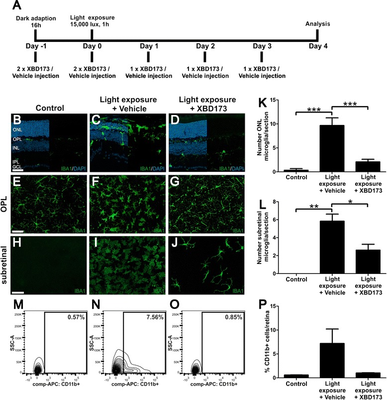
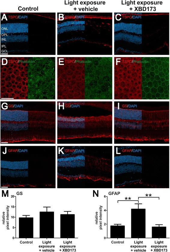
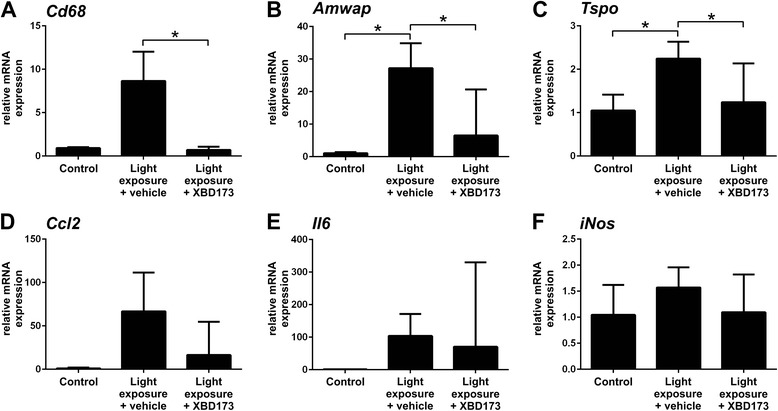
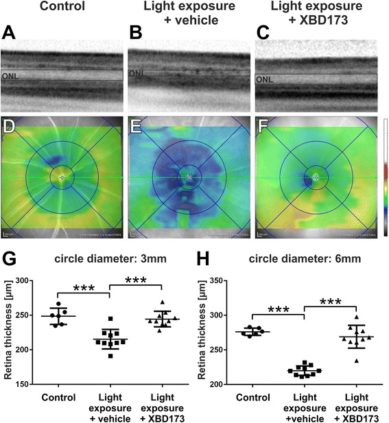
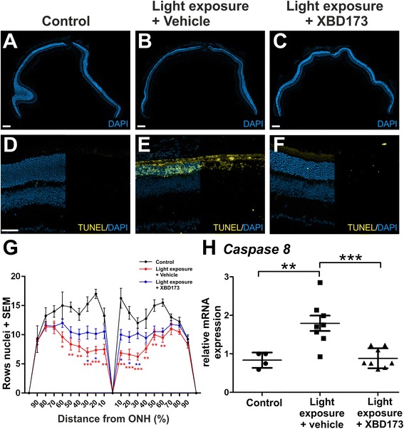
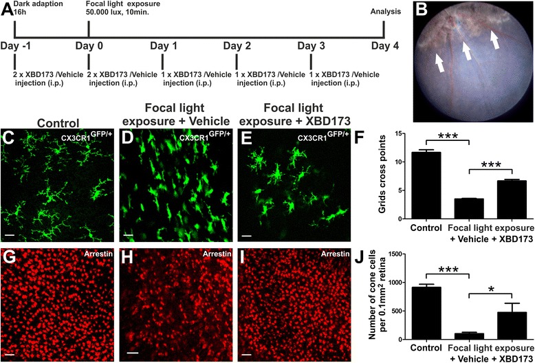
BACKGROUND: Reactive microglia are commonly seen in retinal degenerative diseases, and neurotoxic microglia responses can contribute to photoreceptor cell death. We and others have previously shown that translocator protein (18 kDa) (TSPO) is highly induced in retinal degenerations and that the selective TSPO ligand XBD173 (AC-5216, emapunil) exerts strong anti-inflammatory effects on microglia in vitro and ex vivo. Here, we investigated whether targeting TSPO with XBD173 has immuno-modulatory and neuroprotective functions in two mouse models of acute retinal degeneration using bright white light exposure. METHODS: BALB/cJ and Cx3cr1(GFP/+) mice received intraperitoneal injections of 10 mg/kg XBD173 or vehicle for five consecutive days, starting 1 day prior to exposure to either 15,000 lux white light for 1 h or 50,000 lux focal light for 10 min, respectively. The effects of XBD173 treatment on microglia and Müller cell reactivity were analyzed by immuno-stainings of retinal sections and flat mounts, fluorescence-activated cell sorting (FACS) analysis, and mRNA expression of microglia markers using quantitative real-time PCR (qRT-PCR). Optical coherence tomography (OCT), terminal deoxynucleotidyl transferase dUTP nick end labeling (TUNEL) stainings, and morphometric analyses were used to quantify the extent of retinal degeneration and photoreceptor apoptosis. RESULTS: Four days after the mice were challenged with bright white light, a large number of amoeboid-shaped alerted microglia appeared in the degenerating outer retina, which was nearly completely prevented by treatment with XBD173. This treatment also down-regulated the expression of TSPO protein in microglia but did not change the TSPO levels in the retinal pigment epithelium (RPE). RT-PCR analysis showed that the microglia/macrophage markers Cd68 and activated microglia/macrophage whey acidic protein (Amwap) as well as the pro-inflammatory genes Ccl2 and Il6 were reduced after XBD173 treatment. Light-induced degeneration of the outer retina was nearly fully blocked by XBD173 treatment. We further confirmed these findings in an independent mouse model of focal light damage. Retinas of animals receiving XBD173 therapy displayed significantly more ramified non-reactive microglia and more viable arrestin-positive cone photoreceptors than vehicle controls. CONCLUSIONS: Targeting TSPO with XBD173 effectively counter-regulates microgliosis and ameliorates light-induced retinal damage, highlighting a new pharmacological concept for the treatment of retinal degenerations.
Overview
The claims and statistics of this paper haven’t been checked yet.
Run the check — free, no account needed. It reads the open-access text and checks the sources it cites.
Integrity signals
Registry
Cites
Statistics
Figures
Verification
Claims the paper makes
The claims and statistics of this paper haven’t been checked yet.
Run the check — free, no account needed. It reads the open-access text and checks the sources it cites.
Claims other papers make about this one
Each is checked against this paper’s own text: does it say what the citing paper says it does?
In the citing paper
“Furthermore, increased TSPO expression may not be indicative of the inflammatory phenotype of microglia (Scholz et al, 2015).”
From Microglia Priming with Aging and Stress (2017) · cites this paper as Scholz et al, 2015
Self-check
Percentages, sums and reported differences re-computed from the paper’s own numbers.
The self-check needs the paper’s full text.
Citations
Cites
Resting microglial cells are highly dynamic surveillants of brain parenchyma in vivo
Nimmerjahn, A., Kirchhoff, F., & Helmchen, F. (2005) · Science · doi:10.1126/science.1110647 · Cited by 5,827
Kettenmann, H., Hanisch, U.-K., Noda, M., & Verkhratsky, A. (2011) · Physiological Reviews · doi:10.1152/physrev.00011.2010 · Cited by 3,547
Inflammation in neurodegenerative diseases
Amor, S., Puentes, F., Baker, D., & Van Der Valk, P. (2010) · Immunology · doi:10.1111/j.1365-2567.2009.03225.x · Cited by 1,355
Aloisi, F. (2001) · Glia · doi:10.1002/glia.1106 · Cited by 1,310
Wu, D. C., Jackson-Lewis, V., Vila, M., Tieu, K., Teismann, P., Vadseth, C., Choi, D.-K., Ischiropoulos, H., & Przedborski, S. (2002) · The Journal of Neuroscience · doi:10.1523/jneurosci.22-05-01763.2002 · Cited by 1,183
Rupprecht, R., Papadopoulos, V., Rammes, G., Baghai, T. C., Fan, J., Akula, N., Groyer, G., Adams, D., & Schumacher, M. (2010) · Nature Reviews Drug Discovery · doi:10.1038/nrd3295 · Cited by 930
Microglia derived from aging mice exhibit an altered inflammatory profile
Sierra, A., Gottfried‐Blackmore, A. C., McEwen, B. S., & Bulloch, K. (2007) · Glia · doi:10.1002/glia.20468 · Cited by 688
Para-Inflammation in the Aging Retina
Xu, H., Chen, M., & Forrester, J. V. (2009) · Progress in Retinal and Eye Research · doi:10.1016/j.preteyeres.2009.06.001 · Cited by 659 · cited 1×
“of microglia physiology contribute to degenerative pathologies of the retina and the entire CNS [3, 5–14].”
BackgroundSelf-citationWenzel, A., Grimm, C., Samardzija, M., & Remé, C. E. (2004) · Progress in Retinal and Eye Research · doi:10.1016/j.preteyeres.2004.08.002 · Cited by 655
Self-citationGupta, N., Brown, K. E., & Milam, A. H. (2003) · Experimental Eye Research · doi:10.1016/s0014-4835(02)00332-9 · Cited by 582
Retinal microglia: Just bystander or target for therapy?
Karlstetter, M., Scholz, R., Rutar, M., Wong, W. T., Provis, J. M., & Langmann, T. (2014) · Progress in Retinal and Eye Research · doi:10.1016/j.preteyeres.2014.11.004 · Cited by 570
Self-citationTranslocator protein 18 kDa (TSPO): Molecular sensor of brain injury and repair
Chen, M.-K., & Guilarte, T. R. (2008) · Pharmacology & Therapeutics · doi:10.1016/j.pharmthera.2007.12.004 · Cited by 506
Self-citationAge-related alterations in the dynamic behavior of microglia
Damani, M. R., Zhao, L., Fontainhas, A. M., Amaral, J., Fariss, R. N., & Wong, W. T. (2011) · Aging Cell · doi:10.1111/j.1474-9726.2010.00660.x · Cited by 504
Microglia activation in retinal degeneration
Langmann, T. (2007) · Journal of Leukocyte Biology · doi:10.1189/jlb.0207114 · Cited by 496
Self-citationMicroglial phagocytosis of living photoreceptors contributes to inherited retinal degeneration
Zhao, L., Zabel, M. K., Wang, X., Ma, W., Shah, P., Fariss, R. N., Qian, H., Parkhurst, C. N., Gan, W., & Wong, W. T. (2015) · EMBO Molecular Medicine · doi:10.15252/emmm.201505298 · Cited by 457
Hume, D. A., Perry, V. H., & Gordon, S. (1983) · The Journal of Cell Biology · doi:10.1083/jcb.97.1.253 · Cited by 447
Nuclear imaging of neuroinflammation: a comprehensive review of [11C]PK11195 challengers
Chauveau, F., Boutin, H., Camp, N. V., Dollé, F., & Tavitian, B. (2008) · European Journal of Nuclear Medicine and Molecular Imaging · doi:10.1007/s00259-008-0908-9 · Cited by 419
Sunlight and Age-Related Macular Degeneration
Cruickshanks, K. J. (1993) · Archives of Ophthalmology · doi:10.1001/archopht.1993.01090040106042 · Cited by 401
Animal models of age related macular degeneration
Pennesi, M. E., Neuringer, M., & Courtney, R. J. (2012) · Molecular Aspects of Medicine · doi:10.1016/j.mam.2012.06.003 · Cited by 391 · cited 1×
“This model is also very useful for a quantitative correlation of microglial responses with processes of retinal degeneration [34–36].”
BackgroundDjebaı̈li, M., Guo, Q., Pettus, E. H., Hoffman, S. W., & Stein, D. G. (2005) · Journal of Neurotrauma · doi:10.1089/neu.2005.22.106 · Cited by 387
Parainflammation, Chronic Inflammation and Age-Related Macular Degeneration
Chen, M., & Xu, H. (2015) · Journal of Leukocyte Biology · doi:10.1189/jlb.3ri0615-239r · Cited by 381
Self-citationTranslocator protein (18 kD) as target for anxiolytics without benzodiazepine-like side effects
Rupprecht, R., Rammes, G., Eser, D., Baghai, T. C., Schüle, C., Nothdurfter, C., Troxler, T., Gentsch, C., Kalkman, H. O., Chaperon, F., et al. (2009) · Science · doi:10.1126/science.1175055 · Cited by 328
Sennlaub, F., Auvynet, C., Calippe, B., Lavalette, S., Poupel, L., Hu, S. J., Dominguez, E., Camelo, S., Levy, O., Guyon, E., et al. (2013) · EMBO Molecular Medicine · doi:10.1002/emmm.201302692 · Cited by 325
Youssef, P. N., Sheibani, N., & Albert, D. M. (2010) · Eye · doi:10.1038/eye.2010.149 · Cited by 318
Swaroop, A., Chew, E. Y., Bowes Rickman, C., & Abecasis, G. R. (2009) · Annual Review of Genomics and Human Genetics · doi:10.1146/annurev.genom.9.081307.164350 · Cited by 270
Djebaı̈li, M., Hoffman, S. W., & Stein, D. G. (2003) · Neuroscience · doi:10.1016/j.neuroscience.2003.09.023 · Cited by 256
Marc, R. E., Jones, B. W., Watt, C. B., Vázquez-Chona, F., Vaughan, D. K., & Organisciak, D. T. (2008) · PubMed Central · Cited by 255
Investigating Mitochondria as a Target for Treating Age-Related Macular Degeneration
Terluk, M. R., Kapphahn, R. J., Soukup, L. M., Gong, H., Gallardo, C., Montezuma, S. R., & Ferrington, D. A. (2015) · Journal of Neuroscience · doi:10.1523/jneurosci.0190-15.2015 · Cited by 253
Rhodopsin-mediated blue-light damage to the rat retina: effect of photoreversal of bleaching.
Grimm, C., Wenzel, A., Williams, T. P., Rol, P. O., Hafezi, F., & Remé, C. E. (2001) · PubMed · Cited by 232
Self-citationKarlstetter, M., Nothdurfter, C., Aslanidis, A., Moeller, K., Horn, F., Scholz, R., Neumann, H., Weber, B. H. F., Rupprecht, R., & Langmann, T. (2014) · Journal of Neuroinflammation · doi:10.1186/1742-2094-11-3 · Cited by 224
Self-citationKuhlmann, A. C., & Guilarte, T. R. (2000) · Journal of Neurochemistry · doi:10.1046/j.1471-4159.2000.0741694.x · Cited by 215
Wang, M., Wang, X., Zhao, L., Ma, W., Rodríguez, I. R., Fariss, R. N., & Wong, W. T. (2014) · Journal of Neuroscience · doi:10.1523/jneurosci.3153-13.2014 · Cited by 211
Molecular imaging of microglia/macrophages in the brain
Venneti, S., Lopresti, B. J., & Wiley, C. A. (2012) · Glia · doi:10.1002/glia.22357 · Cited by 194
Ligand for Translocator Protein Reverses Pathology in a Mouse Model of Alzheimer's Disease
Barron, A. M., García‐Segura, L. M., Caruso, D., Jayaraman, A., Lee, J.-W., Melcangi, R. C., & Pike, C. J. (2013) · Journal of Neuroscience · doi:10.1523/jneurosci.1350-13.2013 · Cited by 159
A TSPO ligand is protective in a mouse model of multiple sclerosis
Daugherty, D. J., Selvaraj, V., Chechneva, O. V., Liu, X., Pleasure, D., & Deng, W. (2013) · EMBO Molecular Medicine · doi:10.1002/emmm.201202124 · Cited by 158
Levy, O., Calippe, B., Lavalette, S., Hu, S. J., Raoul, W., Dominguez, E., Housset, M., Pâques, M., Sahel, J., Bemelmans, A., et al. (2015) · EMBO Molecular Medicine · doi:10.15252/emmm.201404524 · Cited by 143
The changing landscape in translocator protein (TSPO) function
Selvaraj, V., & Stocco, D. M. (2015) · Trends in Endocrinology and Metabolism · doi:10.1016/j.tem.2015.02.007 · Cited by 137
Maeda, J., Higuchi, M., Inaji, M., Ji, B., Haneda, E., Okauchi, T., Zhang, M., Suzuki, K., & Suhara, T. (2007) · Brain Research · doi:10.1016/j.brainres.2007.04.054 · Cited by 134
Is there a mitochondrial signaling complex facilitating cholesterol import?
Papadopoulos, V., Liu, J., & Culty, M. (2007) · Molecular and Cellular Endocrinology · doi:10.1016/j.mce.2006.12.004 · Cited by 129
Raoul, W., Auvynet, C., Camelo, S., Guillonneau, X., Feumi, C., Combadière, C., & Sennlaub, F. (2010) · Journal of Neuroinflammation · doi:10.1186/1742-2094-7-87 · Cited by 116
Yao, X.-L., Liu, J., Lee, E., Ling, G. S. F., & McCabe, J. T. (2005) · Journal of Neurotrauma · doi:10.1089/neu.2005.22.656 · Cited by 112
Retina-derived microglial cells induce photoreceptor cell death in vitro
Roque, R. S., Rosales, A. A., Jingjing, L., Agarwal, N., & Al-Ubaidi, M. R. (1999) · Brain Research · doi:10.1016/s0006-8993(99)01625-x · Cited by 101
Neuroprotection of Photoreceptors by Minocycline in Light-Induced Retinal Degeneration
Zhang, C., Lei, B., Lam, T. T., Yang, F. W., Sinha, D., & Tso, M. O. M. (2004) · Investigative Ophthalmology & Visual Science · doi:10.1167/iovs.03-1344 · Cited by 96
The Upregulation of Translocator Protein (18 kDa) Promotes Recovery from Neuropathic Pain in Rats
Wei, X., Wei, X., Chen, F., Zang, Y., Xin, W., Pang, R., Chen, Y., Jun, W., Li, Y.-Y., Shen, K., et al. (2013) · Journal of Neuroscience · doi:10.1523/jneurosci.0324-12.2013 · Cited by 93
Light Damage as a Model of Retinal Degeneration
Grimm, C., & Remé, C. E. (2012) · Methods in molecular biology · doi:10.1007/978-1-62703-080-9_6 · Cited by 75
Self-citationAxonal Regeneration and Neuroinflammation: Roles for the Translocator Protein 18 kDa
Girard, C., Liu, S., Adams, D., Lacroix, C., Sinéus, M., Boucher, C., Papadopoulos, V., Rupprecht, R., Schumacher, M., & Groyer, G. (2012) · Journal of Neuroendocrinology · doi:10.1111/j.1365-2826.2011.02215.x · Cited by 75
Nothdurfter, C., Rammes, G., Baghai, T. C., Schüle, C., Schumacher, M., Papadopoulos, V., & Rupprecht, R. (2011) · Journal of Neuroendocrinology · doi:10.1111/j.1365-2826.2011.02166.x · Cited by 74
Narimatsu, T., Ozawa, Y., Miyake, S., Kubota, S., Hirasawa, M., Nagai, N., Shimmura, S., & Tsubota, K. (2013) · Investigative Ophthalmology & Visual Science · doi:10.1167/iovs.12-11572 · Cited by 73
Lost in translocation: the functions of the 18-kD translocator protein
Gut, P., Zweckstetter, M., & Banati, R. B. (2015) · Trends in Endocrinology and Metabolism · doi:10.1016/j.tem.2015.04.001 · Cited by 72
Karlstetter, M., Walczak, Y., Weigelt, K., Ebert, S., den Brulle, J. V., Schwer, H., Fuchshofer, R., & Langmann, T. (2010) · The Journal of Immunology · doi:10.4049/jimmunol.0903300 · Cited by 70
Self-citationChen, M., Zhao, J., Luo, C., Pandi, S. P. S., Peñalva, R. G., Fitzgerald, D. C., & Xu, H. (2012) · Glia · doi:10.1002/glia.22315 · Cited by 66
Self-citationTranslocator protein (18 kDa) (TSPO) as a therapeutic target for anxiety and neurologic disorders
Nothdurfter, C., Baghai, T. C., Schüle, C., & Rupprecht, R. (2012) · European Archives of Psychiatry and Clinical Neuroscience · doi:10.1007/s00406-012-0352-5 · Cited by 60
Aslanidis, A., Karlstetter, M., Scholz, R., Fauser, S., Neumann, H., Fried, C., Pietsch, M., & Langmann, T. (2015) · Journal of Neuroinflammation · doi:10.1186/s12974-015-0296-6 · Cited by 53
Self-citationNeuroprotective actions of progesterone in an in vivo model of retinitis pigmentosa
Sánchez-Vallejo, V., Benlloch-Navarro, S., López‐Pedrajas, R., Romero, F. J., & Miranda, M. (2015) · Pharmacological Research · doi:10.1016/j.phrs.2015.06.019 · Cited by 49
Pathways of dehydroepiandrosterone formation in rat brain glia
Cascio, C., Brown, R. C., Liu, Y., Han, Z. J., Hales, D. B., & Papadopoulos, V. (2000) · The Journal of Steroid Biochemistry and Molecular Biology · doi:10.1016/s0960-0760(00)00163-1 · Cited by 43
Karlstetter, M., & Langmann, T. (2014) · Advances in experimental medicine and biology · doi:10.1007/978-1-4614-3209-8_27 · Cited by 41
Self-citationVeiga, S. J., Azcoitia, Í., & García‐Segura, L. M. (2005) · Neuroreport · doi:10.1097/01.wnr.0000179081.39659.7d · Cited by 41
Microglia derived IL-6 suppresses neurosphere generation from adult human retinal cell suspensions
Balasubramaniam, B., Carter, D. A., Mayer, E. J., & Dick, A. D. (2009) · Experimental Eye Research · doi:10.1016/j.exer.2009.06.019 · Cited by 39
Microglial phagocytosis of living photoreceptors contributes to inherited retinal degeneration
Zabel, M. K., Zhao, L., Wang, X., Ma, W., Fariss, R. N., Qian, H., Parkhurst, C. N., Gan, W., & Wong, W. T. (2015) · Investigative Ophthalmology & Visual Science · Cited by 18
Schubert, T., Gleiser, C., Heiduschka, P., Franz, C., Nagel‐Wolfrum, K., Sahaboglu, A., Weisschuh, N., Eske, G., Rohbock, K., Rieger, N., et al. (2015) · Cellular and Molecular Life Sciences · doi:10.1007/s00018-015-1913-3 · Cited by 11
Cited by
Sorted by citations
Microglia Priming with Aging and Stress
Niraula, A., Sheridan, J. F., & Godbout, J. P. (2017) · Neuropsychopharmacology · doi:10.1038/npp.2016.185 · Cited by 398
Microglia in Retinal Degeneration
Rashid, K., Akhtar‐Schäfer, I., & Langmann, T. (2019) · Frontiers in Immunology · doi:10.3389/fimmu.2019.01975 · Cited by 365
Self-citationMicroglia and Inflammatory Responses in Diabetic Retinopathy
Kinuthia, U. M., Wolf, A., & Langmann, T. (2020) · Frontiers in Immunology · doi:10.3389/fimmu.2020.564077 · Cited by 332
Self-citationXu, G.-T., Zhang, J., & Tang, L. (2022) · Neural Regeneration Research · doi:10.4103/1673-5374.355743 · Cited by 293
Retinal microglia – A key player in healthy and diseased retina
Rathnasamy, G., Foulds, W. S., Ling, E., & Kaur, C. (2018) · Progress in Neurobiology · doi:10.1016/j.pneurobio.2018.05.006 · Cited by 250
Metadata
- Authors
- Scholz, R., Caramoy, A., Bhuckory, M. B., Rashid, K., Chen, M., Xu, H., Grimm, C., & Langmann, T.
- Affiliations
- University of Cologne (DE)
- Queen's University Belfast (GB)
- University of Zurich (CH)
- Corresponding author
- Langmann, Thomas
- Venue
- Journal of Neuroinflammation
- Published
- 2015-11-02
- Volume
- 12
- Issue
- 1
- Pages
- 201-201
- ISSN
- 1742-2094 (ISSN-L)
- PMC
- PMC4630900
- OpenAlex
- W1944096616
- Cited by
- 111
- References
- 60
- Citations by year
- 2026: 5 · 2025: 5 · 2024: 11 · 2023: 5 · 2022: 12 · 2021: 18 · 2020: 10 · 2019: 11 · 2018: 13 · 2017: 13 · 2016: 7 · 2015: 1
- FWCI
- 4.92
- Type
- journal article
- Language
- en
- Open access
- gold
- doi.org
- Licence
- cc-by
- Locations
- Journal of Neuroinflammation (publishedVersion, cc-by)
- Europe PMC (PubMed Central) (publishedVersion, cc-by)
- PubMed Central (publishedVersion, cc-by)
- PubMed (publishedVersion)
- Kölner Universitäts PublikationsServer (Universität zu Köln) (publishedVersion)
- Zurich Open Repository and Archive (University of Zurich) (acceptedVersion, cc-by)
- Research Portal (Queen's University Belfast) (submittedVersion, cc-by)
- Zurich Open Repository and Archive (University of Zurich) (submittedVersion, cc-by)
- Zurich Open Repository and Archive (University of Zurich)
- Funders
- Deutsche Forschungsgemeinschaft (LA1203/6-2, LA1203/9-1, LA1203/10-1, FOR2240)
- Velux Stiftung (Project 967)
- Hans und Marlies Stoock Foundation (S 061-10.013)
- Pro Retina Foundation (ProRetinaStip LA)
- Bayer (Bayer Graduate Program in Pharmacology)
- Fight for Sight UK (1425/1426)
- Topics
- Health Sciences › Medicine › Ophthalmology › Retinal Development and Disorders
- Life Sciences › Neuroscience › Neurology › Neuroinflammation and Neurodegeneration Mechanisms
- Health Sciences › Medicine › Ophthalmology › Glaucoma and retinal disorders
- Keywords
- translocator protein (TSPO), microglia, retinal degeneration, light-induced retinal damage, photoreceptor apoptosis, neuroprotection, anti-inflammatory activity, Müller cells, CX3CR1, MCP-1, interleukin-6, retinal pigment epithelium, choroidal neovascularization
- MeSH terms
- Animals, Disease Models, Animal, Female, Flow Cytometry, Immunohistochemistry, Inflammation, Male, Mice, Inbred BALB C, Mice, Inbred C57BL, Mice, Transgenic, Purines, Retinal Degeneration, Microglia, Receptors, GABA, Reverse Transcriptase Polymerase Chain Reaction, In Situ Nick-End Labeling, Tomography, Optical Coherence, Mice, Real-Time Polymerase Chain Reaction
About these checks
Registry, retraction, Cites and statistics results are computed, not judged, with one exception: a printed reference that no index lists is read once by a model before it counts as a citation error. Claims are extracted by a model and kept only when their quote appears verbatim in the text; each cited source is then checked, with a model’s help, for whether it says what the claim relies on. Those verdicts can be wrong — read the claim’s quote and the cited source before relying on one.
Statistics and arithmetic are recomputed, not judged.
Confidence bands are shown only for fields calibrated on our annotated test set. None is calibrated yet, so no band is shown.
What counts as a claim ›How we measure confidence ›How we check this ›
Are you an author, or do you see a problem with this page? Request a review
