

The DOI matches its Crossref record; 261 of the 261 references it cites were found in a registry or an index.
The semantics of microglia activation: neuroinflammation, homeostasis, and stress
Open accessAbstract
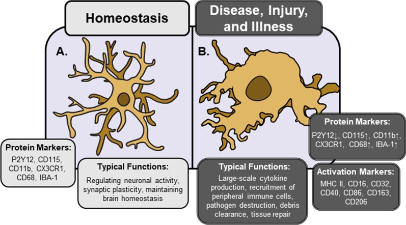
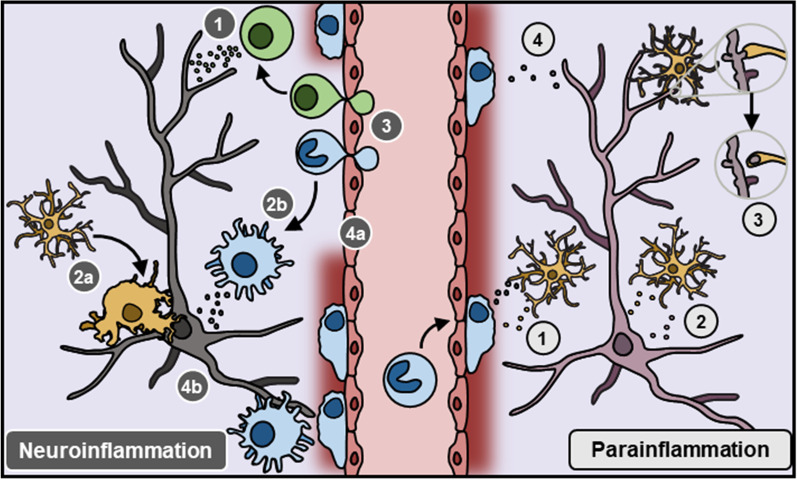
Microglia are emerging as critical regulators of neuronal function and behavior in nearly every area of neuroscience. Initial reports focused on classical immune functions of microglia in pathological contexts, however, immunological concepts from these studies have been applied to describe neuro-immune interactions in the absence of disease, injury, or infection. Indeed, terms such as 'microglia activation' or 'neuroinflammation' are used ubiquitously to describe changes in neuro-immune function in disparate contexts; particularly in stress research, where these terms prompt undue comparisons to pathological conditions. This creates a barrier for investigators new to neuro-immunology and ultimately hinders our understanding of stress effects on microglia. As more studies seek to understand the role of microglia in neurobiology and behavior, it is increasingly important to develop standard methods to study and define microglial phenotype and function. In this review, we summarize primary research on the role of microglia in pathological and physiological contexts. Further, we propose a framework to better describe changes in microglia1 phenotype and function in chronic stress. This approach will enable more precise characterization of microglia in different contexts, which should facilitate development of microglia-directed therapeutics in psychiatric and neurological disease.
Overview
The claims and statistics of this paper haven’t been checked yet.
Run the check — free, no account needed. It reads the open-access text and checks the sources it cites.
Integrity signals
Registry
Cites
Statistics
Figures
Verification
Claims the paper makes
The claims and statistics of this paper haven’t been checked yet.
Run the check — free, no account needed. It reads the open-access text and checks the sources it cites.
Claims other papers make about this one
Each is checked against this paper’s own text: does it say what the citing paper says it does?
In the citing paper
“The role of activated microglial cells is crucial in neuroinflammation since they can exert both neurotoxic and neuroprotective effects depending on the phenotype they acquire upon activation (Woodburn et al., 2021).”
From Exploring the Therapeutic Potential of Phytochemicals in Alzheimer’s Disease: Focus on Polyphenols and Monoterpenes (2022) · cites this paper as Woodburn et al., 2021
Self-check
Percentages, sums and reported differences re-computed from the paper’s own numbers.
The self-check needs the paper’s full text.
Citations
Cites
Amyloid oligomers exacerbate tau pathology in a mouse model of tauopathy
Selenica, M.-L. B., Brownlow, M., Jimenez, J. P., Lee, D. C., Pena, G., Dickey, C. A., Gordon, M. N., & Morgan, D. (2013) · Neurodegenerative Diseases · doi:10.1159/000337230 · Cited by 10,359
Polazzi, E., Mengoni, I., Caprini, M., Peña‐Altamira, E., Kurtys, E., & Monti, B. (2012) · Neurosignals · doi:10.1159/000337115 · Cited by 9,023
Molecular Mechanisms of Depression: Perspectives on New Treatment Strategies
Lang, U. E., & Borgwardt, S. (2013) · Cellular Physiology and Biochemistry · doi:10.1159/000350094 · Cited by 8,526
Glutamate neurotoxicity and diseases of the nervous system
Choi, D. W. (1988) · Neuron · doi:10.1016/0896-6273(88)90162-6 · Cited by 8,188
From inflammation to sickness and depression: when the immune system subjugates the brain
Dantzer, R., O'Connor, J. C., Freund, G. G., Johnson, R. W., & Kelley, K. W. (2008) · Nature Reviews Neuroscience · doi:10.1038/nrn2297 · Cited by 7,395
Origin and physiological roles of inflammation
Medzhitov, R. (2008) · Nature · doi:10.1038/nature07201 · Cited by 6,618
Macrophage activation and polarization: nomenclature and experimental guidelines
Murray, P. J., Allen, J. E., Biswas, S. K., Fisher, E. A., Gilroy, D. W., Goerdt, S., Gordon, S., Hamilton, J. A., Ivashkiv, L. B., Lawrence, T., et al. (2014) · Immunity · doi:10.1016/j.immuni.2014.06.008 · Cited by 6,337
Resting microglial cells are highly dynamic surveillants of brain parenchyma in vivo
Nimmerjahn, A., Kirchhoff, F., & Helmchen, F. (2005) · Science · doi:10.1126/science.1110647 · Cited by 5,827
A Unique Microglia Type Associated with Restricting Development of Alzheimer’s Disease
Keren-Shaul, H., Spinrad, A., Weiner, A., Matcovitch-Natan, O., Dvir-Szternfeld, R., Ulland, T. K., David, E., Baruch, K., Lara-Astaiso, D., Toth, B., et al. (2017) · Cell · doi:10.1016/j.cell.2017.05.018 · Cited by 5,493
Fate Mapping Analysis Reveals That Adult Microglia Derive from Primitive Macrophages
Ginhoux, F., Greter, M., Leboeuf, M., Nandi, S., See, P., Gokhan, S., Mehler, M. F., Conway, S. J., Ng, L. G., Stanley, E. R., et al. (2010) · Science · doi:10.1126/science.1194637 · Cited by 5,175
Cited by
Sorted by citations
Zeng, J., Bao, T., Yang, K., Zhu, X., Wang, S., Xiang, W., Ge, A., Zeng, L., & Ge, J. (2023) · Frontiers in Immunology · doi:10.3389/fimmu.2022.1047550 · Cited by 175
Martins-Ferreira, R., Calafell-Segura, J., Leal, B., Rodríguez-Ubreva, J., Martínez-Saez, E., Mereu, E., Pinho E Costa, P., Laguna, A., & Ballestar, E. (2025) · Nature Communications · doi:10.1038/s41467-025-56124-1 · Cited by 123
Piccialli, I., Tedeschi, V., Caputo, L., D’Errico, S., Ciccone, R., De Feo, V., Secondo, A., & Pannaccione, A. (2022) · Frontiers in Pharmacology · doi:10.3389/fphar.2022.876614 · Cited by 73
Zhao, Q., Chen, J., Wu, M., Yin, X., Jiang, Q., Gao, H., & Zheng, H. (2023) · Chemico-Biological Interactions · doi:10.1016/j.cbi.2023.110638 · Cited by 49
Gut Microbiota Restores Central Neuropeptide Deficits in Germ-Free Mice
Hamamah, S., & Covasa, M. (2022) · International Journal of Molecular Sciences · doi:10.3390/ijms231911756 · Cited by 25
Metadata
- Authors
- Woodburn, S. C., Bollinger, J. L., & Wohleb, E. S.
- Affiliations
- University of Cincinnati (US)
- Corresponding author
- Wohleb, Eric S.
- Venue
- Journal of Neuroinflammation
- Published
- 2021-11-06
- Volume
- 18
- Issue
- 1
- Pages
- 258-258
- ISSN
- 1742-2094 (ISSN-L)
- PMC
- PMC8571840
- OpenAlex
- W3211761706
- Cited by
- 783
- References
- 261
- Citations by year
- 2026: 138 · 2025: 244 · 2024: 204 · 2023: 142 · 2022: 53 · 2021: 1
- FWCI
- 11.80
- Type
- journal article
- Language
- en
- Open access
- gold
- doi.org
- Locations
- Journal of Neuroinflammation (publishedVersion, cc-by)
- Europe PMC (PubMed Central) (publishedVersion, other-oa)
- PubMed Central (publishedVersion, cc-by)
- PubMed (publishedVersion)
- DOAJ (DOAJ: Directory of Open Access Journals) (submittedVersion, cc-by-sa)
- Funders
- foundation for the national institutes of health (R21-MH120614, R01-MH-123545)
- brain and behavior research foundation (NARSAD Young Investigator Grant, 25488)
- college of medicine, university of cincinnati (University of Cincinnati Neurobiology Research Center Pilot Award)
- Topics
- Life Sciences › Neuroscience › Neurology › Neuroinflammation and Neurodegeneration Mechanisms
- Life Sciences › Neuroscience › Biological Psychiatry › Tryptophan and brain disorders
- Life Sciences › Neuroscience › Behavioral Neuroscience › Stress Responses and Cortisol
- Keywords
- microglial activation, neuroinflammation, neuroimmune interactions, chronic stress, microglial phenotype, homeostasis, stress-related psychiatric disorders, terminology standardization
- MeSH terms
- Neuroinflammatory Diseases, Animals, Homeostasis, Humans, Terminology as Topic, Semantics, Stress, Physiological, Microglia
About these checks
Registry, retraction, Cites and statistics results are computed, not judged, with one exception: a printed reference that no index lists is read once by a model before it counts as a citation error. Claims are extracted by a model and kept only when their quote appears verbatim in the text; each cited source is then checked, with a model’s help, for whether it says what the claim relies on. Those verdicts can be wrong — read the claim’s quote and the cited source before relying on one.
Statistics and arithmetic are recomputed, not judged.
Confidence bands are shown only for fields calibrated on our annotated test set. None is calibrated yet, so no band is shown.
What counts as a claim ›How we measure confidence ›How we check this ›
Are you an author, or do you see a problem with this page? Request a review
