

The DOI matches its Crossref record; 33 of the 33 references it cites were found in a registry or an index.
Development and validation of a simplified method to generate human microglia from pluripotent stem cells
Open accessAbstract
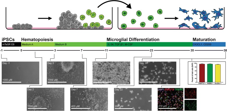
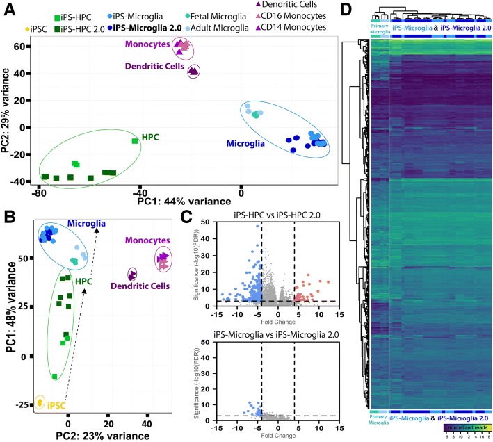
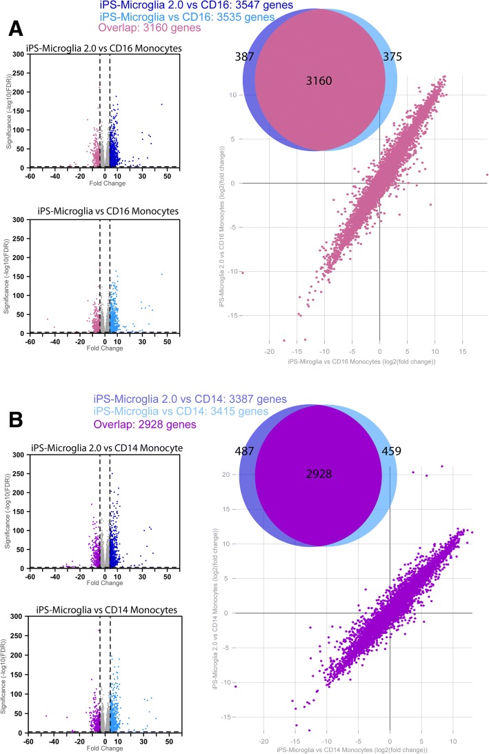
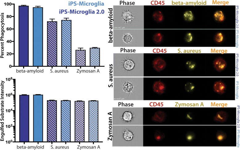
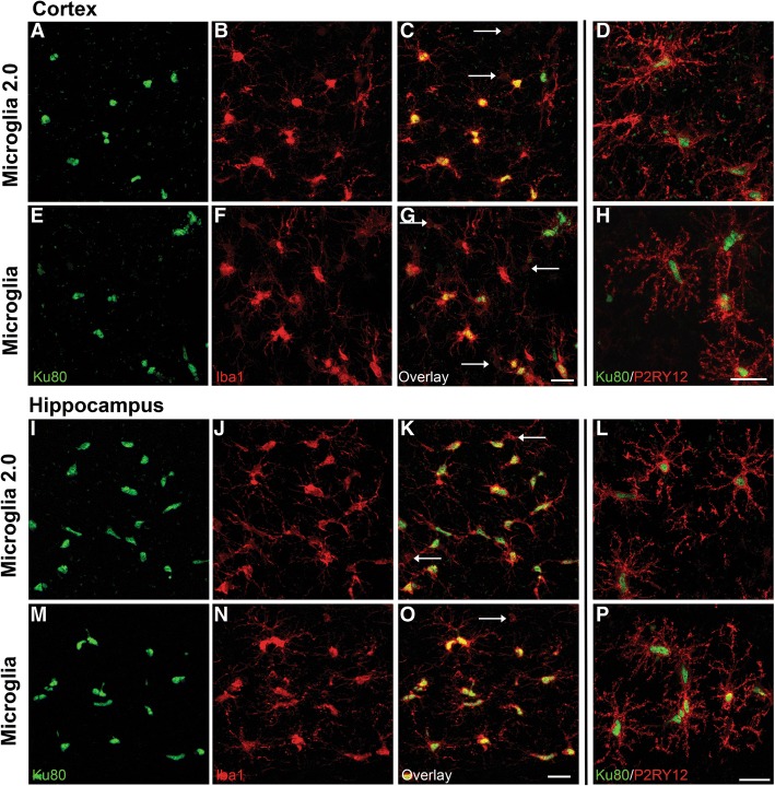
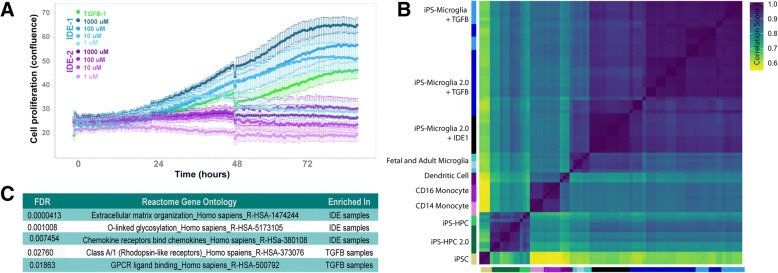
BACKGROUND: Microglia, the principle immune cells of the brain, play important roles in neuronal development, homeostatic function and neurodegenerative disease. Recent genetic studies have further highlighted the importance of microglia in neurodegeneration with the identification of disease risk polymorphisms in many microglial genes. To better understand the role of these genes in microglial biology and disease, we, and others, have developed methods to differentiate microglia from human induced pluripotent stem cells (iPSCs). While the development of these methods has begun to enable important new studies of microglial biology, labs with little prior stem cell experience have sometimes found it challenging to adopt these complex protocols. Therefore, we have now developed a greatly simplified approach to generate large numbers of highly pure human microglia. RESULTS: hematopoietic progenitors are then simply transferred to media that includes three key cytokines (M-CSF, IL-34, and TGFβ-1) that promote differentiation of homeostatic microglia. This updated approach avoids the prior requirement for hypoxic incubation, complex media formulation, FACS sorting, or co-culture, thereby significantly simplifying human microglial generation. To confirm that the resulting cells are equivalent to previously developed iPSC-microglia, we performed RNA-sequencing, functional testing, and transplantation studies. Our findings reveal that microglia generated via this simplified method are virtually identical to iPS-microglia produced via our previously published approach. To also determine whether a small molecule activator of TGFβ signaling (IDE1) can be used to replace recombinant TGFβ1, further reducing costs, we examined growth kinetics and the transcriptome of cells differentiated with IDE1. These data demonstrate that a microglial cell can indeed be produced using this alternative approach, although transcriptional differences do occur that should be considered. CONCLUSION: We anticipate that this new and greatly simplified protocol will enable many interested labs, including those with little prior stem cell or flow cytometry experience, to generate and study human iPS-microglia. By combining this method with other advances such as CRISPR-gene editing and xenotransplantation, the field will continue to improve our understanding of microglial biology and their important roles in human development, homeostasis, and disease.
Overview
The claims and statistics of this paper haven’t been checked yet.
Run the check — free, no account needed. It reads the open-access text and checks the sources it cites.
Integrity signals
Registry
Cites
Statistics
Figures
Verification
Claims the paper makes
The claims and statistics of this paper haven’t been checked yet.
Run the check — free, no account needed. It reads the open-access text and checks the sources it cites.
Claims other papers make about this one
Each is checked against this paper’s own text: does it say what the citing paper says it does?
The source backs part of the claim, not all of it — or backs it for a different population, condition or comparison.
In the citing paper
“Human astrocytes, expressing the astrocytic markers vimentin, S100β, ALDH1L1 and GFAP (Supplementary Figure 3 A), and human microglia, expressing the microglial markers Iba1 and P2Y12 (Supplementary Figure 3 B) were derived from the same hiPSC line [27].”
From Crosstalk between astrocytes and microglia results in increased degradation of α-synuclein and amyloid-β aggregates (2021) · cites this paper as [27]
Experimental — accuracy not yet measuredThe cited source says what the claim attributes to it.
In the citing paper
“E3 and E3S/S iPSCs were differentiated into microglia-like cells (iMGLs)43 (Fig. 6A), confirmed by the expression of microglia markers, IBA1 and TMEM119 (Fig. 6B).”
From APOE3 - R136S mutation confers resilience against tau pathology via cGAS-STING-IFN inhibition (2024) · cites this paper as [43]
The cited source says what the claim attributes to it.
In the citing paper
“HiPSCs were differentiated to hiPSC‐derived microglial cells (iMGL) according to the protocol previously published by McQuade et al., with minor adjustments.22,23”
The check wasn’t confident either way, so it gives no verdict — never disputed.
In the citing paper
“TREM2 deficiency leads to impaired microglial response to β-amyloid plaques, accumulation of the lipid droplets, and increased β-amyloid load in the brain [11, 41].”
From Protective PLCG2 variants associate with a delayed onset of Alzheimer’s disease among heterozygous APOE ε4 carriers (2026) · cites this paper as [41]
Experimental — accuracy not yet measuredThe check wasn’t confident either way, so it gives no verdict — never disputed.
In the citing paper
“AD patients exhibit myelin loss, and higher myelin expression is associated with tau pathology protection in AD43,44.”
From The R136S mutation in the APOE3 gene confers resilience against tau pathology via inhibition of the cGAS-STING-IFN pathway (2025) · cites this paper as [43]
Experimental — accuracy not yet measured
The check wasn’t confident either way, so it gives no verdict — never disputed.
In the citing paper
“higher levels of myelin are associated with protection against tau pathology in AD43,44”
From APOE3 - R136S mutation confers resilience against tau pathology via cGAS-STING-IFN inhibition (2024) · cites this paper as [43]
Experimental — accuracy not yet measuredThe check wasn’t confident either way, so it gives no verdict — never disputed.
In the citing paper
“AD patients exhibit increased myelin loss”
From APOE3 - R136S mutation confers resilience against tau pathology via cGAS-STING-IFN inhibition (2024) · cites this paper as [43]
Experimental — accuracy not yet measuredIn the citing paper
“This has resulted in highly efficient techniques to derive microglia-like cells from human induced pluripotent stem cells (iPSCs) [278, 279].”
From Genetic architecture of Alzheimer's disease (2020) · cites this paper as [278]
Self-check
Percentages, sums and reported differences re-computed from the paper’s own numbers.
The self-check needs the paper’s full text.
Citations
Cites
Moderated estimation of fold change and dispersion for RNA-seq data with DESeq2
Love, M. I., Huber, W., & Anders, S. (2014) · Genome Biology · doi:10.1186/s13059-014-0550-8 · Cited by 95,106
Near-optimal probabilistic RNA-seq quantification
Bray, N. L., Pimentel, H., Melsted, P., & Pachter, L. (2016) · Nature Biotechnology · doi:10.1038/nbt.3519 · Cited by 9,723
Neuroinflammation in Alzheimer's disease
Heneka, M. T., Carson, M. J., Khoury, J. E., Landreth, G. E., Brosseron, F., Feinstein, D. L., Jacobs, A. H., Wyss-Coray, T., Vitorica, J., Ransohoff, R. M., et al. (2015) · The Lancet Neurology · doi:10.1016/s1474-4422(15)70016-5 · Cited by 6,226
Identification of a unique TGF-β–dependent molecular and functional signature in microglia
Butovsky, O., Jedrychowski, M. P., Moore, C. S., Cialic, R., Lanser, A. J., Gabriely, G., Koeglsperger, T., Dake, B., Wu, P. M., Doykan, C. E., et al. (2014) · Nature Neuroscience · doi:10.1038/nn.3599 · Cited by 2,543
Elmore, M. R. P., Najafi, A. R., Koike, M. A., Dagher, N. N., Spangenberg, E. E., Rice, R. A., Kitazawa, M., Matusow, B., Nguyen, H., West, B. L., et al. (2014) · Neuron · doi:10.1016/j.neuron.2014.02.040 · Cited by 1,968
New tools for studying microglia in the mouse and human CNS
Bennett, M. L., Bennett, F. C., Liddelow, S. A., Ajami, B., Zamanian, J. L., Fernhoff, N. B., Mulinyawe, S. B., Bohlen, C. J., Adil, A., Tucker, A., et al. (2016) · Proceedings of the National Academy of Sciences · doi:10.1073/pnas.1525528113 · Cited by 1,804
An environment-dependent transcriptional network specifies human microglia identity
Gosselin, D., Skola, D., Coufal, N. G., Holtman, I. R., Schlachetzki, J. C. M., Sajti, E., Jaeger, B. N., O’Connor, C., Fitzpatrick, C., Pasillas, M. P., et al. (2017) · Science · doi:10.1126/science.aal3222 · Cited by 1,220
iPSC-derived human microglia-like cells to study neurological diseases
Abud, E. M., Ramirez, R. N., Martinez, E. S., Healy, L. M., Nguyen, C. H. H., Newman, S. A., Yeromin, A. V., Scarfone, V. M., Marsh, S. E., Fimbres, C., et al. (2017) · Neuron · doi:10.1016/j.neuron.2017.03.042 · Cited by 1,154
Self-citationDifferential roles of microglia and monocytes in the inflamed central nervous system
Yamasaki, R., Lu, H., Butovsky, O., Ohno, N., Rietsch, A. M., Cialic, R., Wu, P. M., Doykan, C. E., Lin, J., Cotleur, A. C., et al. (2014) · Journal of Experimental Medicine · doi:10.1084/jem.20132477 · Cited by 856
Friedman, B. A., Srinivasan, K., Ayalon, G., Meilandt, W. J., Lin, H., Huntley, M. A., Cao, Y., Lee, S.-H., Haddick, P. C. G., Ngu, H., et al. (2018) · Cell Reports · doi:10.1016/j.celrep.2017.12.066 · Cited by 662
Cited by
Sorted by citations
Items 301–400 of 466
Hu, M., Li, A. Z., Fleming, P., Gralla, J., Terón, K. I. N., Zhou, Y., Miller, E. J., Beyett, T. S., Wen, Z., Du, Y., et al. (2025) · Journal of Medicinal Chemistry · doi:10.1021/acs.jmedchem.5c01425 · Cited by 3
Reis, R. S., Ignatz, L. A., Selvam, S., Wagner, M. C. E., Gianella, S., Chaillon, A., Gomez‐Moreno, V., Martison, J., Rubin, L. H., Haberlen, S., et al. (2025) · Brain Pathology · doi:10.1111/bpa.70064 · Cited by 3
Navigating the frontier of human microglia isolation—standardization and translational insight
Pal, P., Sharma, M., & Gupta, S. K. (2026) · Methods · doi:10.1016/j.ymeth.2026.01.002 · Cited by 4
Lan, Z., Liu, H., Huang, C., Li, J. R., Dai, Z., Yuan, Y. Z., He, C., Qin, S., & Su, Z. (2026) · Neurochemistry International · doi:10.1016/j.neuint.2026.106170 · Cited by 4
Sakthivel, P. S., Scipioni, L., Karam, J. D., Keulen, Z., Blurton-Jones, M. M., Gratton, E., & Anderson, A. J. (2024) · Journal of Neurochemistry · doi:10.1111/jnc.16173 · Cited by 3
Self-citationDerivation of a Human Brain Organoid with Microglia Development
Wang, T., Gastfriend, B. D., McDonald, V., Steiner, J. P., Elkahloun, A. G., & Nath, A. (2025) · Journal of Visualized Experiments · doi:10.3791/67491 · Cited by 3
Juul‐Madsen, K., Rudolph, I., Gomes, J. P., Meyer, K., Ovesen, P. L., Gorniak‐Walas, M., Kokoli, M., Telugu, N. S., Sivertsen, M. V. T., Febbraro, F., et al. (2025) · Alzheimer s & Dementia · doi:10.1002/alz.70591 · Cited by 2
Improving mouse models for the study of Alzheimer's disease
Reagan, A. M., Onos, K. D., Heuer, S. E., Sasner, M., & Howell, G. R. (2022) · Current topics in developmental biology/Current Topics in Developmental Biology · doi:10.1016/bs.ctdb.2021.12.005 · Cited by 5
Siddiqui, A. M., Sabljic, T. F., & Ball, A. K. (2022) · International Journal of Neuroscience · doi:10.1080/00207454.2022.2142579 · Cited by 3
Targeting and eradicating latent CNS reservoirs of HIV-1: Original strategies and new models
Saeb, S., Wallet, C., Rohr, O., Schwartz, C., & Loustau, T. (2023) · Biochemical Pharmacology · doi:10.1016/j.bcp.2023.115679 · Cited by 6
Benoit, C. R., Sattler, L. B., Aylward, A. J., Pembridge, O. G., Kim, B., Muratore, C., Liao, M., He, A., Ashour, N., Fancher, S. B., et al. (2025) · Brain · doi:10.1093/brain/awaf221 · Cited by 3
CD33 and clusterin interact biophysically and genetically to modulate Alzheimer risk
Dodd, R. B., Enomoto, M., Zhou, Y., Satoh, K., Zhang, Y., Chen, F., Acheson, B., Ghaffari, D., Sayn-Wittgenstein, E., Manning, J. J., et al. (2026) · Nature Communications · doi:10.1038/s41467-026-75140-3 · Cited by 4
Aubert, A., Mendoza-Ferri, M. G., Bramoulle, A., Stüder, F., Colombo, B. M., & Mendoza-Parra, M. A. (2023) · bioRxiv (Cold Spring Harbor Laboratory) · doi:10.1101/2023.10.08.561397 · Cited by 3
Cell autonomous microglia defects in a stem cell model of frontotemporal dementiaPreprint
Iyer, A. K., Vermunt, L., Mirfakhar, F. S., Minaya, M., Acquarone, M., Koppisetti, R. K., Renganathan, A., You, S., Danhash, E., Verbeck, A. C., et al. (2024) · medRxiv · doi:10.1101/2024.05.15.24307444 · Cited by 3
Vicidomini, C., Goode, T. D., McAvoy, K. M., Yu, R., Beveridge, C. H., Iyer, S. N., Victor, M. B., Leary, N., Evans, L., Steinbaugh, M. J., et al. (2024) · bioRxiv (Cold Spring Harbor Laboratory) · doi:10.1101/2024.07.26.605338 · Cited by 4
Hammonds, J. E., Moscona, R., Candor, K., Guthier, H., Kumaragurubaran, R., Hagan, T., & Spearman, P. W. (2025) · bioRxiv (Cold Spring Harbor Laboratory) · doi:10.1101/2025.02.13.638028 · Cited by 5
Yeh, H., Cruz, M. A. D., You, Y., Ikezu, S., & Ikezu, T. (2025) · Scientific Reports · doi:10.1038/s41598-025-87543-1 · Cited by 2
Harkin, J., Gomes, C., Afify, R. A. A., Patil, S. V., Hetzer, S. M., Tutrow, K. D., Peña, K. H., Baker, A., Lavekar, S. S., Huang, K.-C., et al. (2026) · Proceedings of the National Academy of Sciences · doi:10.1073/pnas.2522958123 · Cited by 3
Dysregulated Expression of Inflammasome and Extracellular Matrix Genes in C9orf72 -ALS/FTD Microglia
Thiry, L., Pulimood, N. S., Tang, Y. M., & Stifani, S. (2025) · ASN NEURO · doi:10.1080/17590914.2025.2542998 · Cited by 4
Human microglia reduce alpha-synuclein aggregation and are neuroprotective in adult mouse brain
Albert, K., Peltonen, S., Vanne, A., Kälvälä, S., Syvänen, V., Koıstınaho, J., Luk, K. C., & Lehtonen, Š. (2025) · Brain Behavior and Immunity · doi:10.1016/j.bbi.2025.106097 · Cited by 3
Simha, V., LoPiccolo, M. K., Platt, A., Brown, R. J., Johnson, X., Carere, D. A., Donnelly, C. M., Snyder, M. T., Xing, C., Mathews, T. P., et al. (2025) · Journal of Clinical Investigation · doi:10.1172/jci198888 · Cited by 2
Robert, A., McQuade, A., Hertog, S. K.-D., Erlebach, L., Kronenberg‐Versteeg, D., Kampmann, M., Giera, M. A., & van der Kant, R. (2026) · Stem Cell Reports · doi:10.1016/j.stemcr.2025.102779 · Cited by 2
Self-citationQian, L., Rosa, J. S., & Julia, T. C. W. (2026) · STAR Protocols · doi:10.1016/j.xpro.2025.104339 · Cited by 2
Gao, S., Lin, Q., Liu, X., Jia, M., Zhang, A.-Y., Feng, Z., Han, L., Qiu, N., Liu, X., Zhai, H., et al. (2026) · Nature Communications · doi:10.1038/s41467-026-71922-x · Cited by 2
Miserocchi, M., Meyer, P. D., Lattuca, R., Montanari, A., Ferrero, G., Boezio, G. L. M., Stainier, D. Y. R., & Wittamer, V. (2026) · Cell Reports · doi:10.1016/j.celrep.2026.117795 · Cited by 2
Cholesterol dysregulation in APOE4 astrocytes promotes α-synuclein pathology in miBrains
Mesentier-Louro, L. A., Goldman, C., Gaese, S., Buonfiglioli, A., Kyriakis, D., Harlock, A., Ndayisaba, A., Sartori, E. R., Uchitelev, A., Fullard, J. F., et al. (2026) · Cell stem cell · doi:10.1016/j.stem.2026.08.001 · Cited by 6
An aging-sensitive secreted phospholipase confers neuroprotection and cognitive resilience
Vicidomini, C., Goode, T. D., Yu, R., Mehta, I. K., McAvoy, K. M., Beveridge, C. H., Randolph, C. E., Iyer, S. N., Victor, M. B., Leary, N., et al. (2026) · Nature Neuroscience · doi:10.1038/s41593-026-02465-z · Cited by 2
CIRM tools and technologies: Breaking bottlenecks to the development of stem cell therapies
Collins, L. R., & Shepard, K. A. (2020) · Stem Cells Translational Medicine · doi:10.1002/sctm.20-0055 · Cited by 2
Bipolar disorder as a gliopathy
Stertz, L. (2020) · Elsevier eBooks · doi:10.1016/b978-0-12-819182-8.00015-6 · Cited by 2
Glial cell transplant for brain diseases: the supportive saviours?
Albert, K., Goldsteins, G., Kälvälä, S., Jolkkonen, J., & Lehtonen, Š. (2024) · Translational Medicine Communications · doi:10.1186/s41231-024-00182-y · Cited by 3
Protocol to assess engulfment and degradation of synaptosomes by murine microglia in vitro
Matera, A., Compagnion, A., & Paolicelli, R. C. (2025) · STAR Protocols · doi:10.1016/j.xpro.2025.103936 · Cited by 3
Cellular and molecular neurobiology of autism spectrum disorder
Chapman, G., Antony, I., Narasimhan, M., Shen, R., Elman, R., & Kroll, K. L. (2022) · Elsevier eBooks · doi:10.1016/b978-0-12-816393-1.00007-5 · Cited by 2
Loss of TREM2 reduces hyperactivation of progranulin deficient microglia but not lysosomal pathologyPreprint
Reifschneider, A., Robinson, S., van Lengerich, B., Gnörich, J. S., Logan, T., Heindl, S., Vogt, M. A., Weidinger, E., Riedl, L., Wind-Mark, K., et al. (2021) · bioRxiv (Cold Spring Harbor Laboratory) · doi:10.1101/2021.07.08.451574 · Cited by 2
Dorion, M., Yaqubi, M., Murdoch, H. J., Hall, J. A., Dudley, R., Antel, J. P., Durcan, T. M., & Healy, L. M. (2022) · bioRxiv (Cold Spring Harbor Laboratory) · doi:10.1101/2022.07.14.500101 · Cited by 2
The A53T mutation in α-synuclein enhances pro-inflammatory activation in human microgliaPreprint
Krzisch, M., Yuan, B., Chen, W., Osaki, T., Fu, D., Garrett-Engele, C. M., Svoboda, D. S., Andrykovich, K. R., Sur, M., & Jaenisch, R. (2023) · bioRxiv (Cold Spring Harbor Laboratory) · doi:10.1101/2023.08.29.555300 · Cited by 2
ER and SOCE Ca 2+ signals are not required for directed cell migration in human microgliaPreprint
Granzotto, A., McQuade, A., Chadarevian, J. P., Davtyan, H., Sensi, S. L., Parker, I., Blurton-Jones, M. M., & Smith, I. F. (2024) · bioRxiv (Cold Spring Harbor Laboratory) · doi:10.1101/2024.01.18.576126 · Cited by 2
Self-citationLazaro, O., Li, S., Carter, W., Awosika, O., Robertson, S., Hickey, B. E., Angus, S. P., House, A., Clapp, D. W., Qadir, A. S., et al. (2024) · bioRxiv (Cold Spring Harbor Laboratory) · doi:10.1101/2024.05.02.591952 · Cited by 2
Narasipura, S. D., Zayas, J. P., Ash, M. K., Reyes, A. F., Shull, T. L., Gambut, S., Schneider, J. R., Lorenzo-Redondo, R., Al‐Harthi, L., & Mamede, J. I. (2024) · bioRxiv (Cold Spring Harbor Laboratory) · doi:10.1101/2024.06.13.598579 · Cited by 2
A proteogenomic tool uncovers protein markers for human microglial statesPreprint
Haage, V. C., Bautista, A. R., Tuddenham, J. F., Marshe, V. S., Chiu, R., Liu, Y., Lama, T., Kelly, S. S., Parghi, N., Park, J., et al. (2025) · bioRxiv (Cold Spring Harbor Laboratory) · doi:10.1101/2025.03.31.646212 · Cited by 3
Schuldt, B. R., Haworth-Staines, D., Arévalo, A. P., Broekaart, D. W. M., Harlock, A., Wang, L., Bright, A., Gallagher, G., Rabinowitz, G., Goate, A., et al. (2025) · bioRxiv (Cold Spring Harbor Laboratory) · doi:10.1101/2025.09.04.674192 · Cited by 2
Jeskanen, H., Heikkinen, S., Kervinen, I., Lehtisalo, J., Ngandu, T., Willman, R.-M., Rosa, J., Hoffmann, D., FinnGen, Leinonen, V., et al. (2026) · Alzheimer's Research & Therapy · doi:10.1186/s13195-026-01957-1 · Cited by 1
“TREM2 deficiency leads to impaired microglial response to β-amyloid plaques, accumulation of the lipid droplets, and increased β-amyloid load in the brain [11, 41].”
Establishing validity standards for iPSC modeling of neuropsychiatric disorders
Kolsters, N., Vernon, A. C., Kasri, N. N., & Latour, B. L. (2025) · Genomic psychiatry : · doi:10.61373/gp025p.0074 · Cited by 1
C1q-CD44 interactions regulate microglial phagocytosis, proliferation, and migration
Sakthivel, P. S., Villegas, A. J., Lakatos, A., Kaipa, M., Lopez, J. M., Ling, A., Elrachid, Z. H., Karam, J. D., & Anderson, A. J. (2026) · Journal of Neuroinflammation · doi:10.1186/s12974-026-03892-2 · Cited by 1
Protocol for the removal of bound endotoxin from tau protein produced in Escherichia coli
Shultz, A., Mena, D., Roy, J. L., Nulman, J., & Rauch, J. N. (2025) · STAR Protocols · doi:10.1016/j.xpro.2025.104177 · Cited by 1
Gomes, C. M., de Sá, I., Delgado, M., Alves, P. M., & Brito, C. (2025) · Biotechnology and Bioengineering · doi:10.1002/bit.70100 · Cited by 1
iPSC-derived microglia: Decoding roles and therapeutic opportunities in neurodegenerative diseases
Shan, D., Wang, H., Li, J., Tang, Y., Zhan, Z., Yan, C., Xu, J., & Liu, F. (2026) · Brain Research Bulletin · doi:10.1016/j.brainresbull.2026.111779 · Cited by 1
Komleva, Y. K., Satish, A., Berdnikov, A. K., Rozanova, N. A., Kolotyeva, N. A., Tregub, P. P., Saridis, M. R., Yurchenko, S. O., & Salmina, A. B. (2026) · The European Physical Journal Special Topics · doi:10.1140/epjs/s11734-026-02209-x · Cited by 2
MacDougall, E. J., Chen, C. X. ‐., Deneault, É., You, Z., Kalaydjian, D., Abdian, N., Durcan, T. M., Senkevich, K. A., Gan‐Or, Z., & Fon, E. A. (2026) · npj Parkinson s Disease · doi:10.1038/s41531-026-01310-1 · Cited by 3
Chi, L., Tu, H., Li, Z., Qiu, L., Zhang, Z., Chia, S., Tan, J. Y., Ho, I. A. W., Loh, Y., Tan, E., et al. (2026) · Cell Death Discovery · doi:10.1038/s41420-026-02995-2 · Cited by 1
Kim, D., Kondo, T., & Inoue, H. (2026) · Stem Cell Reports · doi:10.1016/j.stemcr.2026.102866 · Cited by 1
Pathogenic myeloid phenotypes drive disease pathology in a novel human neurohistiocytosis model
Rajamanickam, S., Trescott, S., Mak, S., Warden, A. S., Wilpitz, A., Xia, B., Nguyen, C. T., Ding, H., Picarsic, J. L., Glass, C. K., et al. (2026) · Blood · doi:10.1182/blood.2025032207 · Cited by 1
Xin, J., Liu, J., Dong, H.-M., Yu, H., Xiao, Z., Hu, J.-N., Jiang, Q., Zhu, Y.-P., Feng, L., Feng, Y., et al. (2026) · Science China Life Sciences · doi:10.1007/s11427-025-3243-2 · Cited by 2
Saunders, D. M., Kearns, N. A., Ng, B., Sultan, F. A., Vyas, H., Vialle, R. A., Clark, E. M., Tissera, S. L. D., Xu, J., Bennett, D. A. A., et al. (2026) · Stem Cell Research · doi:10.1016/j.scr.2026.104017 · Cited by 1
Disease-associated RNA and protein signatures in iPSC-derived microglia model of Alzheimer’s disease
Wu, W., Choi, E. S., Liu, L., Thamilselvan, V., LI, L., Rippee‐Brooks, M. D., Khatkar, K., Li, D.-Y., McGrath, D., Manning, A. C., et al. (2026) · Frontiers in Neuroscience · doi:10.3389/fnins.2026.1799542 · Cited by 1
HMC3 revealed: how much do these “Microglia” really tell us?
Buccarello, L., Privitera, A., Partsinevelos, K., Serwa, K., Carota, G., Pietro, L. D., Cardaci, V., Mangione, R., Sibbitts, J., Lazzarino, G., et al. (2026) · Frontiers in Immunology · doi:10.3389/fimmu.2026.1778798 · Cited by 2
Özata, G., Wise, R. M., Cardona‐Alberich, A., Mayeen, N. F., Müller, S. A., Lichtenthaler, S. F., Zecca, L., & Burbulla, L. F. (2026) · Scientific Reports · doi:10.1038/s41598-026-55648-w · Cited by 1
Neuroinflammation in glaucoma: a myriad of cellular pathways and players
MacLean, M., Lydon, S. D., Gomes, C., Pizzi, E. M., Diemler, C. A., Yablonski, S. E. R., Howell, G. R., Meyer, J. S., & Libby, R. T. (2026) · Mammalian Genome · doi:10.1007/s00335-026-10253-0 · Cited by 1
Emerging Regenerative Medicine for Spinal Cord Injury: Spinal Cord Organoids-on-a-Chip
Khan, M., Choi, H., Ahmad, S., Lee, S., Park, J., & Han, I. (2026) · International Journal of Molecular Sciences · doi:10.3390/ijms27157060 · Cited by 1
Mordelt, A., Schuurmans, I. M. E., Scheefhals, N., Hommersom, M. P., Slottje, K., Mast, K., Graziani, M., González, C. O., Mulder, K. W., Wingens, L., et al. (2026) · Stem Cell Reports · doi:10.1016/j.stemcr.2026.103053 · Cited by 5
Marques, E. T. A., Demers, M., D’Aiuto, L., Castanha, P. M. S., Yeung, J., Wood, J. A., Chowdari, K. V., Zheng, W., Yolken, R. H., & Nimgaonkar, V. L. (2022) · Current topics in behavioral neurosciences · doi:10.1007/7854_2022_383 · Cited by 1
Human models as new tools for drug development and precision medicine
Dei-Ampeh, A. K., Shah, M. M., Cappuccio, G., Young, D. W., & Maletić‐Savatić, M. (2022) · Elsevier eBooks · doi:10.1016/b978-0-12-822277-5.00004-3 · Cited by 1
Wu, J., & Tang, Y. (2023) · Ageing and Neurodegenerative Diseases · doi:10.20517/and.2023.01 · Cited by 1
Stem cell engineering approaches for investigating glial cues in central nervous system disorders
Vardhan, S., Jordan, T., & Sakiyama‐Elbert, S. E. (2024) · Current Opinion in Biotechnology · doi:10.1016/j.copbio.2024.103131 · Cited by 1
Lazaro, O., Li, S., Smiley, J. S., Carter, W., House, A., Awosika, O., Robertson, S., Haskell, A. K., Hinkel, R. D., Hickey, B. E., et al. (2026) · Scientific Reports · doi:10.1038/s41598-026-69205-y · Cited by 1
Roots of Progress: Uncovering Cerebellar Ataxias Using iPSC Models
Giacich, M., Naef, V., Santorelli, F. M., & Damiani, D. (2025) · Biomedicines · doi:10.3390/biomedicines13092121 · Cited by 2
Gene regulatory networks underlying human microglia maturationPreprint
Han, C. Z., Li, R. Z., Hansen, E. A., Bennett, H. R., Poirion, O., Buchanan, J., Challacombe, J. F., Fixsen, B. R., Trescott, S., Schlachetzki, J. C. M., et al. (2021) · bioRxiv (Cold Spring Harbor Laboratory) · doi:10.1101/2021.06.02.446636 · Cited by 1
MerTK mediates the immunologically silent uptake of alpha-synuclein fibrils by human microgliaPreprint
Dorion, M., Senkevich, K. A., Yaqubi, M., Kieran, N. W., Chen, C. X. ‐., MacDonald, A., Luo, W., Wallis, A., Shlaifer, I., Hall, J. A., et al. (2023) · bioRxiv (Cold Spring Harbor Laboratory) · doi:10.1101/2023.03.04.531108 · Cited by 1
Kwak, C. S., Finan, G. M., Park, Y. R., Garg, A., Harari, O., Mun, J. Y., Rhee, H., & Kim, T. (2023) · bioRxiv (Cold Spring Harbor Laboratory) · doi:10.1101/2023.03.21.533722 · Cited by 1
Niskanen, J., Peltonen, S., Ohtonen, S., Fazaludeen, M. F., Luk, K. C., Giudice, L., Koıstınaho, J., Malm, T., Goldsteins, G., Albert, K., et al. (2024) · Research Square · doi:10.21203/rs.3.rs-4021601/v2 · Cited by 1
GPCR-targeted imaging and manipulation of homeostatic microglia in living systemsPreprint
Chandrasekharan, S. V., Chaubey, P., Khandelwal, S., Medishetti, R., Chakraborty, S., Rajaram, S., Jayakumar, K., Latua, P., Goyal, R., Gopalakrishna, R., et al. (2025) · bioRxiv (Cold Spring Harbor Laboratory) · doi:10.1101/2025.04.01.646501 · Cited by 1
Rodríguez-Nunez, I., Bartley, S. C., Taylor, J. W., Johnston, S. Q., Rogers, B. B., Meadows, S. K., Newberry, K. M., Cochran, J. N., & Myers, R. (2025) · bioRxiv (Cold Spring Harbor Laboratory) · doi:10.1101/2025.05.30.657077 · Cited by 1
Tutrow, K. D., Harkin, J., Varghese, L., Hernandez, M., Huang, K.-C., Fang, Y., Wilcox, P., Bedford, L. M., Lin, T., Zhang, C., et al. (2025) · bioRxiv (Cold Spring Harbor Laboratory) · doi:10.1101/2025.07.01.662163 · Cited by 1
Saunders, D., Sultan, F. A., Vialle, R. A., Kearns, N. A., Ng, B., Clark, E. M., Vyas, H., Tissera, S. L. D., Xu, J., Bennett, D. A., et al. (2025) · bioRxiv (Cold Spring Harbor Laboratory) · doi:10.1101/2025.08.30.673269 · Cited by 1
LRRK2 Kinase Mediates Increased GCase Activity in Microglia in Response to Proinflammatory StimuliPreprint
MacDougall, E. J., Chen, C. X. ‐., Deneault, É., You, Z., Abdian, N., Durcan, T. M., Senkevich, K. A., Gan‐Or, Z., & Fon, E. A. (2025) · bioRxiv (Cold Spring Harbor Laboratory) · doi:10.1101/2025.10.10.681687 · Cited by 1
MEA-LINK identifies the CCL4-CCR5 axis in neuronal hyperactivity control by human microgliaPreprint
Mordelt, A., Scheefhals, N., Schuurmans, I. M. E., Slottje, K., Rivera, M. C., Hommersom, M. P., Huang, A., Bichmann, L., Lewerissa, E., van Hugte, E., et al. (2026) · bioRxiv (Cold Spring Harbor Laboratory) · doi:10.64898/2026.02.04.703799 · Cited by 1
A validated antibody toolbox for ALS researchPreprint
Ayoubi, R., MacDougall, E. J., McDowell, I., Biddle, M., Ferreira, B. T., Zha, C., Dorion, M., Ross, J. P., Bolívar, S. G., Moleón, V. R., et al. (2026) · eLife · doi:10.7554/elife.111349.1 · Cited by 1
Differentiation of iPSC into Microglia-Like Cells (iMGL) v1Preprint
Filipello, F., Marsh, J. A., Martinez, R., & Karch, C. M. M. (2020) · doi:10.17504/protocols.io.be43jgyn
Pessôa, L. V. D. F., Chandrasekaran, A., Thomsen, B. B., Berendt, M., Hyttel, P., & Freude, K. (2021) · Elsevier eBooks · doi:10.1016/b978-0-12-822228-7.00010-2
[Facioacromelic disostosis with metal retardation (author's transl)].
Cantú, J. M., Hernández, A., Nazará-Cazorla, Z., Pacheco, C., & Armendares, S. (1974) · PubMed
Sheridan, S. D., Horng, J. E., Yeh, H., McCrea, L. T., Fu, T., & Perlis, R. H. (2022) · bioRxiv (Cold Spring Harbor Laboratory) · doi:10.1101/2022.10.24.513576
Investigação do papel da Microglia derivada de iPSC no Transtorno do Espectro do Autismo.
Castanha, A. G. (2022) · Digital Library of Theses and Dissertations (Universidade de São Paulo) · doi:10.11606/d.42.2022.tde-18042022-145828
Timmerman, R. (2022) · Utrecht University Repository (Utrecht University) · doi:10.33540/1603
Dorion, M., Casas, D., Yaqubi, M., Fleming, P., Chen, C. X. ‐., Piscopo, V. E. C., Nicouleau, M., Goldsmith, T., Shlaifer, I., MacDonald, A., et al. (2023) · bioRxiv (Cold Spring Harbor Laboratory) · doi:10.1101/2023.09.08.556933
Denman, C. R., Tran, H., Shin, M., Ryu, J., Kühn, B., & Jo, J. (2023) · Stem Cell Research · doi:10.1016/j.scr.2023.103264
Haage, V. C., Tuddenham, J. F., Comandante-Lou, N., Bautista, A. R., Monzel, A. S., Chiu, R., Fujita, M., Garcia, F. G., Bhattarai, P., Patel, R., et al. (2024) · SSRN Electronic Journal · doi:10.2139/ssrn.4755485
Roles of Ipsc in Advancing Treatments for Neurological DisordersPreprint
Zhang, X. (2024) · SSRN Electronic Journal · doi:10.2139/ssrn.4818803
Sahay, A., Vicidomini, C., Goode, T. D., McAvoy, K. M., Yu, R., Beveridge, C. H., Iyer, S. N., Victor, M. B., Leary, N., Steinbaugh, M. J., et al. (2024) · Research Square · doi:10.21203/rs.3.rs-4920738/v1
Takalo, M., Jeskanen, H., Rõlova, T., Kervinen, I., Hellén, M., Heikkinen, S., Koivisto, H., Jokivarsi, K. T., Müller, S. A., Koivumäki, E.-M., et al. (2024) · bioRxiv (Cold Spring Harbor Laboratory) · doi:10.1101/2024.09.12.612207
Podleśny-Drabiniok, A., Romero-Molina, C., Patel, T., Wen, Y.-S., Liu, Y.-Y., Marcora, E., & Goate, A. (2024) · bioRxiv · doi:10.1101/2024.10.24.619910 · Cited by 7
Göttert, R., Vallone, V. B. F., Stachelscheid, H., Metzger, J. J., Caedo, C. C., Buthut, M., Prüß, H., Endres, M., Schilling, S., & Gertz, K. R. (2025) · Stem Cell Research · doi:10.1016/j.scr.2025.103660
SAMHD1 Knockout iPSC model enables high lenti-viral transduction in myeloid cell typesPreprint
Li, H., Afroze, M., Arora, G., Federman, S., Shevade, K., Yang, Y. A., Nguyen, P., Esanov, R., Przybyla, L., Litterman, A. J., et al. (2025) · bioRxiv (Cold Spring Harbor Laboratory) · doi:10.1101/2025.02.04.636295
Dysregulated expression of inflammasome and extracellular matrix genes in C9orf72 -ALS/FTD microgliaPreprint
Pulimood, N. S., Thiry, L., Tang, Y. M., & Stifani, S. (2025) · bioRxiv (Cold Spring Harbor Laboratory) · doi:10.1101/2025.02.13.638162
Hu, B., Shi, Y., Xiong, F., Chen, Y.-T., Zhu, X., Carrillo, E. C., Wen, X., Drolet, N. A., Rajpurohit, C. S., Xu, X., et al. (2025) · bioRxiv (Cold Spring Harbor Laboratory) · doi:10.1101/2025.03.22.644756
SAMHD1 knockout hiPSC model enables high lentiviral transduction efficiency in myeloid cell types
Li, H., Afroze, M., Arora, G., Federman, S., Shevade, K., Yang, Y. A., Nguyen, P., Esanov, R., Przybyla, L., Litterman, A. J., et al. (2025) · Frontiers in Genetics · doi:10.3389/fgene.2025.1574545
Juul‐Madsen, K., Rudolph, I., Gomes, J. P., Meyer, K., Ovesen, P. L., Górniak-Walas, M. J., Kokoli, M., Telugu, N. S., Sivertsen, M. V. T., Sutherland, D. S., et al. (2025) · bioRxiv (Cold Spring Harbor Laboratory) · doi:10.1101/2025.04.22.649924
A proteogenomic tool uncovers protein markers for human microglial statesPreprint
Jager, P. L. D., Haage, V. C., Bautista, A. R., Tuddenham, J. F., Marshe, V. S., Chiu, R., Liu, Y., Lama, T., Kelly, S. S., Parghi, N., et al. (2025) · Research Square · doi:10.21203/rs.3.rs-6380446/v1
Gao, S., Lin, Q., Liu, X., Jia, M., Zhang, A., Feng, Z., Han, L., Qiu, N., Zhai, H., Zhang, X., et al. (2025) · Research Square · doi:10.21203/rs.3.rs-6665030/v1
Emerging Human Choroid Plexus Organoid Models for Blood-CSF Barrier Research
Qiao, H., & Zhao, Z. (2025) · Neuromethods · doi:10.1007/978-1-0716-4474-4_4
Luo, Q., Chen, Y., Li, H., Long, Y., Shan, W., Zeng, X., Cai, S., Meng, Y., Wei, C., Xu, Y., et al. (2025) · Experimental & Molecular Medicine · doi:10.1038/s12276-025-01473-6
Gomes, C. M., Silva, G. P. D. L., Aleixo, M. M., Simão, D. F., Holtkamp, S., Lobo, D. D., Harish, P., Jenkins, R., Dahal, L. N., Nobre, R. J., et al. (2025) · bioRxiv (Cold Spring Harbor Laboratory) · doi:10.1101/2025.07.07.662807
Metadata
- Authors
- McQuade, A., Coburn, M., Tu, C. H., Hasselmann, J., Davtyan, H., & Blurton-Jones, M.
- Affiliations
- University of California, Irvine (US)
- Corresponding author
- Blurton-Jones, Mathew
- ORCID
- Venue
- Molecular Neurodegeneration
- Published
- 2018-12
- Volume
- 13
- Issue
- 1
- Pages
- 67-67
- ISSN
- 1750-1326 (ISSN-L)
- PMC
- PMC6303871
- OpenAlex
- W2905656859
- Cited by
- 449
- References
- 33
- Citations by year
- 2026: 64 · 2025: 98 · 2024: 73 · 2023: 53 · 2022: 54 · 2021: 43 · 2020: 40 · 2019: 23
- FWCI
- 11.92
- Type
- journal article
- Language
- en
- Open access
- gold
- doi.org
- Licence
- Locations
- Molecular Neurodegeneration (publishedVersion, cc-by)
- Europe PMC (PubMed Central) (publishedVersion, other-oa)
- PubMed Central (publishedVersion, cc-by)
- PubMed (publishedVersion)
- eScholarship (California Digital Library) (submittedVersion, cc-by)
- DOAJ (DOAJ: Directory of Open Access Journals) (submittedVersion)
- Funders
- National Institutes of Health (AG048099, AG056303, AG016573)
- California Institute for Regenerative Medicine (RT3-07893)
- National Institute of Neurological Disorders and Stroke (NS082174)
- Topics
- Life Sciences › Neuroscience › Neurology › Neuroinflammation and Neurodegeneration Mechanisms
- Life Sciences › Biochemistry, Genetics and Molecular Biology › Molecular Biology › Pluripotent Stem Cells Research
- Life Sciences › Neuroscience › Developmental Neuroscience › Neurogenesis and neuroplasticity mechanisms
- Keywords
- microglia differentiation, induced pluripotent stem cells, iPSC-derived microglia, hematopoietic progenitor cells, macrophage colony-stimulating factor, IL-34, TGF-β1, TGF-beta signaling, microglial transcriptome, neurodegeneration
- MeSH terms
- Animals, Brain, Cell Differentiation, Humans, Cytokines, Microglia, Pluripotent Stem Cells, Induced Pluripotent Stem Cells
About these checks
Registry, retraction, Cites and statistics results are computed, not judged, with one exception: a printed reference that no index lists is read once by a model before it counts as a citation error. Claims are extracted by a model and kept only when their quote appears verbatim in the text; each cited source is then checked, with a model’s help, for whether it says what the claim relies on. Those verdicts can be wrong — read the claim’s quote and the cited source before relying on one.
Statistics and arithmetic are recomputed, not judged.
Confidence bands are shown only for fields calibrated on our annotated test set. None is calibrated yet, so no band is shown.
What counts as a claim ›How we measure confidence ›How we check this ›
Are you an author, or do you see a problem with this page? Request a review
