

The DOI matches its Crossref record; 33 of the 33 references it cites were found in a registry or an index.
Development and validation of a simplified method to generate human microglia from pluripotent stem cells
Open accessAbstract
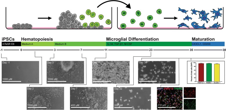
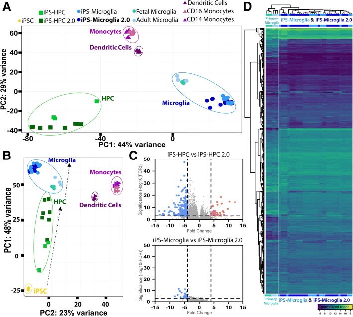
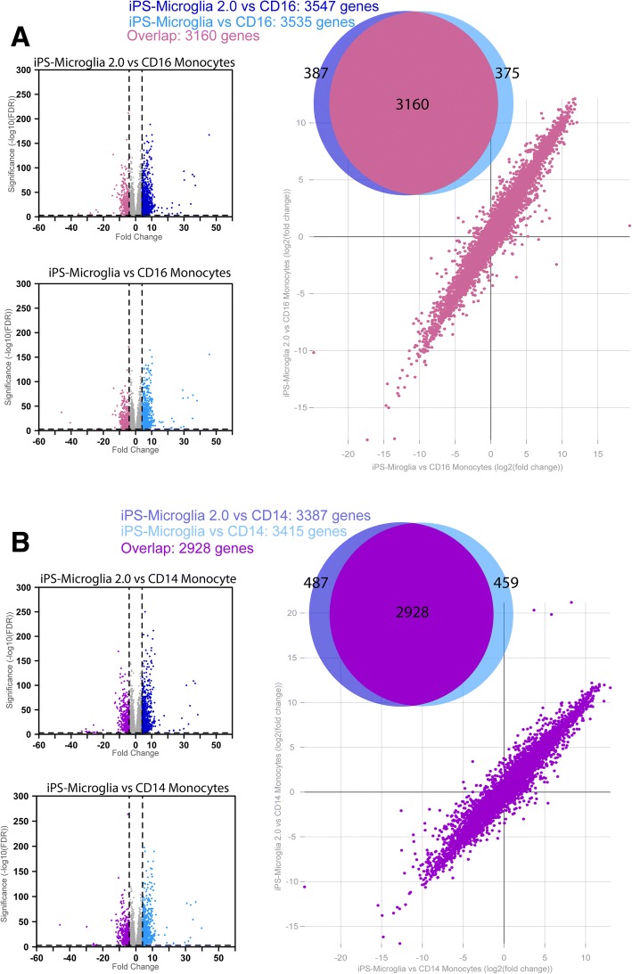
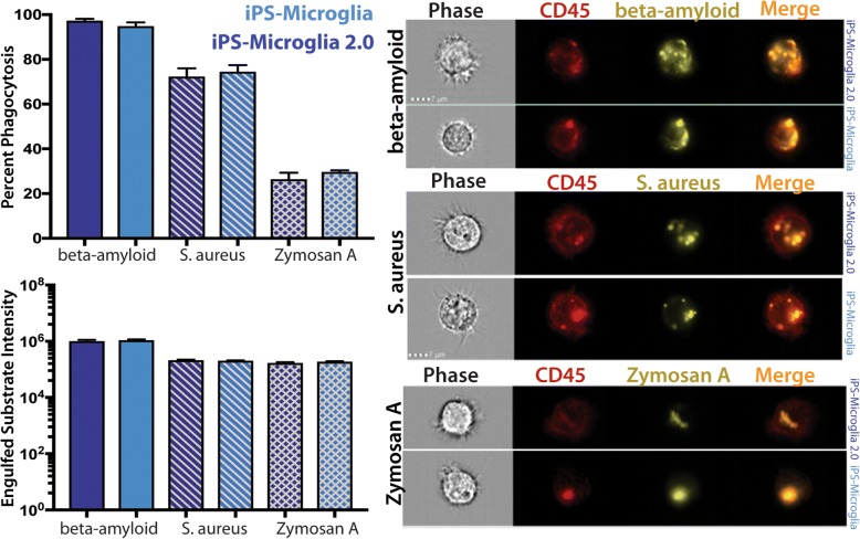
BACKGROUND: Microglia, the principle immune cells of the brain, play important roles in neuronal development, homeostatic function and neurodegenerative disease. Recent genetic studies have further highlighted the importance of microglia in neurodegeneration with the identification of disease risk polymorphisms in many microglial genes. To better understand the role of these genes in microglial biology and disease, we, and others, have developed methods to differentiate microglia from human induced pluripotent stem cells (iPSCs). While the development of these methods has begun to enable important new studies of microglial biology, labs with little prior stem cell experience have sometimes found it challenging to adopt these complex protocols. Therefore, we have now developed a greatly simplified approach to generate large numbers of highly pure human microglia. RESULTS: hematopoietic progenitors are then simply transferred to media that includes three key cytokines (M-CSF, IL-34, and TGFβ-1) that promote differentiation of homeostatic microglia. This updated approach avoids the prior requirement for hypoxic incubation, complex media formulation, FACS sorting, or co-culture, thereby significantly simplifying human microglial generation. To confirm that the resulting cells are equivalent to previously developed iPSC-microglia, we performed RNA-sequencing, functional testing, and transplantation studies. Our findings reveal that microglia generated via this simplified method are virtually identical to iPS-microglia produced via our previously published approach. To also determine whether a small molecule activator of TGFβ signaling (IDE1) can be used to replace recombinant TGFβ1, further reducing costs, we examined growth kinetics and the transcriptome of cells differentiated with IDE1. These data demonstrate that a microglial cell can indeed be produced using this alternative approach, although transcriptional differences do occur that should be considered. CONCLUSION: We anticipate that this new and greatly simplified protocol will enable many interested labs, including those with little prior stem cell or flow cytometry experience, to generate and study human iPS-microglia. By combining this method with other advances such as CRISPR-gene editing and xenotransplantation, the field will continue to improve our understanding of microglial biology and their important roles in human development, homeostasis, and disease.
Overview
The claims and statistics of this paper haven’t been checked yet.
Run the check — free, no account needed. It reads the open-access text and checks the sources it cites.
Integrity signals
Registry
Cites
Statistics
Figures
Verification
Claims the paper makes
The claims and statistics of this paper haven’t been checked yet.
Run the check — free, no account needed. It reads the open-access text and checks the sources it cites.
Claims other papers make about this one
Each is checked against this paper’s own text: does it say what the citing paper says it does?
The source backs part of the claim, not all of it — or backs it for a different population, condition or comparison.
In the citing paper
“Human astrocytes, expressing the astrocytic markers vimentin, S100β, ALDH1L1 and GFAP (Supplementary Figure 3 A), and human microglia, expressing the microglial markers Iba1 and P2Y12 (Supplementary Figure 3 B) were derived from the same hiPSC line [27].”
From Crosstalk between astrocytes and microglia results in increased degradation of α-synuclein and amyloid-β aggregates (2021) · cites this paper as [27]
Experimental — accuracy not yet measuredThe cited source says what the claim attributes to it.
In the citing paper
“E3 and E3S/S iPSCs were differentiated into microglia-like cells (iMGLs)43 (Fig. 6A), confirmed by the expression of microglia markers, IBA1 and TMEM119 (Fig. 6B).”
From APOE3 - R136S mutation confers resilience against tau pathology via cGAS-STING-IFN inhibition (2024) · cites this paper as [43]
The cited source says what the claim attributes to it.
In the citing paper
“HiPSCs were differentiated to hiPSC‐derived microglial cells (iMGL) according to the protocol previously published by McQuade et al., with minor adjustments.22,23”
The check wasn’t confident either way, so it gives no verdict — never disputed.
In the citing paper
“TREM2 deficiency leads to impaired microglial response to β-amyloid plaques, accumulation of the lipid droplets, and increased β-amyloid load in the brain [11, 41].”
From Protective PLCG2 variants associate with a delayed onset of Alzheimer’s disease among heterozygous APOE ε4 carriers (2026) · cites this paper as [41]
Experimental — accuracy not yet measuredThe check wasn’t confident either way, so it gives no verdict — never disputed.
In the citing paper
“AD patients exhibit myelin loss, and higher myelin expression is associated with tau pathology protection in AD43,44.”
From The R136S mutation in the APOE3 gene confers resilience against tau pathology via inhibition of the cGAS-STING-IFN pathway (2025) · cites this paper as [43]
Experimental — accuracy not yet measured
The check wasn’t confident either way, so it gives no verdict — never disputed.
In the citing paper
“higher levels of myelin are associated with protection against tau pathology in AD43,44”
From APOE3 - R136S mutation confers resilience against tau pathology via cGAS-STING-IFN inhibition (2024) · cites this paper as [43]
Experimental — accuracy not yet measuredThe check wasn’t confident either way, so it gives no verdict — never disputed.
In the citing paper
“AD patients exhibit increased myelin loss”
From APOE3 - R136S mutation confers resilience against tau pathology via cGAS-STING-IFN inhibition (2024) · cites this paper as [43]
Experimental — accuracy not yet measuredIn the citing paper
“This has resulted in highly efficient techniques to derive microglia-like cells from human induced pluripotent stem cells (iPSCs) [278, 279].”
From Genetic architecture of Alzheimer's disease (2020) · cites this paper as [278]
Self-check
Percentages, sums and reported differences re-computed from the paper’s own numbers.
The self-check needs the paper’s full text.
Citations
Cites
Moderated estimation of fold change and dispersion for RNA-seq data with DESeq2
Love, M. I., Huber, W., & Anders, S. (2014) · Genome Biology · doi:10.1186/s13059-014-0550-8 · Cited by 95,106
Near-optimal probabilistic RNA-seq quantification
Bray, N. L., Pimentel, H., Melsted, P., & Pachter, L. (2016) · Nature Biotechnology · doi:10.1038/nbt.3519 · Cited by 9,723
Neuroinflammation in Alzheimer's disease
Heneka, M. T., Carson, M. J., Khoury, J. E., Landreth, G. E., Brosseron, F., Feinstein, D. L., Jacobs, A. H., Wyss-Coray, T., Vitorica, J., Ransohoff, R. M., et al. (2015) · The Lancet Neurology · doi:10.1016/s1474-4422(15)70016-5 · Cited by 6,226
Identification of a unique TGF-β–dependent molecular and functional signature in microglia
Butovsky, O., Jedrychowski, M. P., Moore, C. S., Cialic, R., Lanser, A. J., Gabriely, G., Koeglsperger, T., Dake, B., Wu, P. M., Doykan, C. E., et al. (2014) · Nature Neuroscience · doi:10.1038/nn.3599 · Cited by 2,543
Elmore, M. R. P., Najafi, A. R., Koike, M. A., Dagher, N. N., Spangenberg, E. E., Rice, R. A., Kitazawa, M., Matusow, B., Nguyen, H., West, B. L., et al. (2014) · Neuron · doi:10.1016/j.neuron.2014.02.040 · Cited by 1,968
New tools for studying microglia in the mouse and human CNS
Bennett, M. L., Bennett, F. C., Liddelow, S. A., Ajami, B., Zamanian, J. L., Fernhoff, N. B., Mulinyawe, S. B., Bohlen, C. J., Adil, A., Tucker, A., et al. (2016) · Proceedings of the National Academy of Sciences · doi:10.1073/pnas.1525528113 · Cited by 1,804
An environment-dependent transcriptional network specifies human microglia identity
Gosselin, D., Skola, D., Coufal, N. G., Holtman, I. R., Schlachetzki, J. C. M., Sajti, E., Jaeger, B. N., O’Connor, C., Fitzpatrick, C., Pasillas, M. P., et al. (2017) · Science · doi:10.1126/science.aal3222 · Cited by 1,220
iPSC-derived human microglia-like cells to study neurological diseases
Abud, E. M., Ramirez, R. N., Martinez, E. S., Healy, L. M., Nguyen, C. H. H., Newman, S. A., Yeromin, A. V., Scarfone, V. M., Marsh, S. E., Fimbres, C., et al. (2017) · Neuron · doi:10.1016/j.neuron.2017.03.042 · Cited by 1,154
Self-citationDifferential roles of microglia and monocytes in the inflamed central nervous system
Yamasaki, R., Lu, H., Butovsky, O., Ohno, N., Rietsch, A. M., Cialic, R., Wu, P. M., Doykan, C. E., Lin, J., Cotleur, A. C., et al. (2014) · Journal of Experimental Medicine · doi:10.1084/jem.20132477 · Cited by 856
Friedman, B. A., Srinivasan, K., Ayalon, G., Meilandt, W. J., Lin, H., Huntley, M. A., Cao, Y., Lee, S.-H., Haddick, P. C. G., Ngu, H., et al. (2018) · Cell Reports · doi:10.1016/j.celrep.2017.12.066 · Cited by 662
Cited by
Sorted by citations
Items 401–466 of 466
Espinoza, A. P. V., Na, N., Campanati, L., Ngo, P., Baldwin, K. K., & Cline, H. T. (2025) · bioRxiv (Cold Spring Harbor Laboratory) · doi:10.1101/2025.08.03.668224
Özata, G., Wise, R. M., Cardona‐Alberich, A., Mayeen, N. F., Müller, S. A., Lichtenthaler, S. F., Zecca, L., & Burbulla, L. F. (2025) · bioRxiv (Cold Spring Harbor Laboratory) · doi:10.1101/2025.09.04.668126
A validated antibody toolbox for ALS research
Ayoubi, R., MacDougall, E. J., McDowell, I., Biddle, M., Ferreira, B. T., Zha, C., Dorion, M., Ross, J. P., Bolívar, S. G., Moleón, V. R., et al. (2026) · eLife · doi:10.1101/2025.09.14.676084
The protective PLCG2 variants delay Alzheimer's disease onset age in APOE ϵ4 carriersPreprint
Jeskanen, H., Heikkinen, S., Kervinen, I., Lehtisalo, J., Ngandu, T., Willman, R.-M., Rosa, J., Hoffmann, D., FinnGen, Leinonen, V., et al. (2025) · medRxiv · doi:10.1101/2025.09.22.25336304
Tiwari, S. K., Baidwan, G., Trescott, S., Jakhmola, S., Coufal, N. G., & Rana, T. M. (2025) · bioRxiv (Cold Spring Harbor Laboratory) · doi:10.64898/2025.12.06.692767
Alzheimer’s disease risk variant rs11218343 determines functional expression of SORL1 in microgliaPreprint
Gorniak-Walas, M., Telugu, N. S., Rudolph, I., Diecke, S., & Willnow, T. E. (2025) · bioRxiv (Cold Spring Harbor Laboratory) · doi:10.64898/2025.12.15.694337
Mulenge, F., Gern, O. L., Busker, L. M., Aringo, A., Ghita, L., Waltl, I., Pavlou, A., & Kalinke, U. (2024) · Figshare · doi:10.6084/m9.figshare.26778013
Additional file 10 of An integrated toolkit for human microglia functional genomics
Haq, I., Ngo, J. C., Roy, N., Pan, R., Nawsheen, N., Chiu, R., Zhang, Y., Fujita, M., Soni, R. K., Wu, X., et al. (2024) · Figshare · doi:10.6084/m9.figshare.26704734
Mulenge, F., Gern, O. L., Busker, L. M., Aringo, A., Ghita, L., Waltl, I., Pavlou, A., & Kalinke, U. (2024) · Figshare · doi:10.6084/m9.figshare.26778007
Additional file 6 of An integrated toolkit for human microglia functional genomics
Haq, I., Ngo, J. C., Roy, N., Pan, R., Nawsheen, N., Chiu, R., Zhang, Y., Fujita, M., Soni, R. K., Wu, X., et al. (2024) · Figshare · doi:10.6084/m9.figshare.26704722
Mulenge, F., Gern, O. L., Busker, L. M., Aringo, A., Ghita, L., Waltl, I., Pavlou, A., & Kalinke, U. (2024) · Figshare · doi:10.6084/m9.figshare.26778004
Additional file 4 of An integrated toolkit for human microglia functional genomics
Haq, I., Ngo, J. C., Roy, N., Pan, R., Nawsheen, N., Chiu, R., Zhang, Y., Fujita, M., Soni, R. K., Wu, X., et al. (2024) · Figshare · doi:10.6084/m9.figshare.26704716
Additional file 8 of An integrated toolkit for human microglia functional genomics
Haq, I., Ngo, J. C., Roy, N., Pan, R., Nawsheen, N., Chiu, R., Zhang, Y., Fujita, M., Soni, R. K., Wu, X., et al. (2024) · Figshare · doi:10.6084/m9.figshare.26704728
Quiroga, I. Y., Cruikshank, A. E., Bond, M. L., Reed, K. S. M., Evangelista, B., Tseng, J., Ragusa, J. V., Meeker, R. B., Won, H., Cohen, S., et al. (2022) · Figshare · doi:10.6084/m9.figshare.19641880
Additional file 3 of An integrated toolkit for human microglia functional genomics
Haq, I., Ngo, J. C., Roy, N., Pan, R., Nawsheen, N., Chiu, R., Zhang, Y., Fujita, M., Soni, R. K., Wu, X., et al. (2024) · Figshare · doi:10.6084/m9.figshare.26704713
Additional file 1 of An integrated toolkit for human microglia functional genomics
Haq, I., Ngo, J. C., Roy, N., Pan, R., Nawsheen, N., Chiu, R., Zhang, Y., Fujita, M., Soni, R. K., Wu, X., et al. (2024) · Figshare · doi:10.6084/m9.figshare.25583880
Dorion, M.-F., Casas, D., Shlaifer, I., Yaqubi, M., Fleming, P., Karpilovsky, N. C., Chen, C. X. ‐., Nicouleau, M., Piscopo, V. E. C., MacDougall, E. J., et al. (2024) · Figshare · doi:10.6084/m9.figshare.26702383
Dorion, M.-F., Casas, D., Shlaifer, I., Yaqubi, M., Fleming, P., Karpilovsky, N. C., Chen, C. X. ‐., Nicouleau, M., Piscopo, V. E. C., MacDougall, E. J., et al. (2024) · Figshare · doi:10.6084/m9.figshare.25546980
Mulenge, F., Gern, O. L., Busker, L. M., Aringo, A., Ghita, L., Waltl, I., Pavlou, A., & Kalinke, U. (2024) · Figshare · doi:10.6084/m9.figshare.26778010
Dorion, M.-F., Casas, D., Shlaifer, I., Yaqubi, M., Fleming, P., Karpilovsky, N. C., Chen, C. X. ‐., Nicouleau, M., Piscopo, V. E. C., MacDougall, E. J., et al. (2024) · Figshare · doi:10.6084/m9.figshare.26702377
Dorion, M.-F., Casas, D., Shlaifer, I., Yaqubi, M., Fleming, P., Karpilovsky, N. C., Chen, C. X. ‐., Nicouleau, M., Piscopo, V. E. C., MacDougall, E. J., et al. (2024) · Figshare · doi:10.6084/m9.figshare.25547144
Additional file 11 of An integrated toolkit for human microglia functional genomics
Haq, I., Ngo, J. C., Roy, N., Pan, R., Nawsheen, N., Chiu, R., Zhang, Y., Fujita, M., Soni, R. K., Wu, X., et al. (2024) · Figshare · doi:10.6084/m9.figshare.26704737
Additional file 9 of An integrated toolkit for human microglia functional genomics
Haq, I., Ngo, J. C., Roy, N., Pan, R., Nawsheen, N., Chiu, R., Zhang, Y., Fujita, M., Soni, R. K., Wu, X., et al. (2024) · Figshare · doi:10.6084/m9.figshare.26704731
Claes, C., Danhash, E., Hasselmann, J., Chadarevian, J. P., Shabestari, S. K., England, W., Lim, T. E., Hidalgo, J. L. S., Spitale, R. C., Davtyan, H., et al. (2021) · Figshare · doi:10.6084/m9.figshare.15047551
Self-citationDorion, M.-F., Casas, D., Shlaifer, I., Yaqubi, M., Fleming, P., Karpilovsky, N. C., Chen, C. X. ‐., Nicouleau, M., Piscopo, V. E. C., MacDougall, E. J., et al. (2024) · Figshare · doi:10.6084/m9.figshare.26702380
Pesämaa, I., Müller, S. A., Robinson, S., Darcher, A., Paquet, D., Zetterberg, H., Lichtenthaler, S. F., & Haass, C. (2024) · Figshare · doi:10.6084/m9.figshare.24223891
Additional file 5 of An integrated toolkit for human microglia functional genomics
Haq, I., Ngo, J. C., Roy, N., Pan, R., Nawsheen, N., Chiu, R., Zhang, Y., Fujita, M., Soni, R. K., Wu, X., et al. (2024) · Figshare · doi:10.6084/m9.figshare.26704719
Additional file 7 of An integrated toolkit for human microglia functional genomics
Haq, I., Ngo, J. C., Roy, N., Pan, R., Nawsheen, N., Chiu, R., Zhang, Y., Fujita, M., Soni, R. K., Wu, X., et al. (2024) · Figshare · doi:10.6084/m9.figshare.26704725
Sullivan, M. A., Lane, S. D., McKenzie, A. D. J., Ball, S. R., Sunde, M., Neely, G. G., Moreno, C. L., Maximova, A., Werry, E. L., & Kassiou, M. (2024) · Figshare · doi:10.6084/m9.figshare.24946342
Mulenge, F., Gern, O. L., Busker, L. M., Aringo, A., Ghita, L., Waltl, I., Pavlou, A., & Kalinke, U. (2024) · Figshare · doi:10.6084/m9.figshare.26778016
Additional file 2 of An integrated toolkit for human microglia functional genomics
Haq, I., Ngo, J. C., Roy, N., Pan, R., Nawsheen, N., Chiu, R., Zhang, Y., Fujita, M., Soni, R. K., Wu, X., et al. (2024) · Figshare · doi:10.6084/m9.figshare.25584031
Evitts, K. M., Turschak, E., Williams, C. A., Kinoshita, C., Rosner, A., Battista, W., Hui, K., Beck, I. H., Reid, A. N., ZHENG, Y., et al. (2025) · bioRxiv (Cold Spring Harbor Laboratory) · doi:10.64898/2025.12.19.695574
Exploring retinal microglia: development, degeneration, and iPSC-derived model systems
Gao, M.-L., Ju, Y.-Y., & Zhang, J. (2026) · Brain Structure and Function · doi:10.1007/s00429-025-03067-4
Comolli, D., Murari, E., Cerovic, M., Soltuzu, L., Liao, W., Bianchi, A., Seminara, S., Craparotta, I., Fumagalli, S., Balducci, C., et al. (2026) · Research Square · doi:10.21203/rs.3.rs-8549806/v1
Cell type-resolved proteomics reveals intra- and intercellular signaling in Alzheimer’s diseasePreprint
Zhang, X., Yu, K., Shrestha, H. K., Zhou, Y., Yang, J., Wang, Z., Ren, X., Chen, P.-C., Sun, H., Liu, D., et al. (2026) · bioRxiv (Cold Spring Harbor Laboratory) · doi:10.64898/2026.02.02.703357
In vitro and in vivo study of the effects of Levetiracetam on activated microglia during sepsis
Fleurance, R. (2021) · theses.fr (ABES) · doi:10.70675/0a82ed6ez6595z4366z832fz70cbe839c7ff
Vautheny, A. (2019) · theses.fr (ABES) · doi:10.70675/1812d71aze8d9z48d6z87d1z5a23a4e81953
Genome-scale metabolic modeling uncovers cell-type specific signatures associated with APOE variants
Uzuner, D., Stephenson, R. A., Cheng, L., Yang, L. G., Narayan, P. S., Çakır, T., & Thambisetty, M. (2026) · iScience · doi:10.1016/j.isci.2026.115638
Gorniak‐Walas, M., Telugu, N. S., Rudolph, I., Diecke, S., & Willnow, T. E. (2026) · Alzheimer s & Dementia · doi:10.1002/alz.71390
Macrophage heterogeneity influences cellular response to HIV infection and latency modulation
Wong, M. E., Rikard-Bell, L., Ebrahimnezhaddarzi, S., Brunton-O’Sullivan, M., Kouskousis, B., Ziemann, M., Jaworowski, A., & Hearps, A. C. (2026) · Journal of Leukocyte Biology · doi:10.1093/jleuko/qiag053
A validated antibody toolbox for ALS researchPreprint
Ayoubi, R., MacDougall, E. J., McDowell, I., Biddle, M., Ferreira, B. T., Zha, C., Dorion, M., Ross, J. P., Bolívar, S. G., Moleón, V. R., et al. (2026) · eLife · doi:10.7554/elife.111349
Rogers, B. B., Anderson, A. G., Rodríguez-Nunez, I., Bartley, S. C., Johnston, S. Q., Taylor, J. W., Meadows, S. K., Newberry, K. M., Myers, R., & Cochran, J. N. (2026) · iScience · doi:10.1016/j.isci.2026.116412 · Cited by 1
Ilse, V., Barendrecht, S., Hahndorf, J., Geißler, S., Hietel, B., Wagner, D., Staege, M. S., & Cynis, H. (2026) · Scientific Reports · doi:10.1038/s41598-026-61142-0
Reshaping the immune landscape: next-generation microglia-targeted therapies for Alzheimer’s disease
Wu, J., Zhao, J. J., Chen, S. Y., & Xu, F. (2026) · Biological Research · doi:10.1186/s40659-026-00716-8
The Kynurenine Pathway: Unraveling Its Role in Neurological Disorders via Mammalian Cellular Models
Podshivalova, E. S., Kutsev, S. I., & Shestopalov, A. V. (2026) · International Journal of Molecular Sciences · doi:10.3390/ijms27146337
CX3CR1-mediated extracellular Tau internalization and phagosomal proteins in iPSC-derived microglia
Narayanappa, T. S., Ananthanarayana, V., & Chinnathambi, S. (2026) · Methods in enzymology on CD-ROM/Methods in enzymology · doi:10.1016/bs.mie.2026.07.011
Air-Liquid interface midbrain organoids model the pathological features of Parkinson’s disease
Kälvälä, S., Abushik, P. A., Rezaie, M., Dougalis, A., Koıstınaho, J., Gardberg, M., Luk, K. C., Malm, T., Pelkonen, A., & Lehtonen, Š. (2026) · Neuroscience · doi:10.1016/j.neuroscience.2026.07.027
Optimization of a neuron-microglia co-culture model to explore cell-to-cell interaction dynamics
Vogiatzis, S., Severa, M., Pietrantoni, A., Cairo, G., Falco, R. P., Veroni, C., Lanciotti, A., Etna, M. P., Abate, D. A., Nencioni, L., et al. (2026) · Frontiers in Immunology · doi:10.3389/fimmu.2026.1850795
Retinal Pigment Epithelium and Microglia Transplantation in Age-related Macular Degeneration
Surawatsatien, N., Solanky, R. M., van de Werken, R. P., Kong, M., Diaconita, V., & Tsang, S. H. (2026) · American Journal of Ophthalmology · doi:10.1016/j.ajo.2026.07.036
Klimmt, J., Gonçalves, C. C., Montgomery, J. V., Müller, S. A., Bublitz, M., Filser, S., Paeger, L., Nuscher, B., Dannert, A., Roeber, S., et al. (2026) · Nature Neuroscience · doi:10.1038/s41593-026-02367-0 · Cited by 2
Sreeram, S., 陈雅, Bury, L. A. D., Leskov, K., Ye, F., García‐Mesa, Y., Luttge, B. G., Eum, J., 黄军友, Kallianpur, A. R., et al. (2026) · bioRxiv (Cold Spring Harbor Laboratory) · doi:10.64898/2026.08.10.743763
Assir, M. Z. K., Yanakiev, M., Gim, D. H., Valkila, S. S. M., Muscolino, P., Liu, P., Lecante, L. L., Fowler, P., Berg, D. A., & Kang, E. (2026) · Nature Neuroscience · doi:10.1038/s41593-026-02400-2 · Cited by 2
ARID5A RNA-binding coordinates microglial defense and ferroptosis in iPSC-derived models
Schafer, D. M., Blue, S. M., Xu, S., Mizrahi, O., Gosztyla, M. L., Street, L., Rothamel, K. L., Kopalle, H., Landry, S. B., Rosales, E. K. F., et al. (2026) · Nature Communications · doi:10.1038/s41467-026-76131-0
Nikolova, M. T., Freitag, K., Okamoto, R., Catellani, A., Hinke, J., Luginbühl, N., Lin, H., Kaindl, J., He, Z., Wahle, P., et al. (2026) · bioRxiv (Cold Spring Harbor Laboratory) · doi:10.64898/2026.09.25.754357
Peng, Y., Jin, C. J., Sun, Y., Mu, W., & Zheng, X. (2026) · Frontiers in Immunology · doi:10.3389/fimmu.2026.1898176
Ikezu, T., You, Y., Nguyen, S. H., Mann, S., Vadakattu, P., Kanekiyo, T., Zhang, Z., Ray, N. B., Tuck, T., Julia, T. C. W., et al. (2026) · Research Square · doi:10.21203/rs.3.rs-8594804/v1
Haage, V. C., Tuddenham, J. F., Comandante-Lou, N., Bautista, A. R., Monzel, A. S., Garcia, F. G., Chiu, R., Fujita, M., Bhattarai, P., White, C. C., et al. (2026) · Cell Reports · doi:10.1016/j.celrep.2026.118031
A pericyte-to-myofibroblast transition links APOE4 to cerebrovascular degeneration
Schuldt, B. R., Haworth-Staines, D., Arévalo, A. P., Broekaart, D. W. M., Harlock, A., Wang, L., Bright, A., Gallagher, G., Rabinowitz, G., Goate, A., et al. (2026) · Cell · doi:10.1016/j.cell.2026.08.058 · Cited by 2
Wong, H. T., Gleason, C., Boylan, A., Adler, M. J., Kosoy, R., Fullard, J. F., Levendosky, E., Hoffman, G. E., Molina, H., Roussos, P., et al. (2026) · bioRxiv (Cold Spring Harbor Laboratory) · doi:10.64898/2026.09.25.754430
A validated antibody toolbox for ALS researchPreprint
Ayoubi, R., MacDougall, E. J., McDowell, I., Biddle, M., Ferreira, B. T., Zha, C., Dorion, M., Ross, J. P., Bolívar, S. G., Moleón, V. R., et al. (2026) · eLife · doi:10.7554/elife.111349.2
Translational modelling of human microglia from cultures to chimeras
Lee, J., & Yoo, Y. (2026) · Experimental & Molecular Medicine · doi:10.1038/s12276-026-01866-1
Modeling human neurodegeneration with iPSCs: opportunities, limitations, and the challenge of aging
Wang, S., Elia, L., Liu, W., & Finkbeiner, S. M. (2026) · Journal of Clinical Investigation · doi:10.1172/jci209602
Heuer, S. E., Ahsan, F. M., Terzioğlu, G., Misra, A., Duong, D., Katkar, C., Giera, M. A., van der Kant, R., Seyfried, N. T., & Young-Pearse, T. L. (2026) · bioRxiv (Cold Spring Harbor Laboratory) · doi:10.64898/2026.10.16.759681
Harej, A., Rački, V., Belančić, A., Križ, J., Vitezić, D., Mršić‐Pelčić, J., Acharya, G., Varshney, M., & Pilipović, K. (2026) · Stem Cells International · doi:10.1155/sci/1130043
Endogenous versus exogeneous expression of Tau in iPSC-derived immune microglia
Ananthanarayana, V., Narayanappa, T. S., & Chinnathambi, S. (2026) · Advances in pharmacology · doi:10.1016/bs.apha.2026.08.007
Metadata
- Authors
- McQuade, A., Coburn, M., Tu, C. H., Hasselmann, J., Davtyan, H., & Blurton-Jones, M.
- Affiliations
- University of California, Irvine (US)
- Corresponding author
- Blurton-Jones, Mathew
- ORCID
- Venue
- Molecular Neurodegeneration
- Published
- 2018-12
- Volume
- 13
- Issue
- 1
- Pages
- 67-67
- ISSN
- 1750-1326 (ISSN-L)
- PMC
- PMC6303871
- OpenAlex
- W2905656859
- Cited by
- 449
- References
- 33
- Citations by year
- 2026: 64 · 2025: 98 · 2024: 73 · 2023: 53 · 2022: 54 · 2021: 43 · 2020: 40 · 2019: 23
- FWCI
- 11.92
- Type
- journal article
- Language
- en
- Open access
- gold
- doi.org
- Licence
- Locations
- Molecular Neurodegeneration (publishedVersion, cc-by)
- Europe PMC (PubMed Central) (publishedVersion, other-oa)
- PubMed Central (publishedVersion, cc-by)
- PubMed (publishedVersion)
- eScholarship (California Digital Library) (submittedVersion, cc-by)
- DOAJ (DOAJ: Directory of Open Access Journals) (submittedVersion)
- Funders
- National Institutes of Health (AG048099, AG056303, AG016573)
- California Institute for Regenerative Medicine (RT3-07893)
- National Institute of Neurological Disorders and Stroke (NS082174)
- Topics
- Life Sciences › Neuroscience › Neurology › Neuroinflammation and Neurodegeneration Mechanisms
- Life Sciences › Biochemistry, Genetics and Molecular Biology › Molecular Biology › Pluripotent Stem Cells Research
- Life Sciences › Neuroscience › Developmental Neuroscience › Neurogenesis and neuroplasticity mechanisms
- Keywords
- microglia differentiation, induced pluripotent stem cells, iPSC-derived microglia, hematopoietic progenitor cells, macrophage colony-stimulating factor, IL-34, TGF-β1, TGF-beta signaling, microglial transcriptome, neurodegeneration
- MeSH terms
- Animals, Brain, Cell Differentiation, Humans, Cytokines, Microglia, Pluripotent Stem Cells, Induced Pluripotent Stem Cells
About these checks
Registry, retraction, Cites and statistics results are computed, not judged, with one exception: a printed reference that no index lists is read once by a model before it counts as a citation error. Claims are extracted by a model and kept only when their quote appears verbatim in the text; each cited source is then checked, with a model’s help, for whether it says what the claim relies on. Those verdicts can be wrong — read the claim’s quote and the cited source before relying on one.
Statistics and arithmetic are recomputed, not judged.
Confidence bands are shown only for fields calibrated on our annotated test set. None is calibrated yet, so no band is shown.
What counts as a claim ›How we measure confidence ›How we check this ›
Are you an author, or do you see a problem with this page? Request a review
