

The DOI matches its Crossref record; 33 of the 33 references it cites were found in a registry or an index.
Development and validation of a simplified method to generate human microglia from pluripotent stem cells
Open accessAbstract
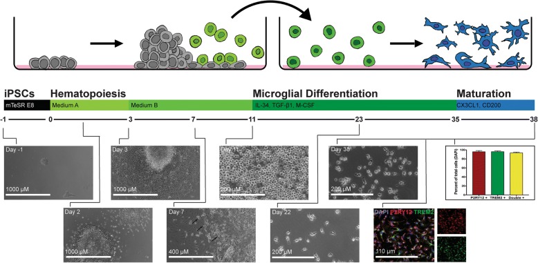
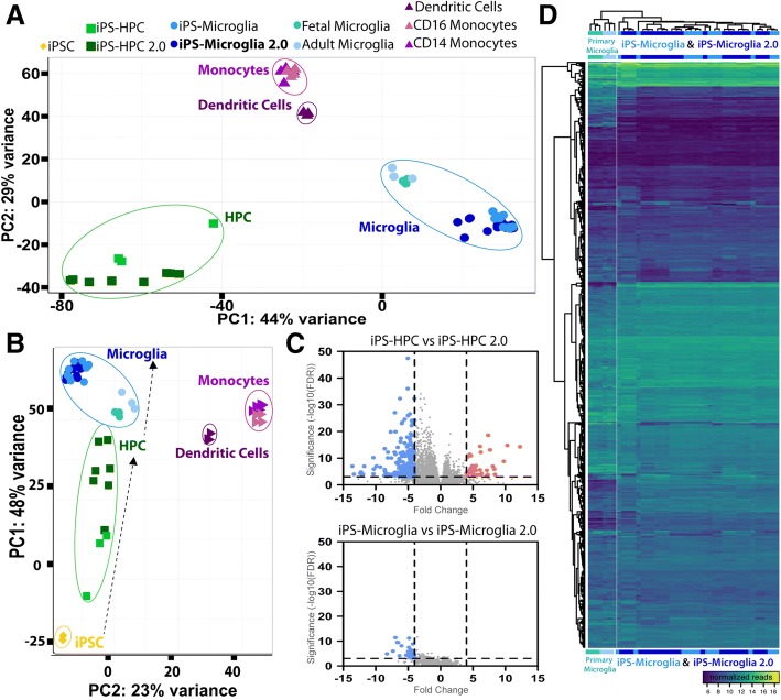
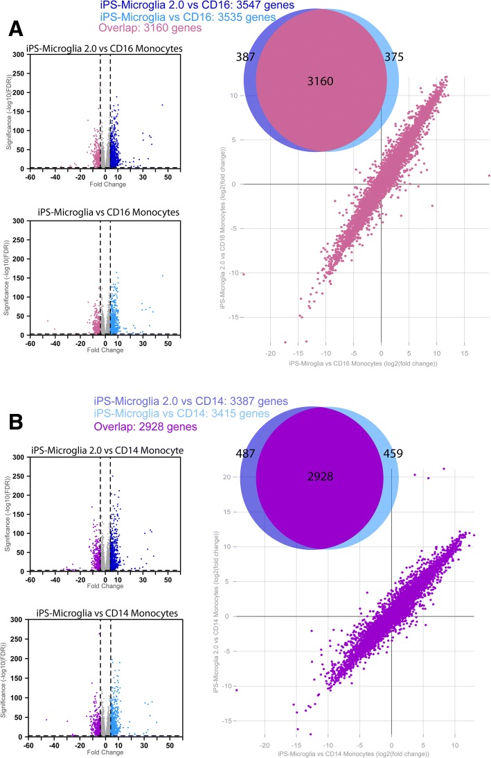
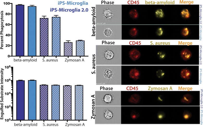
BACKGROUND: Microglia, the principle immune cells of the brain, play important roles in neuronal development, homeostatic function and neurodegenerative disease. Recent genetic studies have further highlighted the importance of microglia in neurodegeneration with the identification of disease risk polymorphisms in many microglial genes. To better understand the role of these genes in microglial biology and disease, we, and others, have developed methods to differentiate microglia from human induced pluripotent stem cells (iPSCs). While the development of these methods has begun to enable important new studies of microglial biology, labs with little prior stem cell experience have sometimes found it challenging to adopt these complex protocols. Therefore, we have now developed a greatly simplified approach to generate large numbers of highly pure human microglia. RESULTS: hematopoietic progenitors are then simply transferred to media that includes three key cytokines (M-CSF, IL-34, and TGFβ-1) that promote differentiation of homeostatic microglia. This updated approach avoids the prior requirement for hypoxic incubation, complex media formulation, FACS sorting, or co-culture, thereby significantly simplifying human microglial generation. To confirm that the resulting cells are equivalent to previously developed iPSC-microglia, we performed RNA-sequencing, functional testing, and transplantation studies. Our findings reveal that microglia generated via this simplified method are virtually identical to iPS-microglia produced via our previously published approach. To also determine whether a small molecule activator of TGFβ signaling (IDE1) can be used to replace recombinant TGFβ1, further reducing costs, we examined growth kinetics and the transcriptome of cells differentiated with IDE1. These data demonstrate that a microglial cell can indeed be produced using this alternative approach, although transcriptional differences do occur that should be considered. CONCLUSION: We anticipate that this new and greatly simplified protocol will enable many interested labs, including those with little prior stem cell or flow cytometry experience, to generate and study human iPS-microglia. By combining this method with other advances such as CRISPR-gene editing and xenotransplantation, the field will continue to improve our understanding of microglial biology and their important roles in human development, homeostasis, and disease.
Overview
The claims and statistics of this paper haven’t been checked yet.
Run the check — free, no account needed. It reads the open-access text and checks the sources it cites.
Integrity signals
Registry
Cites
Statistics
Figures
Verification
Claims the paper makes
The claims and statistics of this paper haven’t been checked yet.
Run the check — free, no account needed. It reads the open-access text and checks the sources it cites.
Claims other papers make about this one
Each is checked against this paper’s own text: does it say what the citing paper says it does?
The source backs part of the claim, not all of it — or backs it for a different population, condition or comparison.
In the citing paper
“Human astrocytes, expressing the astrocytic markers vimentin, S100β, ALDH1L1 and GFAP (Supplementary Figure 3 A), and human microglia, expressing the microglial markers Iba1 and P2Y12 (Supplementary Figure 3 B) were derived from the same hiPSC line [27].”
From Crosstalk between astrocytes and microglia results in increased degradation of α-synuclein and amyloid-β aggregates (2021) · cites this paper as [27]
Experimental — accuracy not yet measuredThe cited source says what the claim attributes to it.
In the citing paper
“E3 and E3S/S iPSCs were differentiated into microglia-like cells (iMGLs)43 (Fig. 6A), confirmed by the expression of microglia markers, IBA1 and TMEM119 (Fig. 6B).”
From APOE3 - R136S mutation confers resilience against tau pathology via cGAS-STING-IFN inhibition (2024) · cites this paper as [43]
The cited source says what the claim attributes to it.
In the citing paper
“HiPSCs were differentiated to hiPSC‐derived microglial cells (iMGL) according to the protocol previously published by McQuade et al., with minor adjustments.22,23”
The check wasn’t confident either way, so it gives no verdict — never disputed.
In the citing paper
“TREM2 deficiency leads to impaired microglial response to β-amyloid plaques, accumulation of the lipid droplets, and increased β-amyloid load in the brain [11, 41].”
From Protective PLCG2 variants associate with a delayed onset of Alzheimer’s disease among heterozygous APOE ε4 carriers (2026) · cites this paper as [41]
Experimental — accuracy not yet measuredThe check wasn’t confident either way, so it gives no verdict — never disputed.
In the citing paper
“AD patients exhibit myelin loss, and higher myelin expression is associated with tau pathology protection in AD43,44.”
From The R136S mutation in the APOE3 gene confers resilience against tau pathology via inhibition of the cGAS-STING-IFN pathway (2025) · cites this paper as [43]
Experimental — accuracy not yet measured
The check wasn’t confident either way, so it gives no verdict — never disputed.
In the citing paper
“higher levels of myelin are associated with protection against tau pathology in AD43,44”
From APOE3 - R136S mutation confers resilience against tau pathology via cGAS-STING-IFN inhibition (2024) · cites this paper as [43]
Experimental — accuracy not yet measuredThe check wasn’t confident either way, so it gives no verdict — never disputed.
In the citing paper
“AD patients exhibit increased myelin loss”
From APOE3 - R136S mutation confers resilience against tau pathology via cGAS-STING-IFN inhibition (2024) · cites this paper as [43]
Experimental — accuracy not yet measuredIn the citing paper
“This has resulted in highly efficient techniques to derive microglia-like cells from human induced pluripotent stem cells (iPSCs) [278, 279].”
From Genetic architecture of Alzheimer's disease (2020) · cites this paper as [278]
Self-check
Percentages, sums and reported differences re-computed from the paper’s own numbers.
The self-check needs the paper’s full text.
Citations
Cites
Moderated estimation of fold change and dispersion for RNA-seq data with DESeq2
Love, M. I., Huber, W., & Anders, S. (2014) · Genome Biology · doi:10.1186/s13059-014-0550-8 · Cited by 95,106
Near-optimal probabilistic RNA-seq quantification
Bray, N. L., Pimentel, H., Melsted, P., & Pachter, L. (2016) · Nature Biotechnology · doi:10.1038/nbt.3519 · Cited by 9,723
Neuroinflammation in Alzheimer's disease
Heneka, M. T., Carson, M. J., Khoury, J. E., Landreth, G. E., Brosseron, F., Feinstein, D. L., Jacobs, A. H., Wyss-Coray, T., Vitorica, J., Ransohoff, R. M., et al. (2015) · The Lancet Neurology · doi:10.1016/s1474-4422(15)70016-5 · Cited by 6,226
Identification of a unique TGF-β–dependent molecular and functional signature in microglia
Butovsky, O., Jedrychowski, M. P., Moore, C. S., Cialic, R., Lanser, A. J., Gabriely, G., Koeglsperger, T., Dake, B., Wu, P. M., Doykan, C. E., et al. (2014) · Nature Neuroscience · doi:10.1038/nn.3599 · Cited by 2,543
Elmore, M. R. P., Najafi, A. R., Koike, M. A., Dagher, N. N., Spangenberg, E. E., Rice, R. A., Kitazawa, M., Matusow, B., Nguyen, H., West, B. L., et al. (2014) · Neuron · doi:10.1016/j.neuron.2014.02.040 · Cited by 1,968
New tools for studying microglia in the mouse and human CNS
Bennett, M. L., Bennett, F. C., Liddelow, S. A., Ajami, B., Zamanian, J. L., Fernhoff, N. B., Mulinyawe, S. B., Bohlen, C. J., Adil, A., Tucker, A., et al. (2016) · Proceedings of the National Academy of Sciences · doi:10.1073/pnas.1525528113 · Cited by 1,804
An environment-dependent transcriptional network specifies human microglia identity
Gosselin, D., Skola, D., Coufal, N. G., Holtman, I. R., Schlachetzki, J. C. M., Sajti, E., Jaeger, B. N., O’Connor, C., Fitzpatrick, C., Pasillas, M. P., et al. (2017) · Science · doi:10.1126/science.aal3222 · Cited by 1,220
iPSC-derived human microglia-like cells to study neurological diseases
Abud, E. M., Ramirez, R. N., Martinez, E. S., Healy, L. M., Nguyen, C. H. H., Newman, S. A., Yeromin, A. V., Scarfone, V. M., Marsh, S. E., Fimbres, C., et al. (2017) · Neuron · doi:10.1016/j.neuron.2017.03.042 · Cited by 1,154
Self-citationDifferential roles of microglia and monocytes in the inflamed central nervous system
Yamasaki, R., Lu, H., Butovsky, O., Ohno, N., Rietsch, A. M., Cialic, R., Wu, P. M., Doykan, C. E., Lin, J., Cotleur, A. C., et al. (2014) · Journal of Experimental Medicine · doi:10.1084/jem.20132477 · Cited by 856
Friedman, B. A., Srinivasan, K., Ayalon, G., Meilandt, W. J., Lin, H., Huntley, M. A., Cao, Y., Lee, S.-H., Haddick, P. C. G., Ngu, H., et al. (2018) · Cell Reports · doi:10.1016/j.celrep.2017.12.066 · Cited by 662
Cited by
Sorted by citations
Items 101–200 of 466
Liaudanskaya, V., Fiore, N. J., Zhang, Y., Milton, Y., Kelly, M. F., Coe, M., Barreiro, A., Rose, V. K., Shapiro, M. R., Mullis, A. S., et al. (2023) · Cell Death and Disease · doi:10.1038/s41419-023-05980-0 · Cited by 38
Self-citationSun, G. G., Wang, C., Mazzarino, R. C., Perez-Corredor, P. A., Davtyan, H., Blurton-Jones, M., Lopera, F., Arboleda-Velasquez, J. F., & Shi, Y. (2024) · Cell Reports · doi:10.1016/j.celrep.2024.114982 · Cited by 36
Self-citationTools for studying human microglia: In vitro and in vivo strategies
Warden, A. S., Han, C. Z., Hansen, E. A., Trescott, S., Nguyen, C. T., Kim, R. Y., Schafer, D. M., Johnson, A., Wright, M., Ramírez, G., et al. (2022) · Brain Behavior and Immunity · doi:10.1016/j.bbi.2022.10.008 · Cited by 38
Microglia Gravitate toward Amyloid Plaques Surrounded by Externalized Phosphatidylserine via TREM2
Park, J., Han, J. W., Lee, W., Kim, J., Lee, S., Lee, D., Choi, H., Han, J., Kang, Y. J., Diep, Y. N., et al. (2024) · Advanced Science · doi:10.1002/advs.202400064 · Cited by 39
In vitro models of microglia: a comparative study
Woolf, Z., Stevenson, T. J., Lee, K., Highet, B., Foliaki, J. M., Ratiu, R., Rustenhoven, J., Correia, J. A., Schweder, P. M., Heppner, P. A., et al. (2025) · Scientific Reports · doi:10.1038/s41598-025-99867-z · Cited by 40
The application of in vitro ‐derived human neurons in neurodegenerative disease modeling
D’Souza, G. X., Rose, S. E., Knupp, A., Nicholson, D. A., Keene, C. D., & Young, J. E. (2020) · Journal of Neuroscience Research · doi:10.1002/jnr.24615 · Cited by 36
García‐León, J. A., Cáceres-Palomo, L., Sánchez‐Mejías, E., Mejias‐Ortega, M., Nuñez‐Diaz, C., Fernandez-Valenzuela, J. J., Sanchez-Varo, R. M., Dávila, J. C., Vitórica, J., & Gutiérrez, A. (2020) · International Journal of Molecular Sciences · doi:10.3390/ijms21186867 · Cited by 35
Parajuli, B., Saito, H., Shinozaki, Y., Shigetomi, E., Miwa, H., Yoneda, S., Tanimura, M., Omachi, S., Asaki, T., Takahashi, K., et al. (2021) · Glia · doi:10.1002/glia.23985 · Cited by 35
Al‐Dalahmah, O., Lam, M., McInvale, J. J., Qu, W., Nguyen, T. T. T., Mun, J.-Y., Kwon, S. H., Ifediora, N., Mahajan, A., Humala, N., et al. (2024) · Cell stem cell · doi:10.1016/j.stem.2024.03.013 · Cited by 37
Matera, A., Compagnion, A.-C., Pedicone, C., Kotah, J. M., Ivanov, A., Monsorno, K., Labouèbe, G., Leggio, L., Pereira-Iglesias, M., Beule, D., et al. (2025) · Immunity · doi:10.1016/j.immuni.2024.11.003 · Cited by 34
Wurm, J., Konttinen, H., Andressen, C., Malm, T., & Spittau, B. (2021) · International Journal of Molecular Sciences · doi:10.3390/ijms22063088 · Cited by 37
Wallach, T. E., Raden, M., Hinkelmann, L., Brehm, M., Rabsch, D., Weidling, H., Krüger, C., Kettenmann, H., Backofen, R., & Lehnardt, S. (2023) · Frontiers in Immunology · doi:10.3389/fimmu.2022.1066456 · Cited by 33
Jacob, F., Schnoll, J. G., Song, H., & Ming, G. (2021) · Current topics in developmental biology/Current Topics in Developmental Biology · doi:10.1016/bs.ctdb.2020.12.011 · Cited by 34
Functional microglia derived from human pluripotent stem cells empower retinal organs
Gao, M.-L., Zhang, X., Han, F., Xu, J. M., Yu, S., Jin, K., & Jin, Z. (2022) · Science China Life Sciences · doi:10.1007/s11427-021-2086-0 · Cited by 33
de Soysa, Y., Therrien, M., Walker, A. C., & Stevens, B. (2022) · Seminars in Immunology · doi:10.1016/j.smim.2022.101651 · Cited by 34
Lymphoid gene expression supports neuroprotective microglia function
Ayata, P., Crowley, J. M., Challman, M. F., Sahasrabuddhe, V., Gratuze, M., Werneburg, S., Ribeiro, D., Hays, E. C., Durán-Laforet, V., Faust, T. E., et al. (2025) · Nature · doi:10.1038/s41586-025-09662-z · Cited by 32
Naguib, S., Lopez-Lee, C., Torres, E. R., Lee, S.-I., Zhu, J., Zhu, D., Ye, P., Norman, K., Zhao, M., Wong, M. Y., et al. (2025) · Immunity · doi:10.1016/j.immuni.2025.05.023 · Cited by 32
Alzheimer’s in a dish – induced pluripotent stem cell-based disease modeling
de Leeuw, S., & Tackenberg, C. (2019) · Translational Neurodegeneration · doi:10.1186/s40035-019-0161-0 · Cited by 31
Advanced in vitro models: Microglia in action
Cakir, B., Kiral, F. R., & Park, I.-H. (2022) · Neuron · doi:10.1016/j.neuron.2022.10.004 · Cited by 32
Integration of iPSC-Derived Microglia into Brain Organoids for Neurological Research
Mrza, M. A., He, J., & Wang, Y. (2024) · International Journal of Molecular Sciences · doi:10.3390/ijms25063148 · Cited by 31
Growing Glia: Cultivating Human Stem Cell Models of Gliogenesis in Health and Disease
Lanjewar, S. N., & Sloan, S. A. (2021) · Frontiers in Cell and Developmental Biology · doi:10.3389/fcell.2021.649538 · Cited by 31
Gumbs, S. B. H., Kübler, R., Gharu, L., Schipper, P. J., Borst, A. L., Snijders, G. J. L., Ormel, P. R., van Berlekom, A. B., Wensing, A. M. J., de Witte, L. D., et al. (2022) · Journal of NeuroVirology · doi:10.1007/s13365-021-01049-w · Cited by 34
A novel molecular class that recruits HDAC/MECP2 complexes to PU.1 motifs reduces neuroinflammation
Ralvenius, W. T., Mungenast, A. E., Woolf, H., Huston, M. M., Gillingham, T. Z., Godin, S. K., Penney, J., Cam, H. P., Gao, F., Fernandez, C. G., et al. (2023) · Journal of Experimental Medicine · doi:10.1084/jem.20222105 · Cited by 29
“Karyotyping was performed by Cell Line Genetics. iPSC and ES cells were differentiated into iMGLs using an optimized version of a published protocol (McQuade et al., 2018), beginning with the STEMdiff Hematopoietic Kit (05310; STEMCELL) reagents for 11 d, followed by maintenance in DMEM/F12, HEPES,…”
Lish, A. M., Ashour, N., Pearse, R. V., Galle, P. C., Orme, G. A., Heuer, S. E., Benoit, C. R., Alexander, K. D., Grogan, E. F. L., Terzioğlu, G., et al. (2025) · Cell Reports · doi:10.1016/j.celrep.2025.115777 · Cited by 35
Amyloid-beta impairs TOM1-mediated IL-1R1 signaling
Martini, A. C., Gómez-Arboledas, Á., Forner, S., Rodríguez‐Ortiz, C. J., McQuade, A., Danhash, E., Phan, J., Javonillo, D. I., Ha, J. V., Tram, M., et al. (2019) · Proceedings of the National Academy of Sciences · doi:10.1073/pnas.1914088116 · Cited by 31
Self-citationAllison, R. L., Welby, E., Khayrullina, G., Burnett, B. G., & Ebert, A. D. (2022) · Glia · doi:10.1002/glia.24153 · Cited by 30
The Alzheimer's Disease Gene SORL1 Regulates Lysosome Function in Human Microglia
Mishra, S., Morshed, N., Sidhu, S. B., Kinoshita, C., Stevens, B., Jayadev, S., & Young, J. E. (2025) · Glia · doi:10.1002/glia.70009 · Cited by 31
Hong, Y., Yang, Q., Song, H., & Ming, G. (2023) · Molecular Psychiatry · doi:10.1038/s41380-023-01990-8 · Cited by 34
Inflammatory responses revealed through HIV infection of microglia-containing cerebral organoids
Narasipura, S. D., Zayas, J. P., Ash, M. K., Reyes, A. F., Shull, T. L., Gambut, S., Szczerkowski, J. L. A., McKee, C., Schneider, J. R., Lorenzo-Redondo, R., et al. (2025) · Journal of Neuroinflammation · doi:10.1186/s12974-025-03353-2 · Cited by 33
MacDougall, G., Brown, L. Y., Kantor, B., & Chiba‐Falek, O. (2021) · Molecular Therapy · doi:10.1016/j.ymthe.2021.01.001 · Cited by 31
Modeling Neurological Disorders in 3D Organoids Using Human-Derived Pluripotent Stem Cells
Bose, R., Banerjee, S., & Dunbar, G. L. (2021) · Frontiers in Cell and Developmental Biology · doi:10.3389/fcell.2021.640212 · Cited by 30
Animal and Cellular Models of Alzheimer’s Disease: Progress, Promise, and Future Approaches
Trujillo‐Estrada, L., Sánchez‐Mejías, E., Sanchez-Varo, R. M., García‐León, J. A., Nuñez‐Diaz, C., Dávila, J. C., Vitórica, J., LaFerla, F. M., Moreno‐González, I., Gutiérrez, A., et al. (2021) · The Neuroscientist · doi:10.1177/10738584211001753 · Cited by 30
Astrocyte priming enhances microglial Aβ clearance and is compromised by APOE4
Lee, S. J., Yu, J. J., Lee, H. G., Kim, B., Jang, M. J., Jo, H., Kim, N. Y., Pak, M. E., Kim, J. K., Cho, S., et al. (2025) · Nature Communications · doi:10.1038/s41467-025-62995-1 · Cited by 28
Honing the Double-Edged Sword: Improving Human iPSC-Microglia Models
Hedegaard, A., Stodolak, S., James, W. S., & Cowley, S. A. (2020) · Frontiers in Immunology · doi:10.3389/fimmu.2020.614972 · Cited by 29
Human Induced Pluripotent Stem Cell-Derived Microglia (hiPSC-Microglia)
McQuade, A., & Blurton-Jones, M. M. (2021) · Methods in molecular biology · doi:10.1007/7651_2021_429 · Cited by 27
Self-citationMulti‐Omics‐Based Autophagy‐Related Untypical Subtypes in Patients with Cerebral Amyloid Pathology
Park, J., Barahona‐Torres, N., Jang, S., Mok, K. Y., Kim, H. J., Han, S., Cho, K., Zhou, X., Fu, A. K. Y., Ip, N. Y., et al. (2022) · Advanced Science · doi:10.1002/advs.202201212 · Cited by 29
Quarta, A., Blon, D. L., D’aes, T., Pieters, Z., Taj, S. H., Miró‐Mur, F., Luyckx, E., Breedam, E. V., Daans, J., Goossens, H., et al. (2019) · Brain Behavior and Immunity · doi:10.1016/j.bbi.2019.09.009 · Cited by 29
Altered Metabolism and DAM-signatures in Female Brains and Microglia with Aging
Cleland, N. R. W., Potter, G. J., Buck, C., Quang, D., Oldham, D., Neal, M., Saviola, A., Niemeyer, C. S., Dobrinskikh, E., & Bruce, K. D. (2024) · Brain Research · doi:10.1016/j.brainres.2024.148772 · Cited by 26
Amyloid-β aggregates activate peripheral monocytes in mild cognitive impairment
Juul‐Madsen, K., Parbo, P., Ismail, R., Ovesen, P. L., Schmidt, V., Madsen, L. S., Thyrsted, J., Gierl, S., Breum, M., Larsen, A., et al. (2024) · Nature Communications · doi:10.1038/s41467-024-45627-y · Cited by 31
Alzheimer’s Disease Research Using Human Microglia
Lue, L., Beach, T. G., & Walker, D. G. (2019) · Cells · doi:10.3390/cells8080838 · Cited by 29
Developmental programming of adult haematopoiesis system
Balistreri, C. R., Garagnani, P., Madonna, R., Vaiserman, A. M., & Melino, G. (2019) · Ageing Research Reviews · doi:10.1016/j.arr.2019.100918 · Cited by 29
APOE3 - R136S mutation confers resilience against tau pathology via cGAS-STING-IFN inhibition
Naguib, S., Lopez-Lee, C., Torres, E. R., Lee, S.-I., Zhu, J., Zhu, D., Ye, P., Norman, K., Zhao, M., Wong, M. Y., et al. (2024) · doi:10.1101/2024.04.25.591140 · Cited by 25
Liu, S., Li, L., Zhang, F., García-Corral, M., Meyer, K., Fortuna, P. R. J., van Sambeek, B., Appleton, E., Ng, A. H. M., Khoshakhlagh, P., et al. (2025) · Nature Communications · doi:10.1038/s41467-025-59596-3 · Cited by 24
Breedam, E. V., & Ponsaerts, P. (2022) · International Journal of Molecular Sciences · doi:10.3390/ijms23137140 · Cited by 26
Hanger, B., Couch, A. C. M., Rajendran, L., Srivastava, D. P., & Vernon, A. C. (2020) · Frontiers in Psychiatry · doi:10.3389/fpsyt.2020.00789 · Cited by 23
Uncovering Plaque-Glia Niches in Human Alzheimer’s Disease Brains Using Spatial Transcriptomics
Avey, D. R., Ng, B., Vialle, R. A., Kearns, N. A., de Paiva Lopes, K., Iatrou, A., De Tissera, S., Vyas, H., Saunders, D. M., Flood, D. J., et al. (2024) · doi:10.1101/2024.09.05.611566 · Cited by 24
Uncovering plaque-glia niches in human Alzheimer’s disease brains using spatial transcriptomics
Avey, D. R., Ng, B., Vialle, R. A., Kearns, N. A., Lopes, K. D. P., Iatrou, A., Tissera, S. L. D., Vyas, H., Saunders, D. M., Flood, D. J., et al. (2025) · Molecular Neurodegeneration Advances · doi:10.1186/s44477-025-00002-z · Cited by 31
Espinoza, A. P. V., Na, N., Campanati, L., Ngo, P., Baldwin, K. K., & Cline, H. T. (2025) · Proceedings of the National Academy of Sciences · doi:10.1073/pnas.2516103122 · Cited by 23
Evolving Models and Tools for Microglial Studies in the Central Nervous System
Zhang, Y., & Cui, D. (2021) · Neuroscience Bulletin · doi:10.1007/s12264-021-00706-8 · Cited by 23
Human microglial models to study host–virus interactions
McMillan, R. E., Wang, E. Y., Carlin, A. F., & Coufal, N. G. (2023) · Experimental Neurology · doi:10.1016/j.expneurol.2023.114375 · Cited by 24
Lavekar, S. S., Harkin, J., Hernandez, M., Gomes, C., Patil, S., Huang, K.-C., Puntambekar, S. S., Lamb, B. T., & Meyer, J. S. (2023) · Scientific Reports · doi:10.1038/s41598-023-40382-4 · Cited by 24
Expression of ALS-PFN1 impairs vesicular degradation in iPSC-derived microglia
Funes, S., Jung, J., Gadd, D. H., Mosqueda, M., Zhong, J., Shankaracharya, Unger, M., Stallworth, K. M., Cameron, D., Rotunno, M. S., et al. (2024) · Nature Communications · doi:10.1038/s41467-024-46695-w · Cited by 28
Grubman, A., Vandekolk, T. H., Schröder, J., Sun, G., Hatwell-Humble, J., Chan, J. M., Oksanen, M., Lehtonen, Š., Hunt, C. P. J., Koıstınaho, J., et al. (2020) · Stem Cell Reports · doi:10.1016/j.stemcr.2020.04.007 · Cited by 26
Matatall, K. A., Wathan, T. K., Nguyen, M., Chen, H., McDonald, A., Qi, G., Belk, J. A., Florez, M. A., Le, D. T., Olarinde, T., et al. (2025) · Cell Stem Cell · doi:10.1016/j.stem.2025.06.006 · Cited by 23
Chen, X., Sun, G., Feng, L., Tian, E., & Shi, Y. (2025) · Communications Biology · doi:10.1038/s42003-024-07401-0 · Cited by 23
Qu, W., Canoll, P., & Hargus, G. (2023) · Neuroscience · doi:10.1016/j.neuroscience.2022.05.006 · Cited by 24
Strategies for Manipulating Microglia to Determine Their Role in the Healthy and Diseased Brain
Parajuli, B., & Koizumi, S. (2022) · Neurochemical Research · doi:10.1007/s11064-022-03742-6 · Cited by 25
Lorenzini, I., Alsop, E. B., Levy, J., Gittings, L. M., Lall, D., Rabichow, B. E., Moore, S. R., Pevey, R. S., Bustos, L. M., Burciu, C., et al. (2023) · Frontiers in Cellular Neuroscience · doi:10.3389/fncel.2023.1179796 · Cited by 34
Self-citationBartalska, K., Hübschmann, V., Korkut-Demirbaş, M., Cubero, R. J. A., Venturino, A., Rössler, K., Czech, T., & Siegert, S. (2022) · iScience · doi:10.1016/j.isci.2022.104580 · Cited by 22
iPSC-derived PSEN2 (N141I) astrocytes and microglia exhibit a primed inflammatory phenotype
Sullivan, M. A., Lane, S. D., McKenzie, A. D. J., Ball, S. R., Sunde, M., Neely, G. G., Moreno, C. L., Maximova, A., Werry, E. L., & Kassiou, M. (2024) · Journal of Neuroinflammation · doi:10.1186/s12974-023-02951-2 · Cited by 23
Lee, H., Pearse, R. V., Lish, A. M., Pan, C., Augur, Z. M., Terzioğlu, G., Gaur, P., Liao, M., Fujita, M., Tio, E. S., et al. (2025) · Glia · doi:10.1002/glia.24677 · Cited by 21
Modeling cell-cell interactions in the brain using cerebral organoids
Oliveira, B., Yahya, A. Ç., & Novarino, G. (2019) · Brain Research · doi:10.1016/j.brainres.2019.146458 · Cited by 20
Applying hiPSCs and Biomaterials Towards an Understanding and Treatment of Traumatic Brain Injury
Lacalle‐Aurioles, M., de Camps, C. C., Zorca, C. E., Beitel, L. K., & Durcan, T. M. (2020) · Frontiers in Cellular Neuroscience · doi:10.3389/fncel.2020.594304 · Cited by 21
Hicks, E. M., Seah, C., Cote, A. C., Marchese, S., Brennand, K., Nestler, E. J., Girgenti, M. J., & Huckins, L. M. (2023) · Translational Psychiatry · doi:10.1038/s41398-023-02412-7 · Cited by 21
Sheridan, S. D., Horng, J. E., Yeh, H., McCrea, L. T., Wang, J. P., Fu, T., & Perlis, R. H. (2022) · Biological Psychiatry · doi:10.1016/j.biopsych.2023.07.022 · Cited by 19
Song, L., Yan, Y., Marzano, M., & Li, Y. (2019) · Cells · doi:10.3390/cells8040299 · Cited by 21
Que, Z., Olivero-Acosta, M. I., Robinson, M. J., Chen, I., Zhang, J., Wettschurack, K. W., Wu, J., Xiao, T., Otterbacher, C. M., Shankar, V., et al. (2024) · Journal of Neuroscience · doi:10.1523/jneurosci.2027-23.2024 · Cited by 18
Quiroga, I. Y., Cruikshank, A. E., Reed, K. S. M., Bond, M. L., Evangelista, B., Tseng, J., Ragusa, J. V., Meeker, R. B., Won, H., Cohen, S., et al. (2022) · Journal of Neuroinflammation · doi:10.1186/s12974-022-02459-1 · Cited by 20
Krzisch, M., Yuan, B., Chen, W., Osaki, T., Fu, D., Garrett-Engele, C. M., Svoboda, D. S., Andrykovich, K. R., Gallagher, M. D., Sur, M., et al. (2024) · Biological Psychiatry · doi:10.1016/j.biopsych.2024.07.011 · Cited by 20
Human macrophages and innate lymphoid cells: Tissue-resident innate immunity in humanized mice
Alisjahbana, A., Mohammad, I., Gao, Y., Evren, E., Ringqvist, E. E., & Willinger, T. (2019) · Biochemical Pharmacology · doi:10.1016/j.bcp.2019.113672 · Cited by 22
Neural In Vitro Models for Studying Substances Acting on the Central Nervous System
Fritsche, E., Tigges, J., Hartmann, J., Kapr, J., Serafini, M. M., & Viviani, B. (2020) · Handbook of experimental pharmacology · doi:10.1007/164_2020_367 · Cited by 21
Human iPSC-Derived Glia as a Tool for Neuropsychiatric Research and Drug Development
Heider, J., Vogel, S., Volkmer, H., & Breitmeyer, R. (2021) · International Journal of Molecular Sciences · doi:10.3390/ijms221910254 · Cited by 23
D’Oliveira Albanus, R., Finan, G. M., Brase, L., Sweeney, N., Kim, T. Y., Chen, S., Ryoo, Y., Park, J., Guo, Q., Iyer, A. K., et al. (2025) · Science Translational Medicine · doi:10.1126/scitranslmed.adx0027 · Cited by 19
Dorion, M., Casas, D., Shlaifer, I., Yaqubi, M., Fleming, P., Karpilovsky, N. C., Chen, C. X. ‐., Nicouleau, M., Piscopo, V. E. C., MacDougall, E. J., et al. (2024) · Molecular Neurodegeneration · doi:10.1186/s13024-024-00723-x · Cited by 22
Sabogal-Guáqueta, A. M., Mitchell-Garcia, T., Hunneman, J., Voshart, D. C., Thiruvalluvan, A., Foijer, F., Kruyt, F. A. E., Trombetta-Lima, M., Eggen, B. J. L., Boddeke, E., et al. (2024) · Neurobiology of Disease · doi:10.1016/j.nbd.2024.106742 · Cited by 19
Zhu, L., Mao, K., Liu, L., Xu, S., Zeng, M., Fu, Y., Huang, J., Li, T., Gao, G., Teng, Z., et al. (2024) · Cell Reports · doi:10.1016/j.celrep.2024.113964 · Cited by 18
iPSC modeling of rare pediatric disorders
Freel, B. A., Sheets, J. N., & Francis, K. R. (2019) · Journal of Neuroscience Methods · doi:10.1016/j.jneumeth.2019.108533 · Cited by 20
Mishra, S., Kinoshita, C., Axtman, A. D., & Young, J. E. (2022) · Frontiers in Molecular Neuroscience · doi:10.3389/fnmol.2022.824956 · Cited by 16
Commander complex regulates lysosomal function and is implicated in Parkinson’s disease risk
Minakaki, G., Safren, N., Bustos, B. I., Lubbe, S. J., Mencacci, N. E., & Krainc, D. (2025) · Science · doi:10.1126/science.adq6650 · Cited by 19
Transcriptional and epigenetic regulation of microglia in substance use disorders
Vilca, S. J., Margetts, A. V., Pollock, T. A., & Tuesta, L. M. (2023) · Molecular and Cellular Neuroscience · doi:10.1016/j.mcn.2023.103838 · Cited by 20
Douvaras, P., Buenaventura, D. F., Sun, B. Q., Lepack, A. E., Baker, E., Simpson, E., Ebel, M. E., Lallos, G., LoSchiavo, D., Stitt, N., et al. (2024) · Nature Communications · doi:10.1038/s41467-024-52400-8 · Cited by 18
Kalpana, K., Rao, C., Semrau, S., Zhang, B., Noggle, S., & Fossati, V. (2024) · Methods in molecular biology · doi:10.1007/7651_2024_554 · Cited by 16
Choi, H., Kim, H. J., Lee, S., Song, H., Kim, J., Han, J., Jeong, J., Lee, D. Y., Chang, S., & Mook‐Jung, I. (2025) · Journal of Neuroinflammation · doi:10.1186/s12974-025-03357-y · Cited by 18
Hu, B., Shi, Y., Xiong, F., Chen, Y.-T., Zhu, X., Carrillo, E. C., Wen, X., Drolet, N. A., Rajpurohit, C. S., Xu, X., et al. (2025) · Nature Communications · doi:10.1038/s41467-025-60378-0 · Cited by 19
Nguyen, C. T., Broersma, E. H., Warden, A. S., Mora, C., Han, C. Z., Keulen, Z., Spann, N. J., Wang, J., Ramírez, G., Mak, S., et al. (2025) · Nature Immunology · doi:10.1038/s41590-025-02299-0 · Cited by 17
Modelling Alzheimer's disease using human brain organoids: current progress and challenges
Yanakiev, M., Soper, O., Berg, D. A., & Kang, E. (2022) · Expert Reviews in Molecular Medicine · doi:10.1017/erm.2022.40 · Cited by 17
Dorion, M., Yaqubi, M., Murdoch, H. J., Hall, J. A., Dudley, R., Antel, J. P., Durcan, T. M., & Healy, L. M. (2023) · Glia · doi:10.1002/glia.24338 · Cited by 17
Stem cells in the treatment of Alzheimer's disease – Promises and pitfalls
Bhatti, J. S., Khullar, N., Mishra, J., Kaur, S., Sehrawat, A., Sharma, E., Bhatti, G. K., Selman, A., & Reddy, P. H. (2023) · Biochimica et Biophysica Acta (BBA) - Molecular Basis of Disease · doi:10.1016/j.bbadis.2023.166712 · Cited by 19
Wenzel, T. J., Le, J., He, J., Alcorn, J., & Mousseau, D. D. (2022) · Journal of Neurochemistry · doi:10.1111/jnc.15741 · Cited by 17
Min, A. K., Javidfar, B., Missall, R. D., Doanman, D., Durens, M., Vil, S. S., Masih, Z., Graziani, M., Mordelt, A., Marro, S. G., et al. (2023) · Journal of Virology · doi:10.1128/jvi.01595-23 · Cited by 16
Human stem cell transplantation models of Alzheimer’s disease
Ifediora, N., Canoll, P., & Hargus, G. (2024) · Frontiers in Aging Neuroscience · doi:10.3389/fnagi.2024.1354164 · Cited by 18
Generation, interrogation, and future applications of microglia-containing brain organoids
Stefano, J. D., Marco, F. D., Cicalini, I., FitzGerald, U., Pieragostino, D., Verhoye, M., Ponsaerts, P., & Breedam, E. V. (2024) · Neural Regeneration Research · doi:10.4103/nrr.nrr-d-24-00921 · Cited by 17
Quarta, A., Berneman, Z. N., & Ponsaerts, P. (2020) · Journal of Leukocyte Biology · doi:10.1002/jlb.3ru0820-536r · Cited by 17
Human Induced Pluripotent Stem Cell Models of Frontotemporal Dementia With Tau Pathology
Kühn, R., Mahajan, A., Canoll, P., & Hargus, G. (2021) · Frontiers in Cell and Developmental Biology · doi:10.3389/fcell.2021.766773 · Cited by 18
A CRISPRi/a platform in iPSC-derived microglia uncovers regulators of disease statesPreprint
Dräger, N. M., Sattler, S. M., Huang, C. T.-L., Teter, O. M., Leng, K., Hashemi, S. H., Hong, J. Y., Aviles, G., Clelland, C. D., Zhan, L., et al. (2021) · bioRxiv (Cold Spring Harbor Laboratory) · doi:10.1101/2021.06.16.448639 · Cited by 16
Haage, V. C., Tuddenham, J. F., Comandante-Lou, N., Bautista, A. R., Monzel, A. S., Chiu, R., Fujita, M., Garcia, F. G., Bhattarai, P., Patel, R., et al. (2024) · bioRxiv (Cold Spring Harbor Laboratory) · doi:10.1101/2024.02.06.579103 · Cited by 15
Takalo, M., Jeskanen, H., Rolova, T., Kervinen, I., Hellén, M., Heikkinen, S., Koivisto, H., Jokivarsi, K., Müller, S. A., Koivumäki, E.-M., et al. (2025) · Journal of Neuroinflammation · doi:10.1186/s12974-025-03387-6 · Cited by 15
“For iMGL differentiation from iPSCs, a protocol described previously was used [37– 39].”
Huang, A. Y., Zhou, Z., Talukdar, M., Enyenihi, L., Miller, M. B., Chhouk, B., Rosen, I., Zheng, M., Zhou, M., Yang, A., et al. (2026) · Cell · doi:10.1016/j.cell.2026.03.040 · Cited by 15
Microglial involvement in autism spectrum disorder: insights from human data and iPSC models
Michels, S., Mali, A. S., Jäntti, H., Rezaie, M., & Malm, T. (2025) · Brain Behavior and Immunity · doi:10.1016/j.bbi.2025.106071 · Cited by 16
Rad, N. K., Zahmatkesh, E., Moeinvaziri, F., Haghparast, N., Baharvand, H., Aghdami, N., & Moghadasali, R. (2021) · Stem Cells and Development · doi:10.1089/scd.2020.0189 · Cited by 15
Metadata
- Authors
- McQuade, A., Coburn, M., Tu, C. H., Hasselmann, J., Davtyan, H., & Blurton-Jones, M.
- Affiliations
- University of California, Irvine (US)
- Corresponding author
- Blurton-Jones, Mathew
- ORCID
- Venue
- Molecular Neurodegeneration
- Published
- 2018-12
- Volume
- 13
- Issue
- 1
- Pages
- 67-67
- ISSN
- 1750-1326 (ISSN-L)
- PMC
- PMC6303871
- OpenAlex
- W2905656859
- Cited by
- 449
- References
- 33
- Citations by year
- 2026: 64 · 2025: 98 · 2024: 73 · 2023: 53 · 2022: 54 · 2021: 43 · 2020: 40 · 2019: 23
- FWCI
- 11.92
- Type
- journal article
- Language
- en
- Open access
- gold
- doi.org
- Licence
- Locations
- Molecular Neurodegeneration (publishedVersion, cc-by)
- Europe PMC (PubMed Central) (publishedVersion, other-oa)
- PubMed Central (publishedVersion, cc-by)
- PubMed (publishedVersion)
- eScholarship (California Digital Library) (submittedVersion, cc-by)
- DOAJ (DOAJ: Directory of Open Access Journals) (submittedVersion)
- Funders
- National Institutes of Health (AG048099, AG056303, AG016573)
- California Institute for Regenerative Medicine (RT3-07893)
- National Institute of Neurological Disorders and Stroke (NS082174)
- Topics
- Life Sciences › Neuroscience › Neurology › Neuroinflammation and Neurodegeneration Mechanisms
- Life Sciences › Biochemistry, Genetics and Molecular Biology › Molecular Biology › Pluripotent Stem Cells Research
- Life Sciences › Neuroscience › Developmental Neuroscience › Neurogenesis and neuroplasticity mechanisms
- Keywords
- microglia differentiation, induced pluripotent stem cells, iPSC-derived microglia, hematopoietic progenitor cells, macrophage colony-stimulating factor, IL-34, TGF-β1, TGF-beta signaling, microglial transcriptome, neurodegeneration
- MeSH terms
- Animals, Brain, Cell Differentiation, Humans, Cytokines, Microglia, Pluripotent Stem Cells, Induced Pluripotent Stem Cells
About these checks
Registry, retraction, Cites and statistics results are computed, not judged, with one exception: a printed reference that no index lists is read once by a model before it counts as a citation error. Claims are extracted by a model and kept only when their quote appears verbatim in the text; each cited source is then checked, with a model’s help, for whether it says what the claim relies on. Those verdicts can be wrong — read the claim’s quote and the cited source before relying on one.
Statistics and arithmetic are recomputed, not judged.
Confidence bands are shown only for fields calibrated on our annotated test set. None is calibrated yet, so no band is shown.
What counts as a claim ›How we measure confidence ›How we check this ›
Are you an author, or do you see a problem with this page? Request a review
