

The DOI matches its Crossref record; 33 of the 33 references it cites were found in a registry or an index.
Development and validation of a simplified method to generate human microglia from pluripotent stem cells
Open accessAbstract
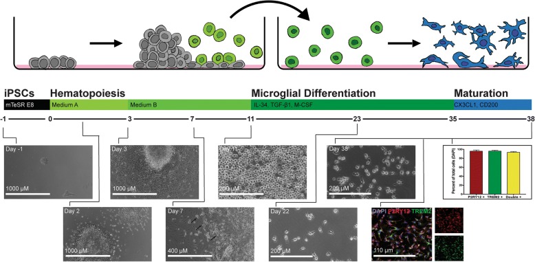
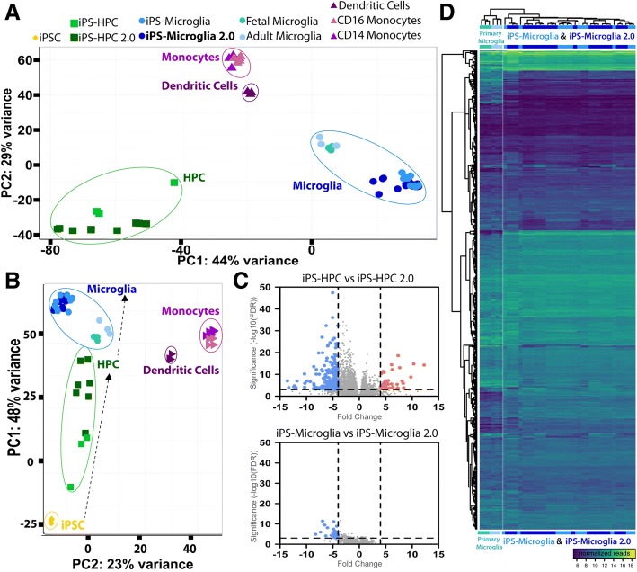
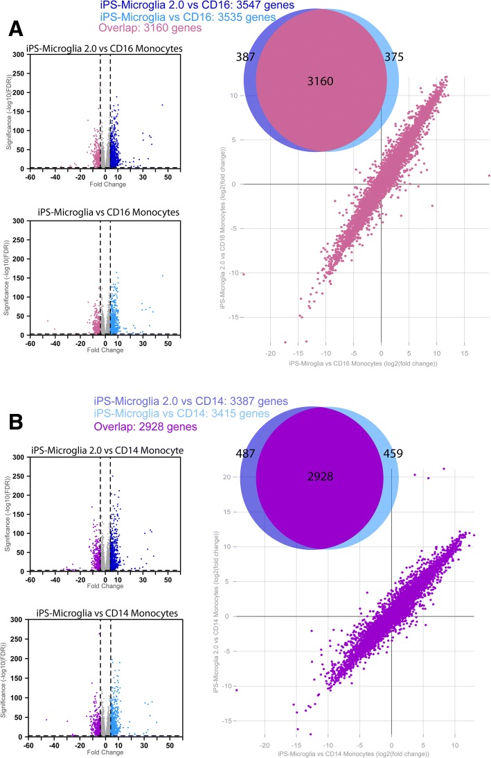
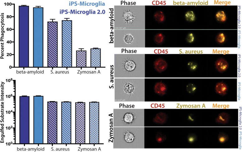
BACKGROUND: Microglia, the principle immune cells of the brain, play important roles in neuronal development, homeostatic function and neurodegenerative disease. Recent genetic studies have further highlighted the importance of microglia in neurodegeneration with the identification of disease risk polymorphisms in many microglial genes. To better understand the role of these genes in microglial biology and disease, we, and others, have developed methods to differentiate microglia from human induced pluripotent stem cells (iPSCs). While the development of these methods has begun to enable important new studies of microglial biology, labs with little prior stem cell experience have sometimes found it challenging to adopt these complex protocols. Therefore, we have now developed a greatly simplified approach to generate large numbers of highly pure human microglia. RESULTS: hematopoietic progenitors are then simply transferred to media that includes three key cytokines (M-CSF, IL-34, and TGFβ-1) that promote differentiation of homeostatic microglia. This updated approach avoids the prior requirement for hypoxic incubation, complex media formulation, FACS sorting, or co-culture, thereby significantly simplifying human microglial generation. To confirm that the resulting cells are equivalent to previously developed iPSC-microglia, we performed RNA-sequencing, functional testing, and transplantation studies. Our findings reveal that microglia generated via this simplified method are virtually identical to iPS-microglia produced via our previously published approach. To also determine whether a small molecule activator of TGFβ signaling (IDE1) can be used to replace recombinant TGFβ1, further reducing costs, we examined growth kinetics and the transcriptome of cells differentiated with IDE1. These data demonstrate that a microglial cell can indeed be produced using this alternative approach, although transcriptional differences do occur that should be considered. CONCLUSION: We anticipate that this new and greatly simplified protocol will enable many interested labs, including those with little prior stem cell or flow cytometry experience, to generate and study human iPS-microglia. By combining this method with other advances such as CRISPR-gene editing and xenotransplantation, the field will continue to improve our understanding of microglial biology and their important roles in human development, homeostasis, and disease.
Overview
The claims and statistics of this paper haven’t been checked yet.
Run the check — free, no account needed. It reads the open-access text and checks the sources it cites.
Integrity signals
Registry
Cites
Statistics
Figures
Verification
Claims the paper makes
The claims and statistics of this paper haven’t been checked yet.
Run the check — free, no account needed. It reads the open-access text and checks the sources it cites.
Claims other papers make about this one
Each is checked against this paper’s own text: does it say what the citing paper says it does?
The source backs part of the claim, not all of it — or backs it for a different population, condition or comparison.
In the citing paper
“Human astrocytes, expressing the astrocytic markers vimentin, S100β, ALDH1L1 and GFAP (Supplementary Figure 3 A), and human microglia, expressing the microglial markers Iba1 and P2Y12 (Supplementary Figure 3 B) were derived from the same hiPSC line [27].”
From Crosstalk between astrocytes and microglia results in increased degradation of α-synuclein and amyloid-β aggregates (2021) · cites this paper as [27]
Experimental — accuracy not yet measuredThe cited source says what the claim attributes to it.
In the citing paper
“E3 and E3S/S iPSCs were differentiated into microglia-like cells (iMGLs)43 (Fig. 6A), confirmed by the expression of microglia markers, IBA1 and TMEM119 (Fig. 6B).”
From APOE3 - R136S mutation confers resilience against tau pathology via cGAS-STING-IFN inhibition (2024) · cites this paper as [43]
The cited source says what the claim attributes to it.
In the citing paper
“HiPSCs were differentiated to hiPSC‐derived microglial cells (iMGL) according to the protocol previously published by McQuade et al., with minor adjustments.22,23”
The check wasn’t confident either way, so it gives no verdict — never disputed.
In the citing paper
“TREM2 deficiency leads to impaired microglial response to β-amyloid plaques, accumulation of the lipid droplets, and increased β-amyloid load in the brain [11, 41].”
From Protective PLCG2 variants associate with a delayed onset of Alzheimer’s disease among heterozygous APOE ε4 carriers (2026) · cites this paper as [41]
Experimental — accuracy not yet measuredThe check wasn’t confident either way, so it gives no verdict — never disputed.
In the citing paper
“AD patients exhibit myelin loss, and higher myelin expression is associated with tau pathology protection in AD43,44.”
From The R136S mutation in the APOE3 gene confers resilience against tau pathology via inhibition of the cGAS-STING-IFN pathway (2025) · cites this paper as [43]
Experimental — accuracy not yet measured
The check wasn’t confident either way, so it gives no verdict — never disputed.
In the citing paper
“higher levels of myelin are associated with protection against tau pathology in AD43,44”
From APOE3 - R136S mutation confers resilience against tau pathology via cGAS-STING-IFN inhibition (2024) · cites this paper as [43]
Experimental — accuracy not yet measuredThe check wasn’t confident either way, so it gives no verdict — never disputed.
In the citing paper
“AD patients exhibit increased myelin loss”
From APOE3 - R136S mutation confers resilience against tau pathology via cGAS-STING-IFN inhibition (2024) · cites this paper as [43]
Experimental — accuracy not yet measuredIn the citing paper
“This has resulted in highly efficient techniques to derive microglia-like cells from human induced pluripotent stem cells (iPSCs) [278, 279].”
From Genetic architecture of Alzheimer's disease (2020) · cites this paper as [278]
Self-check
Percentages, sums and reported differences re-computed from the paper’s own numbers.
The self-check needs the paper’s full text.
Citations
Cites
Moderated estimation of fold change and dispersion for RNA-seq data with DESeq2
Love, M. I., Huber, W., & Anders, S. (2014) · Genome Biology · doi:10.1186/s13059-014-0550-8 · Cited by 95,106
Near-optimal probabilistic RNA-seq quantification
Bray, N. L., Pimentel, H., Melsted, P., & Pachter, L. (2016) · Nature Biotechnology · doi:10.1038/nbt.3519 · Cited by 9,723
Neuroinflammation in Alzheimer's disease
Heneka, M. T., Carson, M. J., Khoury, J. E., Landreth, G. E., Brosseron, F., Feinstein, D. L., Jacobs, A. H., Wyss-Coray, T., Vitorica, J., Ransohoff, R. M., et al. (2015) · The Lancet Neurology · doi:10.1016/s1474-4422(15)70016-5 · Cited by 6,226
Identification of a unique TGF-β–dependent molecular and functional signature in microglia
Butovsky, O., Jedrychowski, M. P., Moore, C. S., Cialic, R., Lanser, A. J., Gabriely, G., Koeglsperger, T., Dake, B., Wu, P. M., Doykan, C. E., et al. (2014) · Nature Neuroscience · doi:10.1038/nn.3599 · Cited by 2,543
Elmore, M. R. P., Najafi, A. R., Koike, M. A., Dagher, N. N., Spangenberg, E. E., Rice, R. A., Kitazawa, M., Matusow, B., Nguyen, H., West, B. L., et al. (2014) · Neuron · doi:10.1016/j.neuron.2014.02.040 · Cited by 1,968
New tools for studying microglia in the mouse and human CNS
Bennett, M. L., Bennett, F. C., Liddelow, S. A., Ajami, B., Zamanian, J. L., Fernhoff, N. B., Mulinyawe, S. B., Bohlen, C. J., Adil, A., Tucker, A., et al. (2016) · Proceedings of the National Academy of Sciences · doi:10.1073/pnas.1525528113 · Cited by 1,804
An environment-dependent transcriptional network specifies human microglia identity
Gosselin, D., Skola, D., Coufal, N. G., Holtman, I. R., Schlachetzki, J. C. M., Sajti, E., Jaeger, B. N., O’Connor, C., Fitzpatrick, C., Pasillas, M. P., et al. (2017) · Science · doi:10.1126/science.aal3222 · Cited by 1,220
iPSC-derived human microglia-like cells to study neurological diseases
Abud, E. M., Ramirez, R. N., Martinez, E. S., Healy, L. M., Nguyen, C. H. H., Newman, S. A., Yeromin, A. V., Scarfone, V. M., Marsh, S. E., Fimbres, C., et al. (2017) · Neuron · doi:10.1016/j.neuron.2017.03.042 · Cited by 1,154
Self-citationDifferential roles of microglia and monocytes in the inflamed central nervous system
Yamasaki, R., Lu, H., Butovsky, O., Ohno, N., Rietsch, A. M., Cialic, R., Wu, P. M., Doykan, C. E., Lin, J., Cotleur, A. C., et al. (2014) · Journal of Experimental Medicine · doi:10.1084/jem.20132477 · Cited by 856
Friedman, B. A., Srinivasan, K., Ayalon, G., Meilandt, W. J., Lin, H., Huntley, M. A., Cao, Y., Lee, S.-H., Haddick, P. C. G., Ngu, H., et al. (2018) · Cell Reports · doi:10.1016/j.celrep.2017.12.066 · Cited by 662
Cited by
Sorted by citations
Brain cell type–specific enhancer–promoter interactome maps and disease - risk association
Nott, A., Holtman, I. R., Coufal, N. G., Schlachetzki, J. C. M., Yu, M., Hu, R., Han, C. Z., Pena, M., Xiao, J., Wu, Y., et al. (2019) · Science · doi:10.1126/science.aay0793 · Cited by 699
Modeling Alzheimer’s disease with iPSC-derived brain cells
Penney, J., Ralvenius, W. T., & Tsai, L.-H. (2020) · Molecular Psychiatry · doi:10.1038/s41380-019-0468-3 · Cited by 471
Human microglial state dynamics in Alzheimer’s disease progression
Sun, N., Victor, M. B., Park, Y. P., Xiong, X., Scannail, A. N., Leary, N., Prosper, S., Viswanathan, S., Luna, X., Boix, C. A., et al. (2023) · Cell · doi:10.1016/j.cell.2023.08.037 · Cited by 462
Development of a Chimeric Model to Study and Manipulate Human Microglia In Vivo
Hasselmann, J., Coburn, M. A., England, W., Figueroa Velez, D. X., Kiani Shabestari, S., Tu, C. H., McQuade, A., Kolahdouzan, M., Echeverria, K., Claes, C., et al. (2019) · Neuron · doi:10.1016/j.neuron.2019.07.002 · Cited by 380
Self-citationA reference human induced pluripotent stem cell line for large-scale collaborative studies
Pantazis, C. B., Yang, A., Lara, E., McDonough, J. A., Blauwendraat, C., Peng, L., Oguro, H., Kanaujiya, J., Zou, J., Sebesta, D., et al. (2022) · Cell Stem Cell · doi:10.1016/j.stem.2022.11.004 · Cited by 310
McQuade, A., Kang, Y. J., Hasselmann, J., Jairaman, A., Sotelo, A., Coburn, M., Shabestari, S. K., Chadarevian, J. P., Fote, G., Tu, C. H., et al. (2020) · Nature Communications · doi:10.1038/s41467-020-19227-5 · Cited by 361
Self-citationTranscriptional signature in microglia associated with Aβ plaque phagocytosis
Grubman, A., Choo, X. Y., Chew, G., Ouyang, J. F., Sun, G., Croft, N. P., Rossello, F. J., Simmons, R., Buckberry, S., Landin, D. V., et al. (2021) · Nature Communications · doi:10.1038/s41467-021-23111-1 · Cited by 357
APOE4 disrupts intracellular lipid homeostasis in human iPSC-derived glia
Sienski, G., Narayan, P., Bonner, J. M., Kory, N., Boland, S., Arczewska, A. A., Ralvenius, W. T., Akay, L., Lockshin, E., He, L., et al. (2021) · Science Translational Medicine · doi:10.1126/scitranslmed.aaz4564 · Cited by 336
Astrocytic interleukin-3 programs microglia and limits Alzheimer’s disease
McAlpine, C. S., Park, J., Griciuc, A., Kim, E., Choi, S. H., Iwamoto, Y., Kiss, M. G., Christie, K. A., Vinegoni, C., Poller, W. C., et al. (2021) · Nature · doi:10.1038/s41586-021-03734-6 · Cited by 322
Lipid accumulation induced by APOE4 impairs microglial surveillance of neuronal-network activity
Victor, M. B., Leary, N., Luna, X., Meharena, H. S., Scannail, A. N., Bozzelli, P. L., Samaan, G., Murdock, M. H., von Maydell, D., Effenberger, A. H., et al. (2022) · Cell Stem Cell · doi:10.1016/j.stem.2022.07.005 · Cited by 316
A CRISPRi/a platform in human iPSC-derived microglia uncovers regulators of disease states
Dräger, N. M., Sattler, S. M., Huang, C. T.-L., Teter, O. M., Leng, K., Hashemi, S. H., Hong, J., Aviles, G., Clelland, C. D., Zhan, L., et al. (2022) · Nature Neuroscience · doi:10.1038/s41593-022-01131-4 · Cited by 258
Microglia in Alzheimer's Disease: Exploring How Genetics and Phenotype Influence Risk
McQuade, A., & Blurton-Jones, M. (2019) · Journal of Molecular Biology · doi:10.1016/j.jmb.2019.01.045 · Cited by 244
Self-citationAndreone, B. J., Przybyla, L., Llapashtica, C., Rana, A., Davis, S. S., van Lengerich, B., Lin, K., Shi, J., Mei, Y., Astarita, G., et al. (2020) · Nature Neuroscience · doi:10.1038/s41593-020-0650-6 · Cited by 233
Rostami, J., Mothes, T., Kolahdouzan, M., Eriksson, O., Moslem, M., Bergström, J., Ingelsson, M., O’Callaghan, P., Healy, L. M., Falk, A., et al. (2021) · Journal of Neuroinflammation · doi:10.1186/s12974-021-02158-3 · Cited by 224
“Generation of hematopoietic progenitor cells (HPCs) and further differentiation to microglia were performed according to the previously published protocol [27], with a few modifications.”
MethodXu, R., Boreland, A. J., Li, X., Erickson, C., Jin, M., Atkins, C., Pang, Z. P., Daniels, B. P., & Jiang, P. (2021) · Stem Cell Reports · doi:10.1016/j.stemcr.2021.06.011 · Cited by 229
Astrocyte-immune cell interactions in physiology and pathology
Han, R. T., Kim, R. D., Molofsky, A. V., & Liddelow, S. A. (2021) · Immunity · doi:10.1016/j.immuni.2021.01.013 · Cited by 223
Jorfi, M., Park, J., Hall, C. K., Lin, C.-C. J., Chen, M., von Maydell, D., Kruskop, J. M., Kang, B., Choi, Y., Prokopenko, D., et al. (2023) · Nature Neuroscience · doi:10.1038/s41593-023-01415-3 · Cited by 214
Advancing cell therapy for neurodegenerative diseases
Temple, S. (2023) · Cell stem cell · doi:10.1016/j.stem.2023.03.017 · Cited by 221
Claes, C., Danhash, E. P., Hasselmann, J., Chadarevian, J. P., Shabestari, S. K., England, W. E., Lim, T. E., Hidalgo, J. L. S., Spitale, R. C., Davtyan, H., et al. (2021) · Molecular Neurodegeneration · doi:10.1186/s13024-021-00473-0 · Cited by 197
Self-citationXu, R., Li, X., Boreland, A. J., Posyton, A., Kwan, K. Y., Hart, R. P., & Jiang, P. (2020) · Nature Communications · doi:10.1038/s41467-020-15411-9 · Cited by 200
PSEN1ΔE9, APPswe, and APOE4 Confer Disparate Phenotypes in Human iPSC-Derived Microglia
Konttinen, H., Cabral-da-Silva, M. E. C., Ohtonen, S., Wojciechowski, S., Shakirzyanova, A., Caligola, S., Giugno, R., Ishchenko, Y., Hernández, D., Fazaludeen, M. F., et al. (2019) · Stem Cell Reports · doi:10.1016/j.stemcr.2019.08.004 · Cited by 191
Microglia-containing human brain organoids for the study of brain development and pathology
Zhang, W., Jiang, J., Xu, Z., Yan, H., Tang, B., Liu, C., Chen, C., & Meng, Q. (2022) · Molecular Psychiatry · doi:10.1038/s41380-022-01892-1 · Cited by 195
Knupp, A., Mishra, S., Martinez, R., Braggin, J. E., Szabo, M., Kinoshita, C., Hailey, D. W., Small, S. A., Jayadev, S., & Young, J. E. (2020) · Cell Reports · doi:10.1016/j.celrep.2020.107719 · Cited by 165
A multi-pronged human microglia response to Alzheimer’s disease Aβ pathology
Mancuso, R., Fattorelli, N., Martinez-Muriana, A., Davis, E., Wolfs, L., Van Den Daele, J., Geric, I., Preman, P., Serneels, L., Poovathingal, S., et al. (2022) · doi:10.1101/2022.07.07.499139 · Cited by 169
Absence of microglia promotes diverse pathologies and early lethality in Alzheimer’s disease mice
Kiani Shabestari, S., Morabito, S., Danhash, E. P., McQuade, A., Sanchez, J. R., Miyoshi, E., Chadarevian, J. P., Claes, C., Coburn, M. A., Hasselmann, J., et al. (2022) · Cell Reports · doi:10.1016/j.celrep.2022.110961 · Cited by 167
Self-citationSabogal-Guáqueta, A. M., Marmolejo-Garza, A., Trombetta-Lima, M., Oun, A., Hunneman, J., Chen, T., Koıstınaho, J., Lehtonen, Š., Kortholt, A., Wolters, J. C., et al. (2023) · Nature Communications · doi:10.1038/s41467-023-42096-7 · Cited by 180
You, Y., Muraoka, S., Jedrychowski, M. P., Hu, J., McQuade, A., Young‐Pearse, T. L., Aslebagh, R., Shaffer, S. A., Gygi, S. P., Blurton-Jones, M. M., et al. (2022) · Journal of Extracellular Vesicles · doi:10.1002/jev2.12183 · Cited by 165
Self-citationLagomarsino, V. N., Pearse, R. V., Liu, L., Hsieh, Y.-C., Fernandez, M. A., Vinton, E. A., Paull, D., Felsky, D., Tasaki, S., Gaiteri, C., et al. (2021) · Neuron · doi:10.1016/j.neuron.2021.08.003 · Cited by 151
Human iPSC‐derived microglia: A growing toolset to study the brain's innate immune cells
Hasselmann, J., & Blurton‐Jones, M. (2020) · Glia · doi:10.1002/glia.23781 · Cited by 145
Self-citation3D Bioprinting of Neural Tissues
Cadena, M., Ning, L., King, A. T., Hwang, B., Jin, L., Serpooshan, V., & Sloan, S. A. (2020) · Advanced Healthcare Materials · doi:10.1002/adhm.202001600 · Cited by 151
Alzheimer's-associated PU.1 expression levels regulate microglial inflammatory response
Pimenova, A. A., Herbinet, M., Gupta, I., Machlovi, S. I., Bowles, K. R., Marcora, E., & Goate, A. M. (2021) · Neurobiology of Disease · doi:10.1016/j.nbd.2020.105217 · Cited by 120
“iPSCs were passaged every week using ReLeSR dissociation reagent (StemCell Technologies) and used for hematopoietic stem cell differentiation with STEMdiff Hematopoietic kit (STEMCELL Technologies #05310) followed by differentiation to induced microglial-like cells (iMGLs) using a previously published protocol by (McQ…”
Large-Scale Production of Human iPSC-Derived Macrophages for Drug Screening
Gutbier, S., Wanke, F., Dahm, N., Rümmelin, A., Zimmermann, S., Christensen, K., Köchl, F., Rautanen, A., Hatje, K., Geering, B., et al. (2020) · International Journal of Molecular Sciences · doi:10.3390/ijms21134808 · Cited by 136
Recent progress in translational engineered in vitro models of the central nervous system
Nikolakopoulou, P., Rauti, R., Voulgaris, D., Shlomy, I., Maoz, B. M., & Herland, A. (2020) · Brain · doi:10.1093/brain/awaa268 · Cited by 133
Genetic architecture of Alzheimer's disease
Neuner, S. M., TCW, J., & Goate, A. M. (2020) · Neurobiology of Disease · doi:10.1016/j.nbd.2020.104976 · Cited by 126
“To this end, innovative transplantation methods have been established to implant hematopoietic precursor cells or microglia themselves directly into the brains of mouse models, where they have been shown to fully integrate and differentiate into mature microglia (McQuade et al., 2018; Abud et al., 2017; Hasselmann et…”
BackgroundMethodSARS-CoV-2 Infection of Microglia Elicits Proinflammatory Activation and Apoptotic Cell Death
Jeong, G. U., Lyu, J., Kim, K., Chung, Y. C., Yoon, G. Y., Lee, S., Hwang, I., Shin, W.-H., Ko, J., Lee, J., et al. (2022) · Microbiology Spectrum · doi:10.1128/spectrum.01091-22 · Cited by 126
Gottschlich, A., Thomas, M., Grünmeier, R., Lesch, S., Rohrbacher, L., Igl, V., Briukhovetska, D., Benmebarek, M.-R., Vick, B., Dede, S., et al. (2023) · Nature Biotechnology · doi:10.1038/s41587-023-01684-0 · Cited by 123
Biomaterial-supported MSC transplantation enhances cell–cell communication for spinal cord injury
Lv, B., Zhang, X., Yuan, J., Chen, Y., Ding, H., Cao, X., & Huang, A. (2021) · Stem Cell Research & Therapy · doi:10.1186/s13287-020-02090-y · Cited by 114
Stem-cell-derived human microglia transplanted into mouse brain to study human disease
Fattorelli, N., Martínez‐Muriana, A., Wolfs, L., Geric, I., Strooper, B. D., & Mancuso, R. (2021) · Nature Protocols · doi:10.1038/s41596-020-00447-4 · Cited by 114
Upgrading the Physiological Relevance of Human Brain Organoids
Dosso, A. D., Urenda, J., Nguyen, T. N., & Quadrato, G. (2020) · Neuron · doi:10.1016/j.neuron.2020.08.029 · Cited by 106
Giacomelli, E., Vahsen, B. F., Calder, E. L., Xu, Y., Scaber, J., Gray, E., Dafinca, R., Talbot, K., & Studer, L. (2022) · Cell stem cell · doi:10.1016/j.stem.2021.12.008 · Cited by 100
Therapeutic Plasticity of Neural Stem Cells
Ottoboni, L., von Wunster, B., & Martino, G. (2020) · Frontiers in Neurology · doi:10.3389/fneur.2020.00148 · Cited by 105
Engineering an inhibitor-resistant human CSF1R variant for microglia replacement
Chadarevian, J. P., Lombroso, S. I., Peet, G. C., Hasselmann, J., Tu, C. H., Marzan, D. E., Capocchi, J. K., Purnell, F. S., Nemec, K. M., Lahian, A., et al. (2022) · The Journal of Experimental Medicine · doi:10.1084/jem.20220857 · Cited by 101
Self-citationSpeicher, A. M., Wiendl, H., Meuth, S. G., & Pawlowski, M. (2019) · Molecular Neurodegeneration · doi:10.1186/s13024-019-0347-z · Cited by 100
Association of a common genetic variant with Parkinson’s disease is mediated by microglia
Langston, R. G., Beilina, A., Reed, X., Kaganovich, A., Singleton, A. B., Blauwendraat, C., Gibbs, J. R., & Cookson, M. R. (2022) · Science Translational Medicine · doi:10.1126/scitranslmed.abp8869 · Cited by 93
Reifschneider, A., Robinson, S., van Lengerich, B., Gnörich, J., Logan, T., Heindl, S., Vogt, M. A., Weidinger, E., Riedl, L., Wind, K., et al. (2022) · The EMBO Journal · doi:10.15252/embj.2021109108 · Cited by 89
Immune checkpoint TIM-3 regulates microglia and Alzheimer’s disease
Kimura, K., Subramanian, A., Yin, Z., Khalilnezhad, A., Wu, Y., He, D., Dixon, K. O., Chitta, U. K., Ding, X., Adhikari, N., et al. (2025) · Nature · doi:10.1038/s41586-025-08852-z · Cited by 85
Self-citationChadarevian, J. P., Hasselmann, J., Lahian, A., Capocchi, J. K., Escobar, A., Lim, T. E., Le, L., Tu, C., Nguyen, J., Kiani Shabestari, S., et al. (2024) · Neuron · doi:10.1016/j.neuron.2024.05.023 · Cited by 83
Self-citationFilipello, F., You, S.-F., Mirfakhar, F. S., Mahali, S., Bollman, B., Acquarone, M., Korvatska, O., Marsh, J. A., Sivaraman, A., Martinez, R., et al. (2023) · Acta Neuropathologica · doi:10.1007/s00401-023-02568-y · Cited by 81
Wu, J., Zhang, J., Chen, X., Wettschurack, K., Que, Z., Deming, B. A., Olivero-Acosta, M. I., Cui, N., Eaton, M., Zhao, Y., et al. (2024) · Molecular Psychiatry · doi:10.1038/s41380-024-02518-4 · Cited by 75
Microglia transcriptional states and their functional significance: Context drives diversity
Depp, C., Doman, J. L., Hingerl, M., Xia, J., & Stevens, B. (2025) · Immunity · doi:10.1016/j.immuni.2025.04.009 · Cited by 84
INPP5D regulates inflammasome activation in human microglia
Chou, V., Pearse, R. V., Aylward, A. J., Ashour, N., Taga, M., Terzioglu, G., Fujita, M., Fancher, S. B., Sigalov, A., Benoit, C. R., et al. (2023) · Nature Communications · doi:10.1038/s41467-023-42819-w · Cited by 79
Attenuation of neuroinflammation reverses Adriamycin-induced cognitive impairments
Allen, B. D., Apodaca, L. A., Syage, A. R., Markarian, M., Baddour, A. A. D., Minasyan, H., Alikhani, L., Lu, C., West, B. L., Giedzinski, E., et al. (2019) · Acta Neuropathologica Communications · doi:10.1186/s40478-019-0838-8 · Cited by 76
Mamais, A., Kluss, J. H., Bonet‐Ponce, L., Landeck, N., Langston, R. G., Smith, N., Beilina, A. G., Kaganovich, A., Ghosh, M. C., Pellegrini, L., et al. (2021) · PLoS Biology · doi:10.1371/journal.pbio.3001480 · Cited by 81
Ralvenius, W. T., Andresen, J. L., Huston, M. M., Penney, J., Bonner, J. M., Fenton, O. S., Langer, R., & Tsai, L. (2024) · Advanced Materials · doi:10.1002/adma.202309225 · Cited by 78
Comparative analysis of human microglial models for studies of HIV replication and pathogenesis
Mohammad, A., Hammonds, J. E., Pujato, M. A., Mayhew, C. N., Roskin, K. M., & Spearman, P. W. (2020) · Retrovirology · doi:10.1186/s12977-020-00544-y · Cited by 78
Yadavalli, N., & Ferguson, S. M. (2023) · Proceedings of the National Academy of Sciences · doi:10.1073/pnas.2303789120 · Cited by 74
Human microglia maturation is underpinned by specific gene regulatory networks
Han, C. Z., Li, R. Z., Hansen, E., Trescott, S., Fixsen, B. R., Nguyen, C. T., Mora, C. M., Spann, N. J., Bennett, H. R., Poirion, O., et al. (2023) · Immunity · doi:10.1016/j.immuni.2023.07.016 · Cited by 84
Self-citationHuman iPSC-derived glia models for the study of neuroinflammation
Stöberl, N., Maguire, E., Salis, E., Shaw, B., & Hall-Roberts, H. (2023) · Journal of Neuroinflammation · doi:10.1186/s12974-023-02919-2 · Cited by 71
Liu, T., Zhu, B., Liu, Y., Zhang, X., Yin, J., Li, X., Jiang, L., Hodges, A. P., Rosenthal, S. B., Zhou, L., et al. (2020) · Journal of Experimental Medicine · doi:10.1084/jem.20200474 · Cited by 64
Self-citationAstrocytes and microglia in neurodegenerative diseases: Lessons from human in vitro models
Franklin, H., Clarke, B. E., & Patani, R. (2021) · Progress in Neurobiology · doi:10.1016/j.pneurobio.2020.101973 · Cited by 66
Jairaman, A., McQuade, A., Granzotto, A., Kang, Y. J., Chadarevian, J. P., Gandhi, S., Parker, I., Smith, I., Cho, H., Sensi, S. L., et al. (2022) · eLife · doi:10.7554/elife.73021 · Cited by 62
Self-citationPan-SHIP1/2 inhibitors promote microglia effector functions essential for CNS homeostasis
Pedicone, C., Fernandes, S., Dungan, O. M., Dormann, S. M., Viernes, D. R., Adhikari, A. A., Choi, L. B., De Jong, E. P., Chisholm, J. D., & Kerr, W. G. (2020) · Journal of Cell Science · doi:10.1242/jcs.238030 · Cited by 61
Sabogal-Guáqueta, A. M., Marmolejo-Garza, A., de Pádua, V. P., Eggen, B. J. L., Boddeke, E., & Dolga, A. M. (2020) · Progress in Neurobiology · doi:10.1016/j.pneurobio.2020.101805 · Cited by 65
Microglia promote anti-tumour immunity and suppress breast cancer brain metastasis
Evans, K. T., Blake, K., Longworth, A. J., Coburn, M., Insua‐Rodríguez, J., McMullen, T. P., Nguyen, Q., Ma, D., Lev, T., Hernandez, G. A., et al. (2023) · Nature Cell Biology · doi:10.1038/s41556-023-01273-y · Cited by 64
Self-citationWasher, S. J., Perez‐Alcantara, M., Chen, Y., Steer, J., James, W., Trynka, G., Bassett, A. R., & Cowley, S. A. (2022) · Scientific Reports · doi:10.1038/s41598-022-23477-2 · Cited by 60
Ni, C., Jiang, W., Wang, Z., Wang, Z., Zhang, J., Zheng, X., Liu, Z., Ou, H., Jiang, T., Liang, W., et al. (2020) · Molecular Psychiatry · doi:10.1038/s41380-020-0662-3 · Cited by 58
Venkataraman, L., Fair, S. R., McElroy, C. A., Hester, M. E., & Fu, H. (2020) · Stem Cell Reviews and Reports · doi:10.1007/s12015-020-10068-9 · Cited by 55
Reich, M., Paris, I., Ebeling, M., Dahm, N., Schweitzer, C., Reinhardt, D., Schmucki, R., Prasad, M., Köchl, F., Leist, M., et al. (2021) · Frontiers in Immunology · doi:10.3389/fimmu.2020.617860 · Cited by 49
Triglyceride metabolism controls inflammation and microglial phenotypes associated with APOE4
Stephenson, R. A., Sepulveda, J., Johnson, K. R., Lita, A., Gopalakrishnan, J., Acri, D. J., Beilina, A., Cheng, L., Yang, L. G., Root, J. T., et al. (2025) · Cell Reports · doi:10.1016/j.celrep.2025.115961 · Cited by 55
Pocock, J., Vasilopoulou, F., Svensson, E., & Cosker, K. (2024) · Neuropharmacology · doi:10.1016/j.neuropharm.2024.110020 · Cited by 54
Haenseler, W., & Rajendran, L. (2019) · Stem Cells · doi:10.1002/stem.2995 · Cited by 58
Cellular complexity in brain organoids: Current progress and unsolved issues
Mansour, A. A., Schafer, S. T., & Gage, F. H. (2020) · Seminars in Cell and Developmental Biology · doi:10.1016/j.semcdb.2020.05.013 · Cited by 52
Claes, C., England, W. E., Danhash, E. P., Kiani Shabestari, S., Jairaman, A., Chadarevian, J. P., Hasselmann, J., Tsai, A. P., Coburn, M. A., Sanchez, J., et al. (2022) · Alzheimer's & Dementia · doi:10.1002/alz.12577 · Cited by 52
Self-citationPodleśny-Drabiniok, A., Novikova, G., Liu, Y., Dunst, J., Temizer, R., Giannarelli, C., Marro, S., Kreslavsky, T., Marcora, E., & Goate, A. M. (2024) · Nature Communications · doi:10.1038/s41467-024-46315-7 · Cited by 52
Lee, H., Aylward, A. J., Pearse, R. V., Lish, A. M., Hsieh, Y.-C., Augur, Z. M., Benoit, C. R., Chou, V., Knupp, A., Pan, C., et al. (2023) · Cell Reports · doi:10.1016/j.celrep.2023.112994 · Cited by 58
Harnessing human iPSC-microglia for CNS-wide delivery of disease-modifying proteins
Chadarevian, J. P., Davtyan, H., Chadarevian, A. L., Nguyen, J., Capocchi, J. K., Le, L., Escobar, A., Chadarevian, T., Mansour, K., Deynega, E., et al. (2025) · Cell Stem Cell · doi:10.1016/j.stem.2025.03.009 · Cited by 60
Self-citationLish, A. M., Grogan, E. F. L., Benoit, C. R., Pearse, R. V., Heuer, S. E., Luquez, T., Orme, G. A., Galle, P. C., Milinkeviciute, G., Green, K. N., et al. (2025) · Neuron · doi:10.1016/j.neuron.2025.03.034 · Cited by 55
Li, C., Ren, J., Zhang, M., Wang, H., Fang, Y., Wu, J., & Tang, Y. (2022) · Cellular and Molecular Life Sciences · doi:10.1007/s00018-022-04536-3 · Cited by 52
Modeling Psychiatric Disorder Biology with Stem Cells
Das, D., Feuer, K. L., Wahbeh, M. H., & Avramopoulos, D. (2020) · Current Psychiatry Reports · doi:10.1007/s11920-020-01148-1 · Cited by 49
Penney, J., Ralvenius, W. T., Loon, A., Cerit, O., Dileep, V., Milo, B., Pao, P., Woolf, H., & Tsai, L. (2024) · Glia · doi:10.1002/glia.24485 · Cited by 52
Phosphoproteomics identifies microglial Siglec‐F inflammatory response during neurodegeneration
Morshed, N., Ralvenius, W. T., Nott, A., Watson, L. A., Rodriguez, F. H., Akay, L. A., Joughin, B. A., Pao, P., Penney, J., LaRocque, L. M., et al. (2020) · Molecular Systems Biology · doi:10.15252/msb.20209819 · Cited by 48
Current tools to interrogate microglial biology
Dumas, A. A., Borst, K., & Prinz, M. (2021) · Neuron · doi:10.1016/j.neuron.2021.07.004 · Cited by 47
Generation of pure monocultures of human microglia-like cells from induced pluripotent stem cells
Banerjee, P., Paza, E., Perkins, E., James, O. G., Kenkhuis, B., Lloyd, A. F., Burr, K., Story, D., Yusuf, D., He, X., et al. (2020) · Stem Cell Research · doi:10.1016/j.scr.2020.102046 · Cited by 48
Huang, Y., Motta, E., Nanvuma, C., Kuhrt, L. D., Yang, Y., Xia, P., Lubas, M., Zhu, S., Schnauss, M., Qazi, N., et al. (2022) · Cell Reports · doi:10.1016/j.celrep.2022.110670 · Cited by 45
MerTK is a mediator of alpha-synuclein fibril uptake by human microglia
Dorion, M., Yaqubi, M., Senkevich, K. A., Kieran, N. W., MacDonald, A., Chen, C. X. ‐., Luo, W., Wallis, A., Shlaifer, I., Hall, J. A., et al. (2023) · Brain · doi:10.1093/brain/awad298 · Cited by 43
Genetic contribution to microglial activation in schizophrenia
Koskuvi, M., Pörsti, E., Hewitt, T., Räsänen, N., Wu, Y., Trontti, K., McQuade, A., Kalyanaraman, S., Ojansuu, I., Vaurio, O., et al. (2024) · Molecular Psychiatry · doi:10.1038/s41380-024-02529-1 · Cited by 46
Self-citationHuman in vitro models for understanding mechanisms of autism spectrum disorder
Gordon, A., & Geschwind, D. H. (2020) · Molecular Autism · doi:10.1186/s13229-020-00332-7 · Cited by 45
Species differences in immune‐mediated CNS tissue injury and repair: A (neuro)inflammatory topic
Healy, L. M., Yaqubi, M., Ludwin, S. K., & Antel, J. P. (2019) · Glia · doi:10.1002/glia.23746 · Cited by 41
Pesämaa, I., Müller, S. A., Robinson, S., Darcher, A., Paquet, D., Zetterberg, H., Lichtenthaler, S. F., & Haass, C. (2023) · Molecular Neurodegeneration · doi:10.1186/s13024-023-00657-w · Cited by 47
Microglial contribution to the pathology of neurodevelopmental disorders in humans
Matulevičiūtė, R., Akinluyi, E. T., Muntslag, T. A. O., Dewing, J. M., Long, K. R., Vernon, A. C., Tremblay, M., & Menassa, D. A. (2023) · Acta Neuropathologica · doi:10.1007/s00401-023-02629-2 · Cited by 41
A microglia-containing cerebral organoid model to study early life immune challenges
Buonfiglioli, A., Kübler, R., Missall, R. D., de Jong, R., Chan, S., Haage, V. C., Wendt, S., Lin, A. J., Mattei, D., Graziani, M., et al. (2024) · Brain Behavior and Immunity · doi:10.1016/j.bbi.2024.11.008 · Cited by 41
Ruíz, J. P., Chen, G., Haro‐Mora, J. J., Keyvanfar, K., Liu, C., Zou, J., Beers, J. K., Bloomer, H., Qanash, H., Uchida, N., et al. (2019) · Stem Cell Research · doi:10.1016/j.scr.2019.101600 · Cited by 41
Investigating Microglia in Health and Disease: Challenges and Opportunities
Miron, V. E., & Priller, J. (2020) · Trends in Immunology · doi:10.1016/j.it.2020.07.002 · Cited by 43
Galectin-3 aggravates microglial activation and tau transmission in tauopathy
Siew, J. J., Chen, H.-M., Chiu, F., Lee, C., Chang, Y., Chen, H.-L., Nguyen, T. N. A., Liao, H.-T., Liu, M., Hagar, H. C., et al. (2023) · Journal of Clinical Investigation · doi:10.1172/jci165523 · Cited by 47
The Emerging Roles and Therapeutic Potential of Soluble TREM2 in Alzheimer’s Disease
Zhong, L., & Chen, X. (2019) · Frontiers in Aging Neuroscience · doi:10.3389/fnagi.2019.00328 · Cited by 40
Takata, K., Ginhoux, F., & Shimohama, S. (2021) · Biochemical Pharmacology · doi:10.1016/j.bcp.2021.114754 · Cited by 40
Pluvinage, J. V., Sun, J., Claes, C., Flynn, R. A., Haney, M. S., Iram, T., Meng, X., Lindemann, R. C., Riley, N. M., Danhash, E., et al. (2021) · Science Translational Medicine · doi:10.1126/scitranslmed.abg2919 · Cited by 40
Self-citationLi, F., Sun, X., Sun, K., Kong, F., Jiang, X., & Kong, Q. (2023) · Neural Regeneration Research · doi:10.4103/1673-5374.389302 · Cited by 44
Chen, D., Wang, C., Chen, X., Li, J., Chen, S., Li, Y., Ma, F., Li, T., Zou, M., Li, X., et al. (2025) · Science Translational Medicine · doi:10.1126/scitranslmed.ads6111 · Cited by 42
Utilising Induced Pluripotent Stem Cells in Neurodegenerative Disease Research: Focus on Glia
Albert, K., Niskanen, J., Kälvälä, S., & Lehtonen, Š. (2021) · International Journal of Molecular Sciences · doi:10.3390/ijms22094334 · Cited by 40
Metadata
- Authors
- McQuade, A., Coburn, M., Tu, C. H., Hasselmann, J., Davtyan, H., & Blurton-Jones, M.
- Affiliations
- University of California, Irvine (US)
- Corresponding author
- Blurton-Jones, Mathew
- ORCID
- Venue
- Molecular Neurodegeneration
- Published
- 2018-12
- Volume
- 13
- Issue
- 1
- Pages
- 67-67
- ISSN
- 1750-1326 (ISSN-L)
- PMC
- PMC6303871
- OpenAlex
- W2905656859
- Cited by
- 449
- References
- 33
- Citations by year
- 2026: 64 · 2025: 98 · 2024: 73 · 2023: 53 · 2022: 54 · 2021: 43 · 2020: 40 · 2019: 23
- FWCI
- 11.92
- Type
- journal article
- Language
- en
- Open access
- gold
- doi.org
- Licence
- Locations
- Molecular Neurodegeneration (publishedVersion, cc-by)
- Europe PMC (PubMed Central) (publishedVersion, other-oa)
- PubMed Central (publishedVersion, cc-by)
- PubMed (publishedVersion)
- eScholarship (California Digital Library) (submittedVersion, cc-by)
- DOAJ (DOAJ: Directory of Open Access Journals) (submittedVersion)
- Funders
- National Institutes of Health (AG048099, AG056303, AG016573)
- California Institute for Regenerative Medicine (RT3-07893)
- National Institute of Neurological Disorders and Stroke (NS082174)
- Topics
- Life Sciences › Neuroscience › Neurology › Neuroinflammation and Neurodegeneration Mechanisms
- Life Sciences › Biochemistry, Genetics and Molecular Biology › Molecular Biology › Pluripotent Stem Cells Research
- Life Sciences › Neuroscience › Developmental Neuroscience › Neurogenesis and neuroplasticity mechanisms
- Keywords
- microglia differentiation, induced pluripotent stem cells, iPSC-derived microglia, hematopoietic progenitor cells, macrophage colony-stimulating factor, IL-34, TGF-β1, TGF-beta signaling, microglial transcriptome, neurodegeneration
- MeSH terms
- Animals, Brain, Cell Differentiation, Humans, Cytokines, Microglia, Pluripotent Stem Cells, Induced Pluripotent Stem Cells
About these checks
Registry, retraction, Cites and statistics results are computed, not judged, with one exception: a printed reference that no index lists is read once by a model before it counts as a citation error. Claims are extracted by a model and kept only when their quote appears verbatim in the text; each cited source is then checked, with a model’s help, for whether it says what the claim relies on. Those verdicts can be wrong — read the claim’s quote and the cited source before relying on one.
Statistics and arithmetic are recomputed, not judged.
Confidence bands are shown only for fields calibrated on our annotated test set. None is calibrated yet, so no band is shown.
What counts as a claim ›How we measure confidence ›How we check this ›
Are you an author, or do you see a problem with this page? Request a review
