

The DOI matches its Crossref record; 426 of the 428 references it cites were found in a registry or an index.
Axonal energy metabolism, and the effects in aging and neurodegenerative diseases
Open accessAbstract
Abstract Human studies consistently identify bioenergetic maladaptations in brains upon aging and neurodegenerative disorders of aging (NDAs), such as Alzheimer’s disease, Parkinson’s disease, Huntington’s disease, and Amyotrophic lateral sclerosis. Glucose is the major brain fuel and glucose hypometabolism has been observed in brain regions vulnerable to aging and NDAs. Many neurodegenerative susceptible regions are in the topological central hub of the brain connectome, linked by densely interconnected long-range axons. Axons, key components of the connectome, have high metabolic needs to support neurotransmission and other essential activities. Long-range axons are particularly vulnerable to injury, neurotoxin exposure, protein stress, lysosomal dysfunction, etc. Axonopathy is often an early sign of neurodegeneration. Recent studies ascribe axonal maintenance failures to local bioenergetic dysregulation. With this review, we aim to stimulate research in exploring metabolically oriented neuroprotection strategies to enhance or normalize bioenergetics in NDA models. Here we start by summarizing evidence from human patients and animal models to reveal the correlation between glucose hypometabolism and connectomic disintegration upon aging/NDAs. To encourage mechanistic investigations on how axonal bioenergetic dysregulation occurs during aging/NDAs, we first review the current literature on axonal bioenergetics in distinct axonal subdomains: axon initial segments, myelinated axonal segments, and axonal arbors harboring pre-synaptic boutons. In each subdomain, we focus on the organization, activity-dependent regulation of the bioenergetic system, and external glial support. Second, we review the mechanisms regulating axonal nicotinamide adenine dinucleotide (NAD + ) homeostasis, an essential molecule for energy metabolism processes, including NAD + biosynthetic, recycling, and consuming pathways. Third, we highlight the innate metabolic vulnerability of the brain connectome and discuss its perturbation during aging and NDAs. As axonal bioenergetic deficits are developing into NDAs, especially in asymptomatic phase, they are likely exaggerated further by impaired NAD + homeostasis, the high energetic cost of neural network hyperactivity, and glial pathology. Future research in interrogating the causal relationship between metabolic vulnerability, axonopathy, amyloid/tau pathology, and cognitive decline will provide fundamental knowledge for developing therapeutic interventions.
Overview
The claims and statistics of this paper haven’t been checked yet.
Run the check — free, no account needed. It reads the open-access text and checks the sources it cites.
Integrity signals
Registry
Cites
Statistics
Figures
Verification
Claims the paper makes
The claims and statistics of this paper haven’t been checked yet.
Run the check — free, no account needed. It reads the open-access text and checks the sources it cites.
Claims other papers make about this one
Each is checked against this paper’s own text: does it say what the citing paper says it does?
The source backs part of the claim, not all of it — or backs it for a different population, condition or comparison.
In the citing paper
“A snRNA‐seq study in human AD also reported downregulation of synaptic cell adhesion–related gene expression and enrichment terms in an oligodendrocyte cluster. 65 , 66”
From Unique transcriptomic alterations in 5XFAD;PS19 mouse model identify glial lipid dysregulation and coordinated microglial–oligodendrocyte responses (2026) · cites this paper as [66]
Experimental — accuracy not yet measured
Self-check
Percentages, sums and reported differences re-computed from the paper’s own numbers.
The self-check needs the paper’s full text.
Citations
Cites
1 retracted
Braidy, N., Poljak, A., Grant, R., Jayasena, T., Mansour, H., Chan‐Ling, T., Guillemin, G. J., Smythe, G. A., & Sachdev, P. S. (2013) · Biogerontology · doi:10.1007/s10522-013-9489-5 · Cited by 123
RetractedMapping the structural core of human cerebral cortex
Hagmann, P., Cammoun, L., Gigandet, X., Meuli, R., Honey, C. J., Wedeen, V. J., & Sporns, O. (2008) · PLoS Biology · doi:10.1371/journal.pbio.0060159 · Cited by 4,173 · cited 1×
“The majority of AD-susceptible regions identified are located in the default mode network (DMN), the topological central hub of the brain connectome [38, 39], containing densely interconnected long-range corticocortical axons [40, 41].”
BackgroundClinical and biomarker changes in dominantly inherited Alzheimer's disease
Bateman, R. J., Xiong, C., Benzinger, T. L. S., Fagan, A. M., Goate, A., Fox, N. C., Marcus, D. S., Cairns, N. J., Xie, X., Blazey, T. M., et al. (2012) · New England Journal of Medicine · doi:10.1056/nejmoa1202753 · Cited by 3,814 · cited 1×
“Imaging: MRI, FDG‐PET, and PiB‐PET for 40 noncarriers and 88 PSEN1, PSEN2, APP mutation carriers Aimed to use data from longitudinal study to identify pathophysiological biomarkers • Mutation carriers 10–15 yrs before AD: Bilateral hippocampal atrophy, Precuneus glucose uptake↓ Episodic memory↓ [45]”
MethodInterplay between Cortical Spreading Depolarization and Seizures
Kramer, D. R., Fujii, T., Ohiorhenuan, I. E., & Liu, C. Y. (2017) · Stereotactic and Functional Neurosurgery · doi:10.1159/000452841 · Cited by 3,721
An Energy Budget for Signaling in the Grey Matter of the Brain
Attwell, D., & Laughlin, S. B. (2001) · Journal of Cerebral Blood Flow & Metabolism · doi:10.1097/00004647-200110000-00001 · Cited by 3,543
Ageing as a risk factor for neurodegenerative disease
Hou, Y., Dan, X., Babbar, M., Wei, Y., Hasselbalch, S. G., Croteau, D. L., & Bohr, V. A. (2019) · Nature Reviews Neurology · doi:10.1038/s41582-019-0244-7 · Cited by 3,310 · cited 1×
“Aging is considered one of the biggest risk factors for dementia and sporadic late-onset neurodegenerative diseases [20].”
BackgroundPellerin, L., & Magistretti, P. J. (1994) · Proceedings of the National Academy of Sciences · doi:10.1073/pnas.91.22.10625 · Cited by 2,927 · cited 1×
“Because LDH-B favors the conversion in reverse direction to LDH-A, that is it consumes NAD+ and produces NADH and pyruvate [291, 292], LDH-B’s activity may counteract LDH-A, thus reducing NAD redox potential and restraining glycolysis.”
BackgroundThe mechanism of eukaryotic translation initiation and principles of its regulation
Jackson, R. J., Hellen, C. U. T., & Pestova, T. V. (2010) · Nature Reviews Molecular Cell Biology · doi:10.1038/nrm2838 · Cited by 2,759
Brain energy metabolism: focus on Astrocyte-neuron metabolic cooperation
Bélanger, M., Allaman, I., & Magistretti, P. J. (2011) · Cell Metabolism · doi:10.1016/j.cmet.2011.08.016 · Cited by 2,313
The role of the posterior cingulate cortex in cognition and disease
Leech, R., & Sharp, D. J. (2014) · Brain · doi:10.1093/brain/awt162 · Cited by 2,247
Research works printed but not linked: 2
- Mc cluskey M. thesis: “Study of vesicular glycolysis in health and Huntington’s Disease” Université Grenoble Alpes [2020]. [GIN] Grenoble Institut des Neurosciences; 2021.
- Magali V, Lena H, Jörn D, Ingvill T, Lars S, Marc N, Camila C-W, van den Barbara H, Øyvind S, Roland S, et al. Chronic depletion of subcellular NAD pools reveals their interconnectivity and a buffering function of mitochondria. Nature Portfolio; 2022.
Cited by
Sorted by citations
White matter damage and degeneration in traumatic brain injury.
Armstrong, R. C., Sullivan, G. M., Perl, D. P., Rosarda, J. D., & Radomski, K. L. (2024) · Trends in Neurosciences · doi:10.1016/j.tins.2024.07.003 · Cited by 62
Mitochondria in Alzheimer’s Disease Pathogenesis
Reiss, A. B., Gulkarov, S., Jacob, B., Srivastava, A., Pinkhasov, A., Gomolin, I. H., Stecker, M. M., Wısnıewskı, T., & Leon, J. D. (2024) · Life · doi:10.3390/life14020196 · Cited by 71
“Upon axonal injury or mitochondrial malfunction, NMNAT2 levels are reduced, and so is the production of NAD+. NMNAT2 loss promotes SARM1 activation, and this combination results in energetic failure in axons [48,49].”
BackgroundVoicu, V. A., Toader, C., Șerban, M., Covache-Busuioc, R.-A., & Ciurea, A. V. (2025) · Biomedicines · doi:10.3390/biomedicines13082025 · Cited by 59
“Neurons with long axonal projections—such as layer V pyramidal neurons, cholinergic brainstem neurons and cerebellar Purkinje cells—are at the greatest risk of cytoskeletal degradation and tau-induced microtubule disassembly [169,170].”
Diabetes-related cognitive impairment: Mechanisms, symptoms, and treatments
Yu, X., He, H., Wen, J., Xu, X., Ruan, Z., Hu, R., Wang, F., & Ju, H. (2025) · Open Medicine · doi:10.1515/med-2024-1091 · Cited by 57
Energy Metabolism and Brain Aging: Strategies to Delay Neuronal Degeneration
Na, D., Zhang, Z., Meng, M., Li, M., Gao, J., Kong, J., Zhang, G., & Guo, Y. (2025) · Cellular and Molecular Neurobiology · doi:10.1007/s10571-025-01555-z · Cited by 44
“Age-related impairments in glucose metabolism have been documented in studies (Yang et al. 2023).”
Iron homeostasis and neurodegeneration in the ageing brain: Insight into ferroptosis pathways
Alrouji, M., Anwar, S., Venkatesan, K., Shahwan, M. J., Hassan, M. I., Islam, A., & Shamsi, A. (2024) · Ageing Research Reviews · doi:10.1016/j.arr.2024.102575 · Cited by 45
Brain Metabolism in Health and Neurodegeneration: The Interplay Among Neurons and Astrocytes
Shichkova, P., Coggan, J. S., Markram, H., & Keller, D. X. (2024) · Cells · doi:10.3390/cells13201714 · Cited by 39
“Many such regions possess long-range axons, which are prone to energy metabolism deficits [68].”
BackgroundDing, Y., Wang, J., Li, J., Cheng, Y., Zhou, S., Zhang, Y., Zhao, Y., & Zhou, M. (2025) · Advanced Science · doi:10.1002/advs.202505009 · Cited by 36
Lian, W., Yang, X., Duan, Q., Li, J., Zhao, Y., Yu, C., He, T., Sun, T., Zhao, Y., & Wang, W. (2024) · Molecules · doi:10.3390/molecules29112516 · Cited by 32
Role of mitophagy in ocular neurodegeneration
Brooks, C. D., Kodati, B., Stankowska, D. L., & Krishnamoorthy, R. R. (2023) · Frontiers in Neuroscience · doi:10.3389/fnins.2023.1299552 · Cited by 31
The role of protein lactylation in brain health and disease: current advances and future directions
Han, M., He, W., Zhu, W., & Guo, L. (2025) · Cell Death Discovery · doi:10.1038/s41420-025-02408-w · Cited by 28
“Glucose metabolism disruptions occur not only in pathology but also with aging, marked by metabolic abnormalities and bioenergetic dysregulation [72].”
Disrupted brain mitochondrial morphology after in vivo hydrogen sulfide exposure
Rumbeiha, W. K., Kim, D., Min, A., Nair, M., & Giulivi, C. (2023) · Scientific Reports · doi:10.1038/s41598-023-44807-y · Cited by 21
“Proper mitochondrial function is essential for sustaining neural activities, while its dysfunction may lead to neurodegenerative diseases and cognitive impairments 7–9 .”
Pathobiochemistry of Aging and Neurodegeneration: Deregulation of NAD+ Metabolism in Brain Cells
Kolotyeva, N. A., Groshkov, A. A., Rozanova, N. A., Berdnikov, A. K., Novikova, S. V., Komleva, Y. K., Salmina, A. B., Illarioshkin, S. N., & Пирадов, М. А. (2024) · Biomolecules · doi:10.3390/biom14121556 · Cited by 24
RNA-Targeting CRISPR/CasRx system relieves disease symptoms in Huntington’s disease models
Lin, Y., Li, C., Chen, Y., Gao, J., Li, J., Huang, C., Liu, Z., Wang, W., Zheng, X., Song, X., et al. (2025) · Molecular Neurodegeneration · doi:10.1186/s13024-024-00794-w · Cited by 26
Frontal Cortex Lipid Alterations During the Onset of Alzheimer’s Disease
Moreno‐Rodríguez, M., Perez, S. E., Martínez‐Gardeazabal, J., Manuel, I., Malek‐Ahmadi, M. H., Rodríguez‐Puertas, R., & Mufson, E. J. (2024) · Journal of Alzheimer s Disease · doi:10.3233/jad-231485 · Cited by 21
Tremendous Fidelity of Vitamin D3 in Age-related Neurological Disorders
Skv, M., Abraham, S. M., Eshwari, O., Golla, K., Jhelum, P., Maity, S., & Komal, P. (2024) · Molecular Neurobiology · doi:10.1007/s12035-024-03989-w · Cited by 18
LeVine, S. M. (2024) · Cells · doi:10.3390/cells13080689 · Cited by 20
Wang, P., Shao, Y., Al‐Nusaif, M., Zhang, J., Yang, H., Yang, Y., Kim, K., Li, S., Liu, C., Cai, H., et al. (2024) · Molecular Neurodegeneration · doi:10.1186/s13024-024-00746-4 · Cited by 20
“Given the anatomical structure of DAergic neurons, the distal axons, typically axonal terminals, are more prone to energy depletion [64].”
BackgroundThe mammalian protein MTCH1 can function as an insertase
Dimogkioka, A. R., Elias, A., & Rapaport, D. (2025) · Journal of Cell Science · doi:10.1242/jcs.263736 · Cited by 13
Genome-wide DNA methylation analysis related to ALS patient progression and survival
Yang, T., Li, C., Wei, Q., Pang, D., Cheng, Y., Huang, J., Lin, J., Xiao, Y., Jiang, Q., Wang, S., et al. (2024) · Journal of Neurology · doi:10.1007/s00415-024-12222-6 · Cited by 14
The Impact of HIV on Early Brain Aging—A Pathophysiological (Re)View
Lazăr, M., Moroti, R., Barbu, E. C., Chițu, C. E., Tilişcan, C., Erculescu, T. M., Rosca, R. R., Frasila, S., Schmilevschi, E. T., Simion, V., et al. (2024) · Journal of Clinical Medicine · doi:10.3390/jcm13237031 · Cited by 12
Synaptic mitochondria in aging and neurodegenerative diseases: Functional decline and vulnerability
Cicali, K. A., Torres, A. K., & Tapia-Rojas, C. (2026) · Neural Regeneration Research · doi:10.4103/nrr.nrr-d-24-01571 · Cited by 16
Robust In Vitro Models for Studying Parkinson’s Disease? LUHMES Cells and SH-SH5Y Cells
Keighron, C. N., Avazzedeh, S., & Quinlan, L. R. (2024) · International Journal of Molecular Sciences · doi:10.3390/ijms252313122 · Cited by 13
Aging through the lens of mitochondrial DNA mutations and inheritance paradoxes
Chen, J., Li, H., Liang, R., Huang, Y., & Tang, Q. (2024) · Biogerontology · doi:10.1007/s10522-024-10175-x · Cited by 12
“The brain, one of the most energy-intensive organs, is highly vulnerable to energy deficits, making mitochondrial dysfunction in neurons a primary contributor to neurodegenerative diseases like Alzheimer’s and Parkinson’s (Błaszczyk 2020; Yang et al. 2023).”
Phosphoglycerate kinase 1 as a therapeutic target in neurological disease
McHale-Owen, H., Faller, K. M. E., Chaytow, H., & Gillingwater, T. H. (2025) · Trends in Molecular Medicine · doi:10.1016/j.molmed.2025.03.008 · Cited by 12
Bioenergetics of Axon Integrity and Its Regulation by Oligodendrocytes and Schwann Cells
Mishra, S. K., & Tiwari, S. P. (2024) · Molecular Neurobiology · doi:10.1007/s12035-024-03950-x · Cited by 10
RNA in axons, dendrites, synapses and beyond
Taylor, R., & Nikolaou, N. (2024) · Frontiers in Molecular Neuroscience · doi:10.3389/fnmol.2024.1397378 · Cited by 9
“As functionally and metabolically demanding cell compartments (Harris et al., 2012; Faria-Pereira and Morais, 2022; Yang et al., 2023), neurites require highly efficient protein production and cycling for their development and maintenance.”
BackgroundIntranuclear TCA and mitochondrial overload: The nascent sprout of tumors metabolism
Yuan, W., Lu, G., Zhao, Y., He, X., Liao, S., Wang, Z., Lei, X., Xie, Z., Yang, X., Tang, S., et al. (2025) · Cancer Letters · doi:10.1016/j.canlet.2025.217527 · Cited by 13
Exceeding the Limits with Nutraceuticals: Looking Towards Parkinson’s Disease and Frailty
Montanari, M., Mercuri, N. B., & Martella, G. (2024) · International Journal of Molecular Sciences · doi:10.3390/ijms26010122 · Cited by 12
Potential Therapeutic Interventions Targeting NAD+ Metabolism for ALS
Lundt, S., & Ding, S. (2024) · Cells · doi:10.3390/cells13171509 · Cited by 12
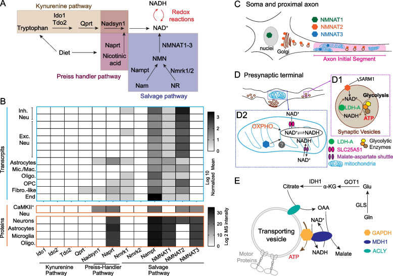
“Autophagy, oxidative stress, mitochondrial dysfunction, and axonal transport have all been found to be positively impacted by improving NAD + metabolism [29–32].”
BackgroundSecond harmonic generation for brain imaging: pathology-related studies
Paveliev, M., Melnikova, A., Samigullin, D., Egorchev, A. A., Андреевна, Т. А., Kiyasov, A. P., Popova, I. Y., Parpura, V., & Aganov, A. V. (2025) · Biophysical Reviews · doi:10.1007/s12551-025-01370-2 · Cited by 8
“…of the live brain and spinal cord (Fenrich et al. 2012; Paveliev et al. 2014; Wei et al. 2021; Lu et al. 2022) could be expanded toward SHG axonal imaging to address the mechanisms of axonopathies, including those associated with neurodegenerative disorders (Berth and Lloyd 2023; Yang et al. 2023).”
Gu, X., Lin, M. M.-W., Xia, Y., Cheng, X., Pan, H., Cai, M., Jiang, M., & Yao, D. (2026) · Biomedicines · doi:10.3390/biomedicines14030636 · Cited by 9
“Studies have revealed that even when blood glucose levels are normal, low-grade inflammation and impaired glucose metabolism accelerate neurological function decline [25,26].”
Neuronal glycolysis: focus on developmental morphogenesis and localized subcellular functions
Gallo, G. (2024) · Communicative & Integrative Biology · doi:10.1080/19420889.2024.2343532 · Cited by 7
Date palm: a potential nutraceutical and phytomedicine in most frequent aging associated diseases
Albishi, T. S. (2024) · Journal of Umm Al-Qura University for Applied Sciences · doi:10.1007/s43994-024-00190-6 · Cited by 8
Meng, L., Kuang, H., Tan, J., Li, X., Liu, Y., Xiang, M., Zhou, Y., Fan, R., & Yu, Y. (2025) · Environment International · doi:10.1016/j.envint.2025.109805 · Cited by 6
Ward, E., Benowitz, L. I., Brunner, T. M., Bu, G., Cayouette, M., Canto-Soler, V., Mesquita, S. D., Polo, A. D., DiAntonio, A., Duan, X., et al. (2025) · Molecular Neurodegeneration · doi:10.1186/s13024-025-00858-5 · Cited by 7
“Additionally, relative to somata, long-range axons are vulnerable to injury, neurotoxin exposure, or issues with protein turnover (e.g., lysosomal dysfunction and plaque formation) [72].”
Axonopathy: mechanisms and potential therapeutic targets for neurodegenerative diseases
Shen, R., Sung, K., Ding, J., & Wu, C. (2026) · Translational Neurodegeneration · doi:10.1186/s40035-026-00543-7 · Cited by 7
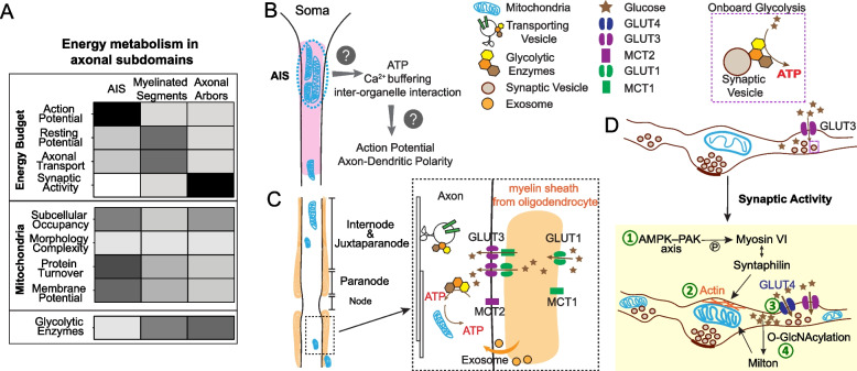
“To optimize energy allocation, axonal metabolism is compartmentalized: the AISs are activity-regulated, the myelinated segments rely on glial metabolic support, and the presynaptic boutons are specialized for synaptic signaling, all under the central regulation of NAD + homeostasis [49].”
InfluentialDisdier, C., Lhotellier, C., Guyot, A.-C., Costa, N., Théodoro, F., Pruvost, A., Skelton, M. R., Joudinaud, T., Mabondzo, A. M., & Bénech, H. (2024) · European Journal of Medicinal Chemistry · doi:10.1016/j.ejmech.2024.117195 · Cited by 8
Emerging nanoplatforms towards microenvironment-responsive glioma therapy
Tripathy, N. S., Sahoo, L., Paikray, S. K., & Dilnawaz, F. (2025) · Medical Oncology · doi:10.1007/s12032-024-02596-y · Cited by 6
“Additionally, NPs have the ability to enter the CNS and pass across the BBB. NPs can travel both anterogradely and retrogradely through neurons' axons and dendrites inside the nervous system [175].”
Fat traffic control: S-acylation in axonal transport
Doerksen, A. H., Herath, N. N., & Sanders, S. S. (2025) · Molecular Pharmacology · doi:10.1016/j.molpha.2025.100039 · Cited by 7
López-Muguruza, E., Peiró-Moreno, C., Pérez‐Cerdá, F., Matute, C., & Ruiz, A. (2025) · Frontiers in Neuroanatomy · doi:10.3389/fnana.2025.1557214 · Cited by 6
“Furthermore, bioenergetic dysfunction and glucose hypometabolism are characteristics common to Alzheimer’s disease (AD), Parkinson’s disease (PD), amyotrophic lateral sclerosis (ALS), and Huntington’s disease (HD), among others (Yang et al., 2023).”
Pabon, A., Bhupana, J. N., & Wong, C. (2024) · Neural Regeneration Research · doi:10.4103/nrr.nrr-d-23-02095 · Cited by 6
Wodrich, A. P. K., Harris, B. T., & Giniger, E. (2024) · Biology Open · doi:10.1242/bio.060335 · Cited by 6
“Although reasons for mitochondrial redistribution into the AIS in the context of injury and disease are becoming more evident, it remains unclear why mitochondria are seemingly excluded from the energetically demanding AIS under normal circumstances (Yang et al., 2023).”
Editorial: Mitochondrial dysfunction as a target in neurodegenerative diseases
Perrone, L. (2023) · Frontiers in Molecular Neuroscience · doi:10.3389/fnmol.2023.1271175 · Cited by 5
“Thus, neurons need to deliver mitochondria through long axons and terminal branches that are characterized by specific energy demands (Yang et al., 2023).”
BackgroundNiou, Z., Yang, S., Enriquez, A., Espinas, N. A., Sri, A., Hines, C. D., Tennessen, J. M., Wu, C., Huang, J.-Y., & Lu, H. (2025) · Advanced Science · doi:10.1002/advs.202509950 · Cited by 4
Self-citationMicrobiota–gut–brain axis and bile acids–driven neuromodulation
Dong, T., Zhang, T., Wang, H., Zhang, J., Abdullah, R., Sun, B., & Peng, G. (2025) · Neural Regeneration Research · doi:10.4103/nrr.nrr-d-25-00927 · Cited by 6
Matrella, M. L., Valletti, A., Gigante, I., Rasmo, D. D., Signorile, A., Russo, S., Lobasso, S., Lobraico, D., Dibattista, M., Pacelli, C., et al. (2024) · Scientific Reports · doi:10.1038/s41598-024-57613-x · Cited by 5
Pyruvate kinase deficiency links metabolic perturbations to neurodegeneration and axonal protection
Waller, T. J., Collins, C. A., & Dus, M. (2025) · Molecular Metabolism · doi:10.1016/j.molmet.2025.102187 · Cited by 4
“In healthy neurons, metabolic homeostasis sustains the high energetic demands of synaptic transmission, ion homeostasis, redox balance, and overall neuronal stability [2,10,146,147].”
Kimura, I., Hayashi, T., & Autio, J. A. (2025) · Imaging Neuroscience · doi:10.1162/imag.a.77 · Cited by 3
Risco-Acevedo, D., Subirós, N., Camacho, H., Ramírez-Sánchez, J., Rodríguez-Virulich, Y., Etchegoyen-Amoros, A., Fuentes-Morales, D., Palenzuela, D. O., Pérez-Saad, H., & García-del-Barco-Herrera, D. (2025) · Experimental Brain Research · doi:10.1007/s00221-025-07183-9 · Cited by 3
“Consistently, postmortem studies of AD patients have revealed a reduction in the number of mature myelinated oligodendrocytes, accompanied by an increase in oligodendrocytes involved in remyelination processes (Yang et al. 2023 ).”
Xu, H., Wang, G., Jiang, Z., Han, Y., Zhao, W., Zhang, H., Liu, H., Liu, H., Li, Z., & Ji, F. (2025) · Advanced Healthcare Materials · doi:10.1002/adhm.202500941 · Cited by 3
Özalay, Ö., Mediavilla, T., Giacobbo, B. L., Marcellino, D. J., Orädd, G., Rieckmann, A., & Sultan, F. R. (2025) · Brain Structure and Function · doi:10.1007/s00429-025-03040-1 · Cited by 2
“…only 1% of all white matter axons (Schuz and Preissl 1996; Schüz and Sultan 2009), their distant presynaptic arbors require a substantial amount of energy, which makes them particularly vulnerable to normal aging and neurodegenerative diseases, such as Alzheimer’s disease (AD) (Yang et al. 2023).”
Transcriptional regulation of human NMNAT2 : insights from 3D genome sequencing and bioinformatics
Chang, Y. C., Yang, S., Cho, M., Nho, K. T., Baizabal, J., & Lu, H. (2025) · FEBS Journal · doi:10.1111/febs.70322 · Cited by 4
Self-citationZhao, H., Lin, S., Chen, H., Yang, L., Gao, K., & Mei, X. (2025) · Materials Today Bio · doi:10.1016/j.mtbio.2025.102624 · Cited by 2
“Among various metabolic pathways, aerobic glucose metabolism is especially critical for neurons, supporting their high energy demand and diverse physiological activities [2].”
Aging-Related Changes in the Injury Response of the Peripheral Nervous System
Cen, Y.-Y., Wang, M.-Y., Song, Q., Gao, X.-L., Zhou, C., Li, C.-J., Liu, F., Zhang, Y.-Y., & Shen, J. (2026) · Neuroscience Bulletin · doi:10.1007/s12264-025-01564-4 · Cited by 2
“However, the age-related impairments in oxidative phosphorylation (OXPHOS), reduced NAD+ levels, and a disrupted TCA cycle collectively cripple ATP production and deprive the cell of essential biosynthetic precursors[247, 277–279].”
Kuznetsov, A. V. (2024) · Journal of Biomechanical Engineering · doi:10.1115/1.4065743 · Cited by 2
Kuznetsov, A. V. (2025) · International Journal of Heat and Mass Transfer · doi:10.1016/j.ijheatmasstransfer.2025.126790 · Cited by 2
Drumond‐Bock, A. L., & Beckstead, M. J. (2025) · European Journal of Neuroscience · doi:10.1111/ejn.70025 · Cited by 1
Parallel Gene Expression Changes in Ventral Midbrain Dopamine and GABA Neurons during Normal Aging
Drumond‐Bock, A. L., Blankenship, H. E., Pham, K. D., Carter, K. A., Freeman, W. M., & Beckstead, M. J. (2025) · eNeuro · doi:10.1523/eneuro.0107-25.2025 · Cited by 3
Zhang, L., Cai, L., Lin, H., Wu, W., Zhu, L., Cai, J., Hu, C., Lin, X., Sun, H., & Wei, X. (2025) · European Journal of Neuroscience · doi:10.1111/ejn.70221 · Cited by 1
Zhang, H., Ya, J., Liao, X., Du, X., Zhao, C., Ren, J., & Qu, X. (2025) · Small · doi:10.1002/smll.202509547 · Cited by 1
SARAK, E. B. G. Ö. G., & Ceylan, Y. (2025) · Bartın University International Journal of Natural and Applied Sciences · doi:10.55930/jonas.1741124 · Cited by 1
Metabolic Symbiosis and Vulnerability in the CNS Axon-Myelin Unit
Miao, K., Meng, M., Liu, L., Li, M., Bai, Y., Guo, Y., Zhang, G., Kong, J., & Mu, J. (2026) · Cellular and Molecular Neurobiology · doi:10.1007/s10571-026-01729-3 · Cited by 1
“Indeed, most of this ATP is consumed by Na⁺/K⁺-ATPase to restore ionic gradients (Yang et al. 2023; Shrivastava et al. 2020 ).”
Jia, Q., Li, Z., Yang, T., Yang, W., & Chi, W. (2026) · Immunity Inflammation and Disease · doi:10.1002/iid3.70469 · Cited by 1
Qian, J., Wang, H., Zhang, Y., Cui, J., Cao, Y., Xie, J., & Shi, L. (2026) · Neuroscience & Biobehavioral Reviews · doi:10.1016/j.neubiorev.2026.106840 · Cited by 1
Early disruptions in brain capillary blood flow control in a mouse model of Alzheimer’s disease
Hariharan, A., Vigderman, A. S., Isaacs, D. P., Weir, N., Park, G., Edwards, S., & Longden, T. A. (2026) · The Journal of General Physiology · doi:10.1085/jgp.202614014 · Cited by 1
Gilloteaux, J., Swert, K. D., Suain, V., & Nicaise, C. (2023) · International Journal of Molecular Sciences · doi:10.3390/ijms242216448 · Cited by 1
Application of proteomics in brain's aging research
Tan, Y., Luo, F., Wei, Y., & Liu, P. (2024) · Human Brain · doi:10.37819/hb.3.1778
Park, J. H., Kim, B., Al‐Amin, M. M., Tate, M. D., Sharify, A. D., John, S. K., Karahan, H., Lu, H., Dabin, L. C., & Kim, J. (2026) · Alzheimer's & Dementia · doi:10.1002/alz.71742
“65,66 Furthermore, CellChat analysis inferred significantly increased communication among the O_3 and M_2 clusters, with additional involvement of the M_1 cluster, in immune-relevant pathways such as PROS and VISTA (Figures 5D and S5C).”
Self-citationUnderstanding Pathophysiological Changes of Traumatic Brain Injury
Zaky, M. Y., & Lamloum, N. S. (2024) · Nutritional neurosciences · doi:10.1007/978-981-97-6341-2_3
Shao, W., Liu, X., Chen, L., Xuedong, Z., Wu, Q., Bu, C., Zheng, F., Yu, G., Hu, H., Guo, Z., et al. (2025) · SSRN Electronic Journal · doi:10.2139/ssrn.5147375
Anatomy of dopamine neuron projections: Axonal arbors, varicosities, and synapses
Trudeau, L., & Denis, R. (2025) · Handbook of behavioral neuroscience · doi:10.1016/b978-0-443-29867-7.00011-6 · Cited by 1
Karkouri, J., Novoselova, M., Miller, S., Zhang, M., Constantinou, A., Graf, C., Atkinson, D., Tramm, B., Schillak, S., Mair, R. J., et al. (2025) · medRxiv · doi:10.1101/2025.10.08.25337571
Yin, C., Kindt, A. S. D., Harms, A. C., Hartman, R., Hankemeier, T., & de Lange, E. C. M. (2025) · Metabolomics · doi:10.1007/s11306-025-02350-z
“These results provide a valuable foundaJon for beper understanding of the interacJon between lipid metabolism, aging, and neurodegeneraJon, as well as for assessing potenJal treatment strategies to address AD progression.”
Wang, P., Shao, Y., Al-Nusaif, M., Zhang, J., Yang, H., Yang, Y., Kim, K., Li, S., Liu, C., Cai, H., et al. (2024) · Figshare · doi:10.6084/m9.figshare.26943114
Wang, P., Shao, Y., Al-Nusaif, M., Zhang, J., Yang, H., Yang, Y., Kim, K., Li, S., Liu, C., Cai, H., et al. (2024) · Figshare · doi:10.6084/m9.figshare.26943117
Wang, P., Shao, Y., Al-Nusaif, M., Zhang, J., Yang, H., Yang, Y., Kim, K., Li, S., Liu, C., Cai, H., et al. (2024) · Figshare · doi:10.6084/m9.figshare.26943120
Shao, W., Liu, X., Chen, L., Zhu, X., Wu, Q., Bu, C., Zheng, F., Yu, G., Hu, H., Guo, Z., et al. (2025) · SSRN Electronic Journal · doi:10.2139/ssrn.5392128
RNA-Seq reveals flight-induced changes in cerebellar transcriptomic profiles of racing pigeons
Stefaniuk‐Szmukier, M., Szmatoła, T., Steg, A., Smołucha, G., & Ropka‐Molik, K. (2025) · Genomics · doi:10.1016/j.ygeno.2025.111188
Neuroprotection and antiaging effects of caloric restriction
Dias, R. A., Mendes, C., Raimundo, N., & Domingues, N. S. (2026) · Elsevier eBooks · doi:10.1016/b978-0-443-33841-0.00023-3
Zhou, H., Xu, G., Chen, Q., Liu, Y., Li, S., Huo, N., Liu, J., Wang, W., Yang, N., & Zhou, N. (2026) · Experimental Neurology · doi:10.1016/j.expneurol.2026.115715
Dai, X., Zhang, Z., Ge, X., Zhou, H., Yu, P., Li, Y., Chen, S., Huang, J.-L., Wu, Q., & Liang, J. (2026) · Research Square · doi:10.21203/rs.3.rs-9024544/v1
Ye, H.-L., Zheng, X.-X., Li, M.-M., Zhang, Z.-Q., Chen, L.-X., Zhang, W.-J., Fan, R. C.-H., He, G., & Xu, A.-C. (2026) · Chemico-Biological Interactions · doi:10.1016/j.cbi.2026.112118
Age-related changes to brain energetics revealed by PET/MRI imaging
Bevington, C. W. J., Dhaliwal, S., McKenzie, J., Stoessl, A. J., & Sossi, V. (2026) · NeuroImage Clinical · doi:10.1016/j.nicl.2026.104014
Wan, X., Lu, W., Liu, S., Zhao, Y., Hu, J., Ding, X., Gu, P., Zou, Y., Jiang, B., & Yang, Y. (2026) · Free Radical Biology and Medicine · doi:10.1016/j.freeradbiomed.2026.05.341
Muhammad, H., Yasin, M. A. B. M., Jaafar, J., Hou, L. J., & Hitam, W. H. W. (2026) · Taiwan Journal of Ophthalmology · doi:10.4103/tjo.tjo-d-25-00165
Tamada, H. (2026) · Frontiers in Neural Circuits · doi:10.3389/fncir.2026.1870424
“Although the mechanisms responsible for mitochondrial exclusion from the healthy AIS remain unclear (Yang et al., 2023), the accumulation of mitochondria under pathological conditions may reflect a protective cellular response.”
Synaptic dysfunction as a major driver in Alzheimer's disease: Beyond the amyloid paradigm
Mierzwa, N., & Wężyk, M. (2026) · Neuroscience & Biobehavioral Reviews · doi:10.1016/j.neubiorev.2026.106858
Ahsan, A., Ou, J., Majumder, P., Chiang, Y. H., Huang, J. K., & Huang, C.-C. S. (2026) · Journal of Neuroimmunology · doi:10.1016/j.jneuroim.2026.579028
Neurons Die Not by One Hit, but by Signaling Convergence
Chauhan, D., Goel, F., & Rajput, M. S. (2026) · Molecular Neurobiology · doi:10.1007/s12035-026-06093-3
Chowdhury, M. R., Jeong, G.-H., & Lee, I. (2026) · Cells · doi:10.3390/cells15151404
“Mitochondrial impairment in neurons can reduce ATP availability, increase mtROS, disturb calcium buffering, and weaken synaptic resilience [49].”
Neuronal Prps is required for synaptic development and locomotor function in Drosophila melanogaster
Dang, T. N., & Yoshida, H. (2026) · Gene · doi:10.1016/j.gene.2026.150361 · Cited by 1
Liu, X., Chen, L., Zhu, X., Wu, Q., Bu, C., Zheng, F., Yu, G., Hu, H., Guo, Z., Zhou, J., et al. (2026) · Chemico-Biological Interactions · doi:10.1016/j.cbi.2026.112318
Devi, D., Rani, A., Singh, J., Quasmi, M. N., Jangra, A., & Kumar, D. (2026) · SN Comprehensive Clinical Medicine · doi:10.1007/s42399-026-02731-y
“Additionally, vascular diseases impact the cognitive functions and contribute to cognitive decline due to decreased cerebral blood flow, which affects the metabolism of glucose [ 12].”
Bone regulates locomotion by sustaining motoneuronal mitochondrial functionPreprint
Romeo-Guitart, D., Tirani, T., Milunov, D., Torres-Juacida, A. K., Lamotte, B., Arenas-Plascencia, C., Durand, S., Nemazanyy, I., Moriceau, S., Saha, S., et al. (2026) · bioRxiv (Cold Spring Harbor Laboratory) · doi:10.64898/2026.10.07.750453
Wang, D., Li, H., Zeng, Y., Gao, J., Xu, M., Bo, B., Pei, M., Liang, Z., Zhou, N., & Thompson, G. J. (2026) · Frontiers in Neuroscience · doi:10.3389/fnins.2026.1949911
“In Alzheimer’s disease models, neuronal injury in affected cortical regions leads to astrocyte damage (Verkhratsky et al., 2017; Toledano-Diaz et al., 2022), contributing to vascular dysfunction and reduced CBF (Amtul et al., 2020), ultimately impacting CMR O2 (Yang et al., 2023).”
Nutrients and Their Role in Autophagy Regulation
Wilson, N., Koçak, G., Sarkar, S., Ruiz, Y. R., & Korolchuk, V. I. (2026) · Biochemistry · doi:10.1021/acs.biochem.6c00482
Metadata
- Authors
- Yang, S., Park, J. H., & Lu, H.-C.
- Affiliations
- Indiana University Bloomington (US)
- Corresponding author
- Lu, Hui-Chen
- Venue
- Molecular Neurodegeneration
- Published
- 2023-07-20
- Volume
- 18
- Issue
- 1
- Pages
- 49-49
- ISSN
- 1750-1326 (ISSN-L)
- PMC
- PMC10357692
- OpenAlex
- W4384925673
- Cited by
- 94
- References
- 428
- Citations by year
- 2026: 25 · 2025: 37 · 2024: 28 · 2023: 4
- FWCI
- 2.54
- Type
- journal article
- Language
- en
- Open access
- gold
- doi.org
- Locations
- Molecular Neurodegeneration (publishedVersion, cc-by)
- PubMed Central (publishedVersion, cc-by)
- PubMed (publishedVersion)
- DOAJ (DOAJ: Directory of Open Access Journals) (submittedVersion, cc-by-sa)
- Funders
- NINDS (NS086794)
- Topics
- Life Sciences › Biochemistry, Genetics and Molecular Biology › Molecular Biology › Mitochondrial Function and Pathology
- Life Sciences › Neuroscience › Cellular and Molecular Neuroscience › Neuroscience and Neuropharmacology Research
- Life Sciences › Neuroscience › Neurology › Neurological Disease Mechanisms and Treatments
- Keywords
- axonal energy metabolism, axonopathy, neurodegenerative diseases, brain aging, glucose hypometabolism, brain connectome, long-range axons, axon initial segment, myelinated axons, axonal arbors, NAD+ homeostasis, NAD biosynthesis, NAD+ recycling, neuroprotection, Alzheimer's disease, Parkinson's disease, Huntington's disease, amyotrophic lateral sclerosis, cognitive decline
- MeSH terms
- Aging, Animals, Axons, Energy Metabolism, Glucose, Humans, NAD, Neurodegenerative Diseases
About these checks
Registry, retraction, Cites and statistics results are computed, not judged, with one exception: a printed reference that no index lists is read once by a model before it counts as a citation error. Claims are extracted by a model and kept only when their quote appears verbatim in the text; each cited source is then checked, with a model’s help, for whether it says what the claim relies on. Those verdicts can be wrong — read the claim’s quote and the cited source before relying on one.
Statistics and arithmetic are recomputed, not judged.
Confidence bands are shown only for fields calibrated on our annotated test set. None is calibrated yet, so no band is shown.
What counts as a claim ›How we measure confidence ›How we check this ›
Are you an author, or do you see a problem with this page? Request a review
