

The DOI matches its Crossref record; 426 of the 428 references it cites were found in a registry or an index.
Axonal energy metabolism, and the effects in aging and neurodegenerative diseases
Open accessAbstract
Abstract Human studies consistently identify bioenergetic maladaptations in brains upon aging and neurodegenerative disorders of aging (NDAs), such as Alzheimer’s disease, Parkinson’s disease, Huntington’s disease, and Amyotrophic lateral sclerosis. Glucose is the major brain fuel and glucose hypometabolism has been observed in brain regions vulnerable to aging and NDAs. Many neurodegenerative susceptible regions are in the topological central hub of the brain connectome, linked by densely interconnected long-range axons. Axons, key components of the connectome, have high metabolic needs to support neurotransmission and other essential activities. Long-range axons are particularly vulnerable to injury, neurotoxin exposure, protein stress, lysosomal dysfunction, etc. Axonopathy is often an early sign of neurodegeneration. Recent studies ascribe axonal maintenance failures to local bioenergetic dysregulation. With this review, we aim to stimulate research in exploring metabolically oriented neuroprotection strategies to enhance or normalize bioenergetics in NDA models. Here we start by summarizing evidence from human patients and animal models to reveal the correlation between glucose hypometabolism and connectomic disintegration upon aging/NDAs. To encourage mechanistic investigations on how axonal bioenergetic dysregulation occurs during aging/NDAs, we first review the current literature on axonal bioenergetics in distinct axonal subdomains: axon initial segments, myelinated axonal segments, and axonal arbors harboring pre-synaptic boutons. In each subdomain, we focus on the organization, activity-dependent regulation of the bioenergetic system, and external glial support. Second, we review the mechanisms regulating axonal nicotinamide adenine dinucleotide (NAD + ) homeostasis, an essential molecule for energy metabolism processes, including NAD + biosynthetic, recycling, and consuming pathways. Third, we highlight the innate metabolic vulnerability of the brain connectome and discuss its perturbation during aging and NDAs. As axonal bioenergetic deficits are developing into NDAs, especially in asymptomatic phase, they are likely exaggerated further by impaired NAD + homeostasis, the high energetic cost of neural network hyperactivity, and glial pathology. Future research in interrogating the causal relationship between metabolic vulnerability, axonopathy, amyloid/tau pathology, and cognitive decline will provide fundamental knowledge for developing therapeutic interventions.
Overview
The claims and statistics of this paper haven’t been checked yet.
Run the check — free, no account needed. It reads the open-access text and checks the sources it cites.
Integrity signals
Registry
Cites
Statistics
Figures
Verification
Claims the paper makes
The claims and statistics of this paper haven’t been checked yet.
Run the check — free, no account needed. It reads the open-access text and checks the sources it cites.
Claims other papers make about this one
Each is checked against this paper’s own text: does it say what the citing paper says it does?
The source backs part of the claim, not all of it — or backs it for a different population, condition or comparison.
In the citing paper
“A snRNA‐seq study in human AD also reported downregulation of synaptic cell adhesion–related gene expression and enrichment terms in an oligodendrocyte cluster. 65 , 66”
From Unique transcriptomic alterations in 5XFAD;PS19 mouse model identify glial lipid dysregulation and coordinated microglial–oligodendrocyte responses (2026) · cites this paper as [66]
Experimental — accuracy not yet measured
Self-check
Percentages, sums and reported differences re-computed from the paper’s own numbers.
The self-check needs the paper’s full text.
Citations
Cites
1 retracted
Braidy, N., Poljak, A., Grant, R., Jayasena, T., Mansour, H., Chan‐Ling, T., Guillemin, G. J., Smythe, G. A., & Sachdev, P. S. (2013) · Biogerontology · doi:10.1007/s10522-013-9489-5 · Cited by 123
RetractedMapping the structural core of human cerebral cortex
Hagmann, P., Cammoun, L., Gigandet, X., Meuli, R., Honey, C. J., Wedeen, V. J., & Sporns, O. (2008) · PLoS Biology · doi:10.1371/journal.pbio.0060159 · Cited by 4,173 · cited 1×
“The majority of AD-susceptible regions identified are located in the default mode network (DMN), the topological central hub of the brain connectome [38, 39], containing densely interconnected long-range corticocortical axons [40, 41].”
BackgroundClinical and biomarker changes in dominantly inherited Alzheimer's disease
Bateman, R. J., Xiong, C., Benzinger, T. L. S., Fagan, A. M., Goate, A., Fox, N. C., Marcus, D. S., Cairns, N. J., Xie, X., Blazey, T. M., et al. (2012) · New England Journal of Medicine · doi:10.1056/nejmoa1202753 · Cited by 3,814 · cited 1×
“Imaging: MRI, FDG‐PET, and PiB‐PET for 40 noncarriers and 88 PSEN1, PSEN2, APP mutation carriers Aimed to use data from longitudinal study to identify pathophysiological biomarkers • Mutation carriers 10–15 yrs before AD: Bilateral hippocampal atrophy, Precuneus glucose uptake↓ Episodic memory↓ [45]”
MethodInterplay between Cortical Spreading Depolarization and Seizures
Kramer, D. R., Fujii, T., Ohiorhenuan, I. E., & Liu, C. Y. (2017) · Stereotactic and Functional Neurosurgery · doi:10.1159/000452841 · Cited by 3,721
An Energy Budget for Signaling in the Grey Matter of the Brain
Attwell, D., & Laughlin, S. B. (2001) · Journal of Cerebral Blood Flow & Metabolism · doi:10.1097/00004647-200110000-00001 · Cited by 3,543
Ageing as a risk factor for neurodegenerative disease
Hou, Y., Dan, X., Babbar, M., Wei, Y., Hasselbalch, S. G., Croteau, D. L., & Bohr, V. A. (2019) · Nature Reviews Neurology · doi:10.1038/s41582-019-0244-7 · Cited by 3,310 · cited 1×
“Aging is considered one of the biggest risk factors for dementia and sporadic late-onset neurodegenerative diseases [20].”
BackgroundPellerin, L., & Magistretti, P. J. (1994) · Proceedings of the National Academy of Sciences · doi:10.1073/pnas.91.22.10625 · Cited by 2,927 · cited 1×
“Because LDH-B favors the conversion in reverse direction to LDH-A, that is it consumes NAD+ and produces NADH and pyruvate [291, 292], LDH-B’s activity may counteract LDH-A, thus reducing NAD redox potential and restraining glycolysis.”
BackgroundThe mechanism of eukaryotic translation initiation and principles of its regulation
Jackson, R. J., Hellen, C. U. T., & Pestova, T. V. (2010) · Nature Reviews Molecular Cell Biology · doi:10.1038/nrm2838 · Cited by 2,759
Brain energy metabolism: focus on Astrocyte-neuron metabolic cooperation
Bélanger, M., Allaman, I., & Magistretti, P. J. (2011) · Cell Metabolism · doi:10.1016/j.cmet.2011.08.016 · Cited by 2,313
The role of the posterior cingulate cortex in cognition and disease
Leech, R., & Sharp, D. J. (2014) · Brain · doi:10.1093/brain/awt162 · Cited by 2,247
Sirtuins as regulators of metabolism and healthspan
Houtkooper, R. H., Pirinen, E., & Auwerx, J. (2012) · Nature Reviews Molecular Cell Biology · doi:10.1038/nrm3293 · Cited by 2,008
Metabolic reduction in the posterior cingulate cortex in very early Alzheimer's disease
Minoshima, S., Giordani, B., Berent, S., Frey, K. A., Foster, N. L., & Kuhl, D. E. (1997) · Annals of Neurology · doi:10.1002/ana.410420114 · Cited by 1,802 · cited 1×
“MCI/AD Imaging: FDG‐PET for 66 AD, 23 early AD, 22 ctrl subjects Aimed to investigate cerebral glucose metabolism in early AD • Glucose uptake in PCC↓ MMSE scores↓ [42]”
MethodSynaptic Energy Use and Supply
Harris, J. J., Jolivet, R., & Attwell, D. (2012) · Neuron · doi:10.1016/j.neuron.2012.08.019 · Cited by 1,747 · cited 1×
“Axonal activities require substantial energy supply [2, 66] and recent studies ascribe axonal maintenance failures to the dysregulation of axonal bioenergetics [67, 68].”
BackgroundSugar for the brain: the role of glucose in physiological and pathological brain function
Mergenthaler, P., Lindauer, U., Dienel, G. A., & Meisel, A. (2013) · Trends in Neurosciences · doi:10.1016/j.tins.2013.07.001 · Cited by 1,612
Molecular diversity and specializations among the cells of the adult mouse brain
Saunders, A., Macosko, E. Z., Wysoker, A., Goldman, M., Krienen, F. M., de Rivera, H., Bien, E., Baum, M., Bortolin, L., Wang, S., et al. (2018) · Cell · doi:10.1016/j.cell.2018.07.028 · Cited by 1,531
Transfer of mitochondria from astrocytes to neurons after stroke
Hayakawa, K., Esposito, E., Wang, X., Terasaki, Y., Liu, Y., Xing, C., Ji, X., & Lo, E. H. (2016) · Nature · doi:10.1038/nature18928 · Cited by 1,423 · cited 1×
“evidence for the transfer of mitochondria from astrocytes to neurons to increase neuronal ATP level and viability upon cerebral ischemia in an astrocytic CD38-dependent manner [184].”
BackgroundAmyotrophic lateral sclerosis is a distal axonopathy: evidence in mice and man
Fischer, L. R., Culver, D. G., Tennant, P., Davis, A. A., Wang, M.-S., Castellano-Sanchez, A. A., Khan, J., Polak, M. A., & Glass, J. D. (2004) · Experimental Neurology · doi:10.1016/j.expneurol.2003.10.004 · Cited by 1,383
Reiman, E. M., Caselli, R. J., Yun, L. S., Chen, K., Bandy, D., Minoshima, S., Thibodeau, S. N., & Osborne, D. (1996) · New England Journal of Medicine · doi:10.1056/nejm199603213341202 · Cited by 1,366
NAD + in aging, metabolism, and neurodegeneration
Verdin, E. (2015) · Science · doi:10.1126/science.aac4854 · Cited by 1,319
Lactate in the brain: from metabolic end-product to signalling molecule
Magistretti, P. J., & Allaman, I. (2018) · Nature Reviews Neuroscience · doi:10.1038/nrn.2018.19 · Cited by 1,298 · cited 1×
“Some evidence suggest that lactate secreted from astrocytes modulates neuronal excitability as a signaling molecule by activating hydroxycarboxylic acid receptor 1 (HCAR1), a putative lactate receptor, in excitatory synapses [249].”
BackgroundA Cellular Perspective on Brain Energy Metabolism and Functional Imaging
Magistretti, P. J., & Allaman, I. (2015) · Neuron · doi:10.1016/j.neuron.2015.03.035 · Cited by 1,285
Normal aging induces A1-like astrocyte reactivity
Clarke, L. E., Liddelow, S. A., Chakraborty, C., Münch, A. E., Heiman, M., & Barres, B. A. (2018) · Proceedings of the National Academy of Sciences · doi:10.1073/pnas.1800165115 · Cited by 1,237 · cited 1×
“Neurotoxic astrocyte subtypes found in aged mouse hippocampus and striatum exhibit downregulated genes in mitochondrial function, antioxidant defense, and cholesterol synthesis [400, 401].”
BackgroundThe axonal transport of mitochondria
Saxton, W. M., & Hollenbeck, P. J. (2012) · Journal of Cell Science · doi:10.1242/jcs.053850 · Cited by 1,218
Axonopathy and Transport Deficits Early in the Pathogenesis of Alzheimer’s Disease
Stokin, G. B., Lillo, C., Falzone, T. L., Brusch, R. G., Rockenstein, E., Mount, S. L., Raman, R., Davies, P., Masliah, E., Williams, D. S., et al. (2005) · Science · doi:10.1126/science.1105681 · Cited by 1,214
The hubs of the human connectome are generally implicated in the anatomy of brain disorders
Crossley, N. A., Mechelli, A., Scott, J., Carletti, F., Fox, P. T., McGuire, P., & Bullmore, E. T. (2014) · Brain · doi:10.1093/brain/awu132 · Cited by 1,166 · cited 1×
“The majority of AD-susceptible regions identified are located in the default mode network (DMN), the topological central hub of the brain connectome [38, 39], containing densely interconnected long-range corticocortical axons [40, 41].”
BackgroundMicroglial brain region-dependent diversity and selective regional sensitivities to aging
Grabert, K., Michoel, T., Karavolos, M. H., Clohisey, S., Baillie, J. K., Stevens, M. P., Freeman, T. C., Summers, K. M., & McColl, B. W. (2016) · Nature Neuroscience · doi:10.1038/nn.4222 · Cited by 1,151
Zhou, Y., Song, W. M., Andhey, P. S., Swain, A., Levy, T., Miller, K. R., Poliani, P. L., Cominelli, M., Grover, S., Gilfillan, S., et al. (2020) · Nature Medicine · doi:10.1038/s41591-019-0695-9 · Cited by 1,150 · cited 1×
“Transcriptionally, astrocytes from human AD prefrontal cortex also exhibit remarkably differences from mouse 5xFAD cortex, featured by the downregulation of genes coordinating free-fatty-acid transport, lipid droplet storage and ROS detoxification [424].”
BackgroundOligodendrocyte heterogeneity in the mouse juvenile and adult central nervous system
Marques, S., Zeisel, A., Codeluppi, S., van Bruggen, D., Mendanha Falcão, A., Xiao, L., Li, H., Häring, M., Hochgerner, H., Romanov, R. A., et al. (2016) · Science · doi:10.1126/science.aaf6463 · Cited by 1,132
Saturated Reconstruction of a Volume of Neocortex
Kasthuri, N., Hayworth, K. J., Berger, D. R., Schalek, R. L., Conchello, J. A., Knowles-Barley, S., Lee, D., Vázquez-Reina, A., Kaynig, V., Jones, T. R., et al. (2015) · Cell · doi:10.1016/j.cell.2015.06.054 · Cited by 1,044 · cited 1×
“EM data from neocortex and hippocampus showed that mitochondria are rarely present in dendritic spine heads or adjacent to the post-synaptic densities [71, 181, 205].”
BackgroundBrain energy rescue: an emerging therapeutic concept for neurodegenerative disorders of ageing
Cunnane, S. C., Trushina, E., Morland, C., Prigione, A., Casadesus, G., Andrews, Z. B., Beal, M. F., Bergersen, L. H., Brinton, R. D., de la Monte, S., et al. (2020) · Nature Reviews Drug Discovery · doi:10.1038/s41573-020-0072-x · Cited by 1,003
Johnson, E. C. B., Dammer, E. B., Duong, D. M., Ping, L., Zhou, M., Yin, L., Higginbotham, L. A., Guajardo, A., White, B., Troncoso, J. C., et al. (2020) · Nature Medicine · doi:10.1038/s41591-020-0815-6 · Cited by 1,001 · cited 1×
“However, the large-scale proteomic analysis of human AD postmortem brains suggests enhanced sugar metabolism in astrocytes and microglia [423].”
BackgroundNutrient-Sensitive Mitochondrial NAD+ Levels Dictate Cell Survival
Yang, H., Yang, T., Baur, J. A., Perez, E., Matsui, T., Carmona, J. J., Lamming, D. W., Souza-Pinto, N. C., Bohr, V. A., Rosenzweig, A., et al. (2007) · Cell · doi:10.1016/j.cell.2007.07.035 · Cited by 950
Yao, J., Irwin, R. W., Zhao, L., Nilsen, J., Hamilton, R. T., & Brinton, R. D. (2009) · Proceedings of the National Academy of Sciences · doi:10.1073/pnas.0903563106 · Cited by 940
Brain Glucose Metabolism: Integration of Energetics with Function
Dienel, G. A. (2018) · Physiological Reviews · doi:10.1152/physrev.00062.2017 · Cited by 911 · cited 1×
“Thus, the brain mainly relies on glucose catabolism to generate adenosine triphosphate (ATP) and cannot afford any disruption of glucose and oxygen delivery [4].”
BackgroundPolydendrocytes (NG2 cells): multifunctional cells with lineage plasticity
Nishiyama, A., Komitova, M., Suzuki, R., & Zhu, X. (2009) · Nature Reviews Neuroscience · doi:10.1038/nrn2495 · Cited by 865 · cited 1×
“Interestingly, OPCs, the unique glia cells forming synapse-like contacts with axons, abundantly express NMNAT2 mRNA [281, 282].”
BackgroundHerrero-Mendez, A., Almeida, A., Fernández, E., Maestre, C., Moncada, S., & Bolaños, J. P. (2009) · Nature Cell Biology · doi:10.1038/ncb1881 · Cited by 844 · cited 1×
“However, its abundance in adult brain neurons remains low due to its constant degradation via the ubiquitin proteasomal pathway [237].”
BackgroundA Single-Cell Transcriptome Atlas of the Aging Drosophila Brain
Davie, K., Janssens, J., Koldere, D., De Waegeneer, M., Pech, U., Kreft, Ł., Aibar, S., Makhzami, S., Christiaens, V., Bravo González-Blas, C., et al. (2018) · Cell · doi:10.1016/j.cell.2018.05.057 · Cited by 840
Activity-driven local ATP synthesis is required for synaptic function
Rangaraju, V., Calloway, N., & Ryan, T. A. (2014) · Cell · doi:10.1016/j.cell.2013.12.042 · Cited by 836 · cited 4×
“During this active state, many calcium-driven processes and SV cycling are the major consumers of ATP, while Na+/K+ ATPase still consumes little [188].”
BackgroundInfluentialCerebral metabolic and cognitive decline in persons at genetic risk for Alzheimer’s disease
Small, G. W., Ercoli, L. M., Silverman, D. H. S., Huang, S.-C., Komo, S., Bookheimer, S. Y., Lavretsky, H., Miller, K., Siddarth, P., Rasgon, N. L., et al. (2000) · Proceedings of the National Academy of Sciences · doi:10.1073/pnas.090106797 · Cited by 790 · cited 2×
“MCI/AD Imaging: FDG‑PET for 66 AD, 23 early AD, 22 ctrl subjects Aimed to investigate cerebral glucose metabolism in early AD • Glucose uptake in PCC↓ MMSE scores↓ [42] Imaging: FDG‑PET and DTI with 20 early AD, 18 ctrl subjects Aimed to explore the association among hippocampal structural integrity, whole brain gluco…”
BackgroundNAD+ in Brain Aging and Neurodegenerative Disorders
Lautrup, S., Sinclair, D. A., Mattson, M. P., & Fang, E. F. (2019) · Cell Metabolism · doi:10.1016/j.cmet.2019.09.001 · Cited by 790
Cell type- and brain region-resolved mouse brain proteome
Sharma, K., Schmitt, S., Bergner, C. G., Tyanova, S., Kannaiyan, N., Manrique-Hoyos, N., Kongi, K., Cantuti, L., Hanisch, U.-K., Philips, M.-A., et al. (2015) · Nature Neuroscience · doi:10.1038/nn.4160 · Cited by 785 · cited 3×
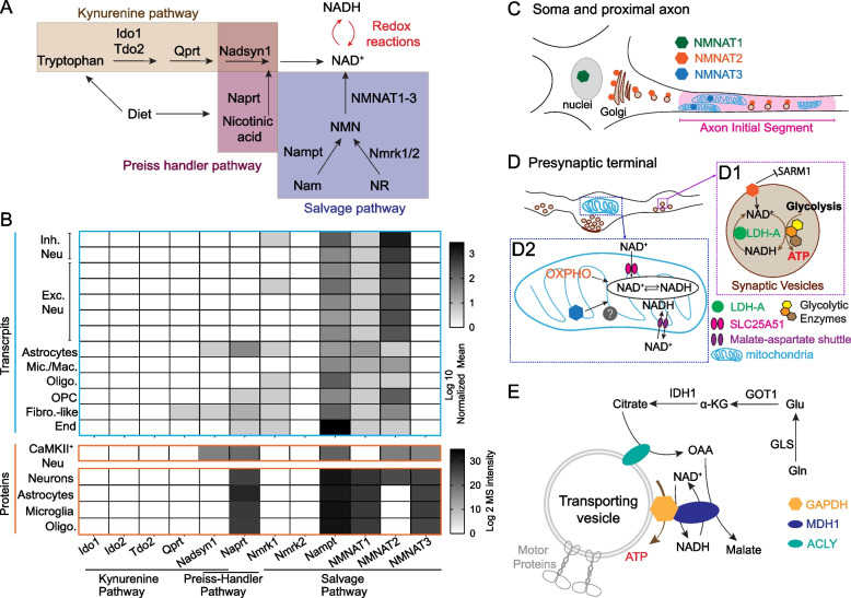
“Single cell transcriptomic and proteomic data suggest the expressions of SLC25A51 and NMNAT3 in neurons (Allen brain single cell transcriptomics explorer and [260]).”
BackgroundThe case for rejecting the amyloid cascade hypothesis
Herrup, K. (2015) · Nature Neuroscience · doi:10.1038/nn.4017 · Cited by 768 · cited 1×
“For instance, the amyloid cascade hypothesis has been popular for decades in explaining AD, however the negative clinical outcomes to amyloiddirected therapies call for consideration of alternative hypothesis and models [355].”
BackgroundSingle-cell transcriptomic profiling of the aging mouse brain
Ximerakis, M., Lipnick, S. L., Innes, B. T., Simmons, S. K., Adiconis, X., Dionne, D., Mayweather, B. A., Nguyen, L., Niziolek, Z., Ozek, C., et al. (2019) · Nature Neuroscience · doi:10.1038/s41593-019-0491-3 · Cited by 736 · cited 2×
“Single-cell transcriptomic analysis identified the decreased expression of mitochondrial OXPHO genes as a consistent signature of brain aging, prevalent across neurons and non-neuronal cell types [21, 22].”
BackgroundWhite matter changes in Alzheimer’s disease: A focus on myelin and oligodendrocytes
Nasrabady, S. E., Rizvi, B., Goldman, J. E., & Brickman, A. M. (2018) · Acta Neuropathologica Communications · doi:10.1186/s40478-018-0515-3 · Cited by 723
Early deficits in synaptic mitochondria in an Alzheimer's disease mouse model
Du, H., Guo, L., Yan, S., Sosunov, A. A., McKhann, G. M., & ShiDu Yan, S. (2010) · Proceedings of the National Academy of Sciences · doi:10.1073/pnas.1006586107 · Cited by 704
The aging astrocyte transcriptome from multiple regions of the mouse brain.
Boisvert, M. M., Erikson, G. A., Shokhirev, M. N., & Allen, N. J. (2018) · Cell Reports · doi:10.1016/j.celrep.2017.12.039 · Cited by 702 · cited 1×
“Neurotoxic astrocyte subtypes found in aged mouse hippocampus and striatum exhibit downregulated genes in mitochondrial function, antioxidant defense, and cholesterol synthesis [400, 401].”
BackgroundAxonal transport: cargo-specific mechanisms of motility and regulation
Maday, S., Twelvetrees, A. E., Moughamian, A. J., & Holzbaur, E. L. F. (2014) · Neuron · doi:10.1016/j.neuron.2014.10.019 · Cited by 683
The genetic epidemiology of neurodegenerative disease
Bertram, L. (2005) · Journal of Clinical Investigation · doi:10.1172/jci24761 · Cited by 680 · cited 1×
“unhealthy lifestyle [26], free radical or neurotoxicants exposures [27]), disease-associated genetic mutations [28], or epigenetic modifications [29] result in NDAs.”
BackgroundTranscellular degradation of axonal mitochondria
Davis, C.-H. O., Kim, K.-Y., Bushong, E. A., Mills, E. A., Boassa, D., Shih, T., Kinebuchi, M., Phan, S., Zhou, Y., Bihlmeyer, N. A., et al. (2014) · Proceedings of the National Academy of Sciences · doi:10.1073/pnas.1404651111 · Cited by 673 · cited 1×
“This has been observed in healthy adult optic axons and is implicated to occur similarly in superficial layers of cerebral cortex [185].”
BackgroundSirtuin regulation of mitochondria: energy production, apoptosis, and signaling
Verdin, E., Hirschey, M. D., Finley, L. W. S., & Haigis, M. C. (2010) · Trends in Biochemical Sciences · doi:10.1016/j.tibs.2010.07.003 · Cited by 661
Jones, D. P., & Sies, H. (2015) · Antioxidants & Redox Signaling · doi:10.1089/ars.2015.6247 · Cited by 647 · cited 23×
“Pivotally, the NAD redox potential (NAD+/NADH) determines the thermodynamic driving force for the NAD+ or NADH consuming steps in both glycolysis and OXPHO [255].”
BackgroundInfluentialMitochondria at the neuronal presynapse in health and disease
Devine, M. J., & Kittler, J. T. (2018) · Nature reviews. Neuroscience · doi:10.1038/nrn.2017.170 · Cited by 636
Wallerian degeneration: an emerging axon death pathway linking injury and disease
Conforti, L., Gilley, J., & Coleman, M. P. (2014) · Nature reviews. Neuroscience · doi:10.1038/nrn3680 · Cited by 630
NAD+ homeostasis in health and disease
Katsyuba, E., Romani, M., Hofer, D., & Auwerx, J. (2020) · Nature Metabolism · doi:10.1038/s42255-019-0161-5 · Cited by 627
Small, G. W. (1995) · JAMA: The Journal of the American Medical Association · doi:10.1001/jama.1995.03520360056039 · Cited by 604 · cited 1×
“Imaging: FDG‐PET of 12 relatives with APOE4, 19 relatives with‐ out APOE4, 7 AD subjects Aimed to determine if APOE4 is associated with brain function decline in relatives at risk for familial AD • APOE4 carriers at risk for AD: Parietal glucose uptake↓ Left– right metabolic asymmetry↑ • Dementia patients: Parietal gl…”
BackgroundDistinct Profiles of Myelin Distribution Along Single Axons of Pyramidal Neurons in the Neocortex
Tomassy, G. S., Berger, D. R., Chen, H.-H., Kasthuri, N., Hayworth, K. J., Vercelli, A., Seung, H. S., Lichtman, J. W., & Arlotta, P. (2014) · Science · doi:10.1126/science.1249766 · Cited by 600
Kang, S. H., Li, Y., Fukaya, M., Lorenzini, I., Cleveland, D. W., Ostrow, L. W., Rothstein, J. D., & Bergles, D. E. (2013) · Nature Neuroscience · doi:10.1038/nn.3357 · Cited by 599
SARM1 activation triggers axon degeneration locally via NAD + destruction
Gerdts, J., Brace, E. J., Sasaki, Y., DiAntonio, A., & Milbrandt, J. (2015) · Science · doi:10.1126/science.1258366 · Cited by 593
Oligodendroglial NMDA receptors regulate glucose import and axonal energy metabolism
Saab, A. S., Tzvetavona, I. D., Trevisiol, A., Baltan, S., Dibaj, P., Kusch, K., Möbius, W., Goetze, B., Jahn, H. M., Huang, W., et al. (2016) · Neuron · doi:10.1016/j.neuron.2016.05.016 · Cited by 590 · cited 6×
“Then, glucose is transported into neurons through GLUT3 and GLUT4 [11–14], or imported into oligodendrocytes and microglia through GLUT1 [15, 16]”
BackgroundInfluentialSmajić, S., Prada-Medina, C. A., Landoulsi, Z., Ghelfi, J., Delcambre, S., Dietrich, C., Jarazo, J., Henck, J., Balachandran, S., Pachchek, S., et al. (2022) · Brain · doi:10.1093/brain/awab446 · Cited by 590 · cited 1×
“Consistently, the population of myelinating oligodendrocyte is reduced in human PD postmortem midbrains, accompanied by the transcriptomic upregulation of unfolded protein response and downregulation of neuro-supportive pathways [412].”
BackgroundMitochondrial CB1 receptors regulate neuronal energy metabolism
Bénard, G., Massa, F., Puente, N., Lourenço, J., Bellocchio, L., Soria-Gómez, E., Matias, I., Delamarre, A., Metna-Laurent, M., Cannich, A., et al. (2012) · Nature Neuroscience · doi:10.1038/nn.3053 · Cited by 588 · cited 1×
“Intriguingly, the recent discovery of the roles of endocannabinoids, a well-known retrograde messenger for synaptic transmission [251], in regulating mitochondrial metabolism in both neurons and astrocytes through mitochondrial type-1 cannabinoid receptor (mtCB1) [252, 253], suggests the potential involvement of the e…”
BackgroundSelective vulnerability in neurodegenerative diseases
Fu, H., Hardy, J., & Duff, K. E. (2018) · Nature Neuroscience · doi:10.1038/s41593-018-0221-2 · Cited by 585 · cited 1×
“Recognizing the distinct excitability, energetic demands, mitochondrial regulatory units, proteome composition, and Ca2+ homeostasis in various neuronal subtypes [396, 397], we encourage the revisit of the axon protective effect offered by NMNAT2 and SARM1 manipulation in the most vulnerable neuronal subtypes in indiv…”
BackgroundIn vivo evidence for a lactate gradient from astrocytes to neurons
Mächler, P., Wyss, M. T., Elsayed, M., Stobart, J., Gutierrez, R., von Faber-Castell, A., Kaelin, V., Zuend, M., San Martín, A., Romero-Gómez, I., et al. (2016) · Cell Metabolism · doi:10.1016/j.cmet.2015.10.010 · Cited by 571
Deep Multilayer Brain Proteomics Identifies Molecular Networks in Alzheimer’s Disease Progression
Bai, B., Wang, X., Li, Y., Chen, P.-C., Yu, K., Dey, K. K., Yarbro, J. M., Han, X., Lutz, B. M., Rao, S., et al. (2020) · Neuron · doi:10.1016/j.neuron.2019.12.015 · Cited by 565 · cited 1×
“Despite extremely low NMNAT3 mRNA levels in mouse brain, NMNAT3 protein has been detected in cultured cortical neurons [265], adult mouse brain [260], and human postmortem brain [285].”
BackgroundThe dynamic regulation of NAD metabolism in mitochondria
Stein, L. R., & Imai, S. (2012) · Trends in Endocrinology and Metabolism · doi:10.1016/j.tem.2012.06.005 · Cited by 558
Winnubst, J., Bas, E., Ferreira, T. A., Wu, Z., Economo, M. N., Edson, P., Arthur, B. J., Bruns, C., Rokicki, K., Schauder, D., et al. (2019) · Cell · doi:10.1016/j.cell.2019.07.042 · Cited by 538
Pacelli, C., Giguère, N., Bourque, M.-J., Lévesque, M., Slack, R. S., & Trudeau, L. (2015) · Current Biology · doi:10.1016/j.cub.2015.07.050 · Cited by 530
Douaud, G., Jbabdi, S., Behrens, T. E. J., Menke, R. A., Gass, A., Monsch, A. U., Rao, A., Whitcher, B., Kindlmann, G., Matthews, P. M., et al. (2011) · NeuroImage · doi:10.1016/j.neuroimage.2010.12.008 · Cited by 514 · cited 1×
“Imaging: DTI with 61 ctrls, 56 MCI, 53 probable AD patients with‐ out a vascular component Aimed to report a comprehensive whole-brain study of diffusion tensor indices and probabilistic tractography obtained from healthy controls, MCI and probable AD subjects • Affected white matter in AD (vs ctrl): Cingulum bundle,…”
MethodBerger, F., Lau, C., Dahlmann, M., & Ziegler, M. (2005) · Journal of Biological Chemistry · doi:10.1074/jbc.m508660200 · Cited by 508 · cited 4×
“2C) and is relatively uniformly expressed across different cell types [268, 269].”
BackgroundInfluentialThe energetics of CNS white matter
Harris, J. J., & Attwell, D. (2012) · The Journal of Neuroscience · doi:10.1523/jneurosci.3430-11.2012 · Cited by 505 · cited 7×
“Myelination greatly improves the energetic efficiency of AP propagation, costing less than 1% of white matter’s energy budget [2, 169].”
BackgroundInfluentialSynaptic degeneration in Alzheimer disease
Tzioras, M., McGeachan, R. I., Durrant, C. S., & Spires-Jones, T. L. (2023) · Nature Reviews Neurology · doi:10.1038/s41582-022-00749-z · Cited by 504
Shepherd, G. M. G., & Harris, K. M. (1998) · The Journal of Neuroscience · doi:10.1523/jneurosci.18-20-08300.1998 · Cited by 502
Vesicular Glycolysis Provides On-Board Energy for Fast Axonal Transport
Zala, D., Hinckelmann, M.-V., Yu, H., da Cunha, M. M. L., Liot, G., Cordelières, F. P., Marco, S., & Saudou, F. (2013) · Cell · doi:10.1016/j.cell.2012.12.029 · Cited by 494 · cited 3×
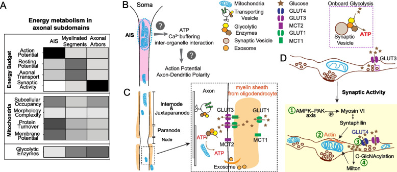
“Studies have shown that glycolytic enzymes are physically associated with fast-moving vesicles through their interaction with Huntingtin and provide onboard ATP synthesis to propel vesicular transport [154, 155] (Fig.”
BackgroundLau, S.-F., Cao, H., Fu, A. K. Y., & Ip, N. Y. (2020) · Proceedings of the National Academy of Sciences · doi:10.1073/pnas.2008762117 · Cited by 476
The Role of Environmental Exposures in Neurodegeneration and Neurodegenerative Diseases
Cannon, J. R., & Greenamyre, J. T. (2011) · Toxicological Sciences · doi:10.1093/toxsci/kfr239 · Cited by 475 · cited 1×
“unhealthy lifestyle [26], free radical or neurotoxicants exposures [27]), disease-associated genetic mutations [28], or epigenetic modifications [29] result in NDAs.”
BackgroundEndogenous Nmnat2 Is an Essential Survival Factor for Maintenance of Healthy Axons
Gilley, J., & Coleman, M. P. (2010) · PLoS Biology · doi:10.1371/journal.pbio.1000300 · Cited by 472
Zhu, X.-H., Lu, M., Lee, B.-Y., Ugurbil, K., & Chen, W. (2015) · Proceedings of the National Academy of Sciences · doi:10.1073/pnas.1417921112 · Cited by 468 · cited 1×
“NAD redox homeostasis disruption NAD+ redox potentials decline in aged brains [371, 372].”
BackgroundLifelong cortical myelin plasticity and age-related degeneration in the live mammalian brain
Hill, R. A., Li, A. M., & Grutzendler, J. (2018) · Nature Neuroscience · doi:10.1038/s41593-018-0120-6 · Cited by 467 · cited 2×
“Additionally, oligodendrocyte numbers and myelin density are decreased in aged frontal cortex, concomitant with the appearance of myelin spheroids and debris [224, 399].”
BackgroundNeuronal Stimulation Triggers Neuronal Glycolysis and Not Lactate Uptake
Díaz-García, C. M., Mongeon, R., Lahmann, C., Koveal, D., Zucker, H., & Yellen, G. (2017) · Cell Metabolism · doi:10.1016/j.cmet.2017.06.021 · Cited by 466
Schönfeld, P., & Reiser, G. (2013) · Journal of Cerebral Blood Flow & Metabolism · doi:10.1038/jcbfm.2013.128 · Cited by 457
Bidirectional cargo transport: moving beyond tug of war
Hancock, W. O. (2014) · Nature Reviews Molecular Cell Biology · doi:10.1038/nrm3853 · Cited by 447 · cited 2×
“Evidence shows that even though kinesin and dynein drive transport in opposite directions, they attach to the same vesicular cargo through modulation by a variety of cargo-specific adaptors and coordinators [180].”
BackgroundThe axon initial segment and the maintenance of neuronal polarity
Rasband, M. N. (2010) · Nature Reviews Neuroscience · doi:10.1038/nrn2852 · Cited by 442 · cited 2×
“The AIS also plays a critical role in establishing and maintaining axon-dendrite polarity, in which the AIS actively guides selective and polarized protein trafficking to axons, and prevents protein diffusion from the somatodendritic area through the physical barrier formed by AIS-anchored membrane proteins and variou…”
BackgroundLRRK2 regulates mitochondrial dynamics and function through direct interaction with DLP1
Wang, X., Ma, Y., Fujioka, H., Liu, J., Wilson‐Delfosse, A. L., Chen, S. G., Perry, G. L., Casadesús, G., & Zhu, X. (2012) · Human Molecular Genetics · doi:10.1093/hmg/dds003 · Cited by 437
Local protein synthesis is a ubiquitous feature of neuronal pre- and postsynaptic compartments
Hafner, A.-S., Donlin-Asp, P. G., Leitch, B., Herzog, E., & Schuman, E. M. (2019) · Science · doi:10.1126/science.aau3644 · Cited by 431
Major Shifts in Glial Regional Identity Are a Transcriptional Hallmark of Human Brain Aging
Soreq, L., Rose, J., Soreq, E., Hardy, J., Trabzuni, D., Cookson, M. R., Smith, C., Ryten, M., Patani, R., & Ule, J. (2017) · Cell Reports · doi:10.1016/j.celrep.2016.12.011 · Cited by 419
Morphological diversity of single neurons in molecularly defined cell types
Peng, H., Xie, P., Liu, L., Kuang, X., Wang, Y., Qu, L., Gong, H., Jiang, S., Li, A., Ruan, Z., et al. (2021) · Nature · doi:10.1038/s41586-021-03941-1 · Cited by 417
Sadick, J. S., O’Dea, M. R., Hasel, P., Dykstra, T., Faustin, A., & Liddelow, S. A. (2022) · Neuron · doi:10.1016/j.neuron.2022.03.008 · Cited by 415
Epigenetic Regulation in Neurodegenerative Diseases
Berson, A., Nativio, R., Berger, S. L., & Bonini, N. M. (2018) · Trends in Neurosciences · doi:10.1016/j.tins.2018.05.005 · Cited by 408 · cited 1×
“unhealthy lifestyle [26], free radical or neurotoxicants exposures [27]), disease-associated genetic mutations [28], or epigenetic modifications [29] result in NDAs.”
BackgroundBiosensor reveals multiple sources for mitochondrial NAD +
Cambronne, X. A., Stewart, M. L., Kim, D., Jones-Brunette, A. M., Morgan, R. K., Farrens, D. L., Cohen, M. S., & Goodman, R. H. (2016) · Science · doi:10.1126/science.aad5168 · Cited by 407
Extracellular vesicle-contained eNAMPT delays aging and extends lifespan in mice
Yoshida, M., Satoh, A., Lin, J. B., Mills, K. F., Sasaki, Y., Rensing, N., Wong, M., Apte, R. S., & Imai, S.-I. (2019) · Cell Metabolism · doi:10.1016/j.cmet.2019.05.015 · Cited by 407 · cited 1×
“Notably, NAMPT can be exchanged transcellularly between cells through extracellular vesicles [266, 267].”
BackgroundGowrishankar, S., Yuan, P., Wu, Y., Schrag, M., Paradise, S., Grutzendler, J., De Camilli, P., & Ferguson, S. M. (2015) · Proceedings of the National Academy of Sciences · doi:10.1073/pnas.1510329112 · Cited by 406
Chung, C. Y., Koprich, J. B., Siddiqi, H. K., & Ole Isacson, A. (2009) · Journal of Neuroscience · doi:10.1523/jneurosci.5427-08.2009 · Cited by 406
Targa Dias Anastacio, H., Matosin, N., & Ooi, L. (2022) · Translational Psychiatry · doi:10.1038/s41398-022-02024-7 · Cited by 401
Voxel-based mapping of brain gray matter volume and glucose metabolism profiles in normal aging
Kalpouzos, G., Chételat, G., Baron, J.-C., Landeau, B., Mevel, K., Godeau, C., Barré, L., Constans, J.-M., Viader, F., Eustache, F., et al. (2009) · Neurobiology of Aging · doi:10.1016/j.neurobiolaging.2007.05.019 · Cited by 392
Functions and effects of creatine in the central nervous system
Andres, R. H., Ducray, A., Schlattner, U., Wallimann, T., & Widmer, H. R. (2008) · Brain Research Bulletin · doi:10.1016/j.brainresbull.2008.02.035 · Cited by 389 · cited 1×
“Facilitated by the creatine kinase/phosphocreatine system [207], ATP produced from mitochondria can rapidly diffuse over a certain range, therefore constantly fulfilling local bioenergetic needs in resident boutons, but also transiently compensating for the needs of nearby boutons lacking mitochondria [200, 208].”
BackgroundDrzezga, A. E., Becker, J. A., Dijk, K. R. A. V., Sreenivasan, A., Talukdar, T., Sullivan, C., Schultz, A. P., Sepulcre, J., Putcha, D., Greve, D. N., et al. (2011) · Brain · doi:10.1093/brain/awr066 · Cited by 385
Oligodendroglia: metabolic supporters of neurons
Philips, T., & Rothstein, J. D. (2017) · Journal of Clinical Investigation · doi:10.1172/jci90610 · Cited by 379 · cited 23×
“Pivotally, the NAD redox potential (NAD+/NADH) determines the thermodynamic driving force for the NAD+ or NADH consuming steps in both glycolysis and OXPHO [255].”
InfluentialMechanism suppressing glycogen synthesis in neurons and its demise in progressive myoclonus epilepsy
Vı́lchez, D., R�os, S., Cifuentes, D., Pujadas, L., Vallès, J., García-Fojeda, B., Criado‐García, O., Fernández-Sánchez, E., Medraño-Fernández, I., Domínguez, J. E., et al. (2007) · Nature Neuroscience · doi:10.1038/nn1998 · Cited by 374 · cited 1×
“Neurons cannot store high energy molecules, such as glycogen and fat, and thus they must synthesize ATP on demand [69, 70].”
BackgroundFueling thought: Management of glycolysis and oxidative phosphorylation in neuronal metabolism
Yellen, G. (2018) · Journal of Cell Biology · doi:10.1083/jcb.201803152 · Cited by 371 · cited 1×
“Glucose is the major brain fuel under normal circumstances [2, 3].”
BackgroundCheng, A., Yang, Y., Zhou, Y., Maharana, C., Lu, D., Peng, W., Liu, Y., Wan, R., Marosi, K., Misiak, M., et al. (2016) · Cell Metabolism · doi:10.1016/j.cmet.2015.10.013 · Cited by 371 · cited 1×
“SIRT3 signaling has been shown to protect neuronal mitochondria against metabolic and excitatory stress [373].”
BackgroundSpatially Stable Mitochondrial Compartments Fuel Local Translation during Plasticity
Rangaraju, V., Lauterbach, M. A., & Schuman, E. M. (2019) · Cell · doi:10.1016/j.cell.2018.12.013 · Cited by 367 · cited 1×
“mitochondrial compartments, consisting of single or multiple mitochondrial filaments with temporal stability of up to 80 min, are exclusively found in dendrites and soma, but not in axons [81].”
BackgroundMagrané, J., Cortez, C. A., Gan, W., & Manfredi, G. (2013) · Human Molecular Genetics · doi:10.1093/hmg/ddt528 · Cited by 359
Axonal transport: Driving synaptic function
Guedes-Dias, P., & Holzbaur, E. L. F. (2019) · Science · doi:10.1126/science.aaw9997 · Cited by 355
Glucose transporter 1 critically controls microglial activation through facilitating glycolysis
Wang, L., Pavlou, S., Du, X., Bhuckory, M. B., Xu, H., & Chen, M. (2019) · Molecular Neurodegeneration · doi:10.1186/s13024-019-0305-9 · Cited by 339 · cited 6×
“Then, glucose is transported into neurons through GLUT3 and GLUT4 [11–14], or imported into oligodendrocytes and microglia through GLUT1 [15, 16]”
InfluentialProgrammed axon degeneration: from mouse to mechanism to medicine
Coleman, M. P., & Höke, A. (2020) · Nature reviews. Neuroscience · doi:10.1038/s41583-020-0269-3 · Cited by 329 · cited 1×
“Though NMNAT overexpressions can offer axonal protections [269, 278], studies also showed that excess amount of NMNAT can cause adverse consequence on presynaptic function in Drosophila [279] and visual circuit plasticity in mice [280].”
BackgroundFacilitation of axon regeneration by enhancing mitochondrial transport and rescuing energy deficits
Zhou, B., Yu, P., Lin, M.-Y., Sun, T., Chen, Y., & Sheng, Z. (2016) · The Journal of Cell Biology · doi:10.1083/jcb.201605101 · Cited by 328
GLUT4 Mobilization Supports Energetic Demands of Active Synapses
Ashrafi, G., Wu, Z., Farrell, R. J., & Ryan, T. A. (2017) · Neuron · doi:10.1016/j.neuron.2016.12.020 · Cited by 327
Actin has a molecular scaffolding, not propulsive, role in presynaptic function
Sankaranarayanan, S., Atluri, P. P., & Ryan, T. A. (2003) · Nature Neuroscience · doi:10.1038/nn1002 · Cited by 324
SARM1 is a metabolic sensor activated by an increased NMN/NAD+ ratio to trigger axon degeneration
Figley, M. D., Gu, W., Nanson, J. D., Shi, Y., Sasaki, Y., Cunnea, K., Malde, A. K., Jia, X., Luo, Z., Saikot, F. K., et al. (2021) · Neuron · doi:10.1016/j.neuron.2021.02.009 · Cited by 324
The Role of Mitochondrially Derived ATP in Synaptic Vesicle Recycling
Pathak, D., Shields, L. Y., Mendelsohn, B. A., Haddad, D., Lin, W., Gerencser, A. A., Kim, H., Brand, M. D., Edwards, R. H., & Nakamura, K. (2015) · Journal of Biological Chemistry · doi:10.1074/jbc.m115.656405 · Cited by 311 · cited 1×
“Facilitated by the creatine kinase/phosphocreatine system [207], ATP produced from mitochondria can rapidly diffuse over a certain range, therefore constantly fulfilling local bioenergetic needs in resident boutons, but also transiently compensating for the needs of nearby boutons lacking mitochondria [200, 208].”
BackgroundSLC25A51 is a mammalian mitochondrial NAD(+) transporter
Luongo, T. S., Eller, J. M., Lu, M.-J., Niere, M., Raith, F., Perry, C., Bornstein, M. R., Oliphint, P., Wang, L., McReynolds, M. R., et al. (2020) · Nature · doi:10.1038/s41586-020-2741-7 · Cited by 308
Genetically Encoded Fluorescent Sensors for Intracellular NADH Detection
Zhao, Y., Jin, J., Hu, Q., Zhou, H.-M., Yi, J., Yu, Z., Xu, L., Wang, X., Yang, Y., & Loscalzo, J. (2011) · Cell Metabolism · doi:10.1016/j.cmet.2011.09.004 · Cited by 302
Pathological Axonal Death through a MAPK Cascade that Triggers a Local Energy Deficit
Yang, J. H., Wu, Z., Renier, N., Simon, D. J., Uryu, K., Park, D. S., Greer, P. A., Tournier, C., Davis, R. J., & Tessier‐Lavigne, M. (2015) · Cell · doi:10.1016/j.cell.2014.11.053 · Cited by 302
Synaptic Mitochondria Are More Susceptible to Ca2+Overload than Nonsynaptic Mitochondria
Brown, M. R., Sullivan, P. G., & Geddes, J. W. (2006) · Journal of Biological Chemistry · doi:10.1074/jbc.m510303200 · Cited by 301
Unappreciated Role of LDHA and LDHB to Control Apoptosis and Autophagy in Tumor Cells
Urbańska, K., & Orzechowski, A. (2019) · International Journal of Molecular Sciences · doi:10.3390/ijms20092085 · Cited by 295 · cited 1×
“During this enzymatic reaction, it regenerates NAD+ by oxidizing glycolysis derived NADH and therefore facilitates the vesicular glycolysis that is required for fast axonal transport [289, 290].”
BackgroundSadleir, K. R., Kandalepas, P. C., Buggia-Prévot, V., Nicholson, D. A., Thinakaran, G., & Vassar, R. (2016) · Acta Neuropathologica · doi:10.1007/s00401-016-1558-9 · Cited by 294
de Wilde, M. C., Overk, C., Sijben, J., & Masliah, E. (2016) · Alzheimer s & Dementia · doi:10.1016/j.jalz.2015.12.005 · Cited by 287 · cited 1×
“Meta-analysis of 417 studies focusing on synaptopathy in postmortem AD brains reveals a consistent loss of synapses in hippocampus and frontal cortex, in which the presynaptic markers are more affected than the postsynaptic markers [328].”
BackgroundD’Acunzo, P., Pérez‐González, R., Kim, Y., Hargash, T., Miller, C., Alldred, M. J., Erdjument‐Bromage, H., Penikalapati, S. C., Pawlik, M., Saito, M., et al. (2021) · Science Advances · doi:10.1126/sciadv.abe5085 · Cited by 287
Glucose Regulates Mitochondrial Motility via Milton Modification by O-GlcNAc Transferase
Pekkurnaz, G., Trinidad, J. C., Wang, X., Kong, D., & Schwarz, T. L. (2014) · Cell · doi:10.1016/j.cell.2014.06.007 · Cited by 283 · cited 1×
“the O-linked-N-acetylglucosaminylation (O-GlcNAcylation) of the mitochondrial adaptor protein, Milton, which allows its interaction with four and a half LIM domains protein 2 (FHL2) to anchor mitochondria onto presynaptic actin filaments, therefore arresting mitochondria in the boutons [217, 218].”
BackgroundEmerging roles of exosomes in neuron-glia communication
Frühbeis, C., Fröhlich, D., & Krämer-Albers, E.-M. (2012) · Frontiers in Physiology · doi:10.3389/fphys.2012.00119 · Cited by 275 · cited 1×
“shed light on the role of oligodendrocyte-secreted exosomes in sustaining axonal transport upon nutritional deprivation [182] (Fig.”
BackgroundRemodeling of Synaptic Actin Induced by Photoconductive Stimulation
Colicos, M. A., Collins, B., Sailor, M. J., & Goda, Y. (2001) · Cell · doi:10.1016/s0092-8674(01)00579-7 · Cited by 275 · cited 1×
“Rearrangement of cytoskeletal components such as actin and microtubule occurs within minutes of electrical stimulation [195, 196] or long-term potentiation (LTP) [197].”
BackgroundGlucose metabolism links astroglial mitochondria to cannabinoid effects
Jiménez-Blasco, D., Busquets-García, A., Hébert-Chatelain, É., Serrat, R., Vicente‐Gutiérrez, C., Ioannidou, C., Gómez-Sotres, P., López-Fabuel, I., Resch-Beusher, M., Resel, E., et al. (2020) · Nature · doi:10.1038/s41586-020-2470-y · Cited by 272 · cited 1×
“Intriguingly, the recent discovery of the roles of endocannabinoids, a well-known retrograde messenger for synaptic transmission [251], in regulating mitochondrial metabolism in both neurons and astrocytes through mitochondrial type-1 cannabinoid receptor (mtCB1) [252, 253], suggests the potential involvement of the e…”
BackgroundMutant A53T α-Synuclein Induces Neuronal Death by Increasing Mitochondrial Autophagy
Choubey, V., Safiulina, D., Vaarmann, A., Cagalinec, M., Warȩski, P., Kuum, M., Zharkovsky, A., & Kaasik, A. (2011) · Journal of Biological Chemistry · doi:10.1074/jbc.m110.132514 · Cited by 271
Pleiotropic Mitochondria: The Influence of Mitochondria on Neuronal Development and Disease
Rangaraju, V., Lewis, T. L., Hirabayashi, Y., Bergami, M., Motori, E., Cartoni, R., Kwon, S., & Courchet, J. (2019) · Journal of Neuroscience · doi:10.1523/jneurosci.1157-19.2019 · Cited by 271 · cited 1×
“It is also possible that clustered mitochondria provide Ca2+ buffering and/or the interactions with endoplasmic reticulum/endosomes to maintain the axon-dendrite polarity [163].”
BackgroundGlycolytic enzymes localize to synapses under energy stress to support synaptic function
Jang, S., Nelson, J. C., Bend, E. G., Rodríguez-Laureano, L., Tueros, F. G., Cartagenova, L., Underwood, K., Jorgensen, E. M., & Colón-Ramos, D. A. (2016) · Neuron · doi:10.1016/j.neuron.2016.03.011 · Cited by 270
Neuronal hyperactivity--A key defect in Alzheimer’s disease?
Busche, M. A., & Konnerth, A. (2015) · BioEssays · doi:10.1002/bies.201500004 · Cited by 266
Desai, M. K., Sudol, K. L., Janelsins, M. C., Mastrangelo, M. A., Frazer, M. E., & Bowers, W. J. (2009) · Glia · doi:10.1002/glia.20734 · Cited by 266
Andersen, J. V., Schousboe, A., & Verkhratsky, A. (2022) · Progress in Neurobiology · doi:10.1016/j.pneurobio.2022.102331 · Cited by 263
Motile axonal mitochondria contribute to the variability of presynaptic strength
Sun, T., Qiao, H., Pan, P.-Y., Chen, Y., & Sheng, Z.-H. (2013) · Cell Reports · doi:10.1016/j.celrep.2013.06.040 · Cited by 263
Pathological structural conversion of α-synuclein at the mitochondria induces neuronal toxicity
Choi, M. L., Chappard, A., Singh, B., Maclachlan, C., Rodrigues, M., Fedotova, E. I., Berezhnov, A. V., De, S., Peddie, C. J., Athauda, D. S., et al. (2022) · Nature Neuroscience · doi:10.1038/s41593-022-01140-3 · Cited by 263
Oligodendroglia: metabolic supporters of axons
Morrison, B. M., Lee, Y., & Rothstein, J. D. (2013) · Trends in Cell Biology · doi:10.1016/j.tcb.2013.07.007 · Cited by 262
Lewis, T. L., Kwon, S.-K., Lee, A., Shaw, R., & Polleux, F. (2018) · Nature Communications · doi:10.1038/s41467-018-07416-2 · Cited by 260
Oxygen and glucose consumption related to Na+-K+ transport in canine brain.
Astrup, J., Sørensen, P. M., & Sørensen, H. R. (1981) · Stroke · doi:10.1161/01.str.12.6.726 · Cited by 251
Mitochondrial sirtuins, metabolism, and aging
Ji, Z., Liu, G., & Qu, J. T. (2021) · Journal of genetics and genomics/Journal of Genetics and Genomics · doi:10.1016/j.jgg.2021.11.005 · Cited by 250 · cited 1×
“SIRTs are well known in governing mitochondrial biology and metabolic homeostasis [305, 308, 309].”
BackgroundChen, Y., & Sheng, Z.-H. (2013) · Journal of Cell Biology · doi:10.1083/jcb.201302040 · Cited by 250 · cited 1×
“[214] showed that Ca2+ release triggered by action potentials binds to Miro, which then releases the C-terminal tail of KIF5 to bind syntaphilin (SNPH).”
BackgroundHara, Y., Yuk, F., Puri, R., Janssen, W. G. M., Rapp, P. R., & Morrison, J. H. (2013) · Proceedings of the National Academy of Sciences · doi:10.1073/pnas.1311310110 · Cited by 244 · cited 3×
“Surgical menopause, causing abrupt estrogen loss, results in an increase of boutons containing donut shaped mitochondria and small active zones, and worsened working memory [220].”
BackgroundInfluentialNon‐signalling energy use in the brain
Engl, E., & Attwell, D. (2015) · The Journal of Physiology · doi:10.1113/jphysiol.2014.282517 · Cited by 243 · cited 3×
“Both actin and tubulin polymerization require ATP and GTP hydrolysis, respectively [148].”
BackgroundAstrocytic processes: from tripartite synapses to the active milieu
Semyanov, A. V., & Verkhratsky, A. (2021) · Trends in Neurosciences · doi:10.1016/j.tins.2021.07.006 · Cited by 243
A connectomic study of a petascale fragment of human cerebral cortex
Shapson-Coe, A., Januszewski, M., Berger, D. R., Pope, A., Wu, Y., Blakely, T., Schalek, R. L., Li, P. H., Wang, S., Maitin-Shepard, J., et al. (2021) · doi:10.1101/2021.05.29.446289 · Cited by 243 · cited 1×
“Inhibitory axo-axonic synapses are selectively enriched onto the AIS region in excitatory pyramidal neurons [165, 166], while ~ 1% of AIS in cortex are associated with microglia [167].”
BackgroundUnravelling and Exploiting Astrocyte Dysfunction in Huntington’s Disease
Khakh, B. S., Beaumont, V., Cachope, R., Muñoz-Sanjuán, I., Goldman, S. A., & Grantyn, R. (2017) · Trends in Neurosciences · doi:10.1016/j.tins.2017.05.002 · Cited by 238
Morgello, S., Uson, R. R., Schwartz, E. J., & Haber, R. S. (1995) · Glia · doi:10.1002/glia.440140107 · Cited by 236 · cited 1×
“Glucose transport: Blood glucose is transferred across the blood–brain barrier through glucose transporter 1 (GLUT1) expressed in endothelial cells and astrocytes [10].”
BackgroundGaude, E., Schmidt, C., Gammage, P. A., Dugourd, A. J. F., Blacker, T. S., Peak‐Chew, S., Sáez-Rodríguez, J., O'Neill, J. S., Szabadkai, G., Minczuk, M. A., et al. (2018) · Molecular Cell · doi:10.1016/j.molcel.2018.01.034 · Cited by 235 · cited 1×
“employed a mammalian cell model of tunable mitochondrial dysfunction to shut down mitochondrial-dependent NAD+ recycling and uncovered that the cytosolic reductive carboxylation of glutamine as an alternative route for NAD+ recycling [293].”
BackgroundAshrafi, G., de Juan‐Sanz, J., Farrell, R. J., & Ryan, T. A. (2019) · Neuron · doi:10.1016/j.neuron.2019.11.020 · Cited by 235
Amyloid-β Peptides Are Cytotoxic to Oligodendrocytes
Xu, J., Chen, S., Ahmed, S. H., Chen, H., Ku, G., Goldberg, M. P., & Hsu, C. Y. (2001) · The Journal of Neuroscience · doi:10.1523/jneurosci.21-01-j0001.2001 · Cited by 230
Inhibition of mitochondrial protein import by mutant huntingtin
Yano, H., Baranov, S. V., Baranova, O. V., Kim, J., Pan, Y., Yablonska, S., Carlisle, D. L., Ferrante, R. J., Kim, A. H., & Friedlander, R. M. (2014) · Nature Neuroscience · doi:10.1038/nn.3721 · Cited by 226 · cited 1×
“During the pre-symptomatic stage of HD mouse brains, the mitochondrial protein import deficit also occurs exclusively in the synaptosome, preceding any observable OXPHO impairment [351].”
BackgroundA metabolome atlas of the aging mouse brain
Ding, J., Ji, J., Rabow, Z., Shen, T., Folz, J. S., Brydges, C. R., Fan, S., Lu, X., Mehta, S. S., Showalter, M. R., et al. (2021) · Nature Communications · doi:10.1038/s41467-021-26310-y · Cited by 224
Metabolic control by sirtuins and other enzymes that sense NAD+, NADH, or their ratio
Anderson, K. A., Madsen, A. S., Olsen, C. A., & Hirschey, M. D. (2017) · Biochimica et Biophysica Acta (BBA) - Bioenergetics · doi:10.1016/j.bbabio.2017.09.005 · Cited by 217
The phosphate makes a difference: cellular functions of NADP
Agledal, L., Niere, M., & Ziegler, M. (2010) · Redox Report · doi:10.1179/174329210x12650506623122 · Cited by 216 · cited 1×
“It is yet to be determined whether NADP+ synthesis by NADK is required to suppress SARM1 activity and if SARM1 also mediated NADP+ degradation to exacerbate oxidative stress [324].”
BackgroundThe physiology of the axon initial segment
Bender, K. J., & Trussell, L. O. (2012) · Annual Review of Neuroscience · doi:10.1146/annurev-neuro-062111-150339 · Cited by 214 · cited 1×
“To form the focal point to initiate AP spikes, voltage gated sodium channels are densely anchored in the AIS to lower the voltage threshold for AP generation [159].”
BackgroundEvaluating the gray and white matter energy budgets of human brain function
Yu, Y., Herman, P., Rothman, D. L., Agarwal, D., & Hyder, F. (2018) · Journal of Cerebral Blood Flow & Metabolism · doi:10.1177/0271678x17708691 · Cited by 211 · cited 1×
“Myelination greatly improves the energetic efficiency of AP propagation, costing less than 1% of white matter’s energy budget [2, 169].”
BackgroundSlowing and Loss of Complexity in Alzheimer′s EEG: Two Sides of the Same Coin?
Dauwels, J., Srinivasan, K., Reddy, M. R., Musha, T., Vialatte, F., Latchoumane, C.-F. V., Jeong, J., & Cichocki, A. S. (2011) · International Journal of Alzheimer s Disease · doi:10.4061/2011/539621 · Cited by 209 · cited 1×
“Computational modeling suggests microscale hyperexcitability can increase the power of the lower frequency oscillatory network [362, 363], which is frequently observed in AD patients [363] and may underlie cognitive dysfunction [364].”
BackgroundThe NAD+-mediated self-inhibition mechanism of pro-neurodegenerative SARM1
Jiang, Y., Liu, T., Lee, C., Chang, Q., Yang, J. H., & Zhang, Z. (2020) · Nature · doi:10.1038/s41586-020-2862-z · Cited by 207
Synaptic Vulnerability in Neurodegenerative Disease
Wishart, T. M., Parson, S. H., & Gillingwater, T. H. (2006) · Journal of Neuropathology & Experimental Neurology · doi:10.1097/01.jnen.0000228202.35163.c4 · Cited by 206
Reconstruction of neocortex: Organelles, compartments, cells, circuits, and activity
Turner, N. L., Macrina, T., Bae, J. A., Yang, R., Wilson, A. M., Schneider-Mizell, C., Lee, K., Lu, R., Wu, J., Bodor, A. L., et al. (2022) · Cell · doi:10.1016/j.cell.2022.01.023 · Cited by 206 · cited 4×
“EM data from neocortex and hippocampus showed that mitochondria are rarely present in dendritic spine heads or adjacent to the post-synaptic densities [71, 181, 205].”
BackgroundInfluentialThe energetic brain—A review from students to students
Bordone, M. P., Salman, M. M., Titus, H. E., Amini, E., Andersen, J. V., Chakraborti, B., Diuba, A. V., Dubouskaya, T. G., Ehrke, E., Espindola de Freitas, A., et al. (2019) · Journal of Neurochemistry · doi:10.1111/jnc.14829 · Cited by 205 · cited 1×
“Housekeeping activities comprise axonal transport, cytoskeleton rearrangement, protein/lipid synthesis, and mitochondrial proton leakage across the inner membrane [148, 176, 177].”
BackgroundMCART1/SLC25A51 is required for mitochondrial NAD transport
Kory, N., uit de Bos, J., van der Rijt, S., Jankovic, N., Güra, M., Arp, N., Pena, I. A., Prakash, G., Chan, S. H., Kunchok, T., et al. (2020) · Science Advances · doi:10.1126/sciadv.abe5310 · Cited by 205
Mitochondrial heterogeneity and homeostasis through the lens of a neuron
Pekkurnaz, G., & Wang, X. (2022) · Nature Metabolism · doi:10.1038/s42255-022-00594-w · Cited by 201
Organelle motility and metabolism in axons vs dendrites of cultured hippocampal neurons
Overly, C. C., Rieff, H. I., & Hollenbeck, P. J. (1996) · Journal of Cell Science · doi:10.1242/jcs.109.5.971 · Cited by 200
Li, J., Pan, L., Pembroke, W. G., Rexach, J. E., Godoy, M. I., Condro, M. C., Alvarado, A. G., Harteni, M., Chen, Y., Stiles, L., et al. (2021) · Nature Communications · doi:10.1038/s41467-021-24232-3 · Cited by 196 · cited 1×
“However, due to the evolutionarily divergent gene expression profiles, human astrocytes appear distinct from rodent astrocytes in Ca2+ signaling and metabolic activities [421, 422].”
BackgroundQuantitative 3D Mapping of the Human Skeletal Muscle Mitochondrial Network
Vincent, A. E., White, K., Davey, T., Philips, J., Ogden, R. T., Lawless, C., Warren, C., Hall, M. G., Ng, Y. S., Falkous, G., et al. (2019) · Cell Reports · doi:10.1016/j.celrep.2019.01.010 · Cited by 195 · cited 1×
“a Complexity index is quantified as described in [146] Species Cell type/Brain region Age Soma Dendrite Axon Reference”
MethodOhno, N., Kidd, G. J., Mahad, D., Kiryu‐Seo, S., Avishai, A., Komuro, H., & Trapp, B. D. (2011) · Journal of Neuroscience · doi:10.1523/jneurosci.0095-11.2011 · Cited by 194 · cited 1×
“However, a study in ex vivo cerebellar Purkinje cells shows that mitochondria reduce their motility in the nodes and paranodes in response to the potentiated electrical activity [174].”
BackgroundKapfhammer, J. P., & Schwab, M. E. (1994) · The Journal of Comparative Neurology · doi:10.1002/cne.903400206 · Cited by 194 · cited 1×
“For example, serotonergic, dopaminergic, and adrenergic axons which are barely myelinated [149, 150].”
BackgroundChen, L., Xie, Z., Turkson, S., & Zhuang, X. (2015) · Journal of Neuroscience · doi:10.1523/jneurosci.0089-14.2015 · Cited by 194
Glycolysis and Glutamate Accumulation into Synaptic Vesicles
Ikemoto, A., Bole, D. G., & Ueda, T. (2003) · Journal of Biological Chemistry · doi:10.1074/jbc.m211617200 · Cited by 193
Dietary habits, lifestyle factors and neurodegenerative diseases
Popa‐Wagner, A., Dumbrava, D., Dumitrascu, D., Căpitănescu, B., Petcu, E. B., Surugiu, R., & Fang, W.-H. (2019) · Neural Regeneration Research · doi:10.4103/1673-5374.266045 · Cited by 190
Zheng, J., Xie, Y., Ren, L., Qi, L., Wu, L., Pan, X., Zhou, J., Zhou, C., & Liu, L. (2021) · Molecular Metabolism · doi:10.1016/j.molmet.2021.101180 · Cited by 190
The Nodes of Ranvier: Molecular Assembly and Maintenance
Rasband, M. N., & Peles, E. (2016) · Cold Spring Harbor Perspectives in Biology · doi:10.1101/cshperspect.a020495 · Cited by 190 · cited 1×
“PNS nodes are solely contacted by the microvilli emanating from Schwann cells [170], while CNS nodes are mostly covered by astrocytic processes and sometimes contacted by oligodendrocyte precursor cells (OPCs) [175].”
BackgroundHenninger, N., Bouley, J., Şıkoğlu, E. M., An, J., Moore, C. M., King, J. A., Bowser, R. P., Freeman, M. R., & Brown, R. H. (2016) · Brain · doi:10.1093/brain/aww001 · Cited by 189
NAMPT-Mediated NAD+ Biosynthesis Is Essential for Vision In Mice
Lin, J. B., Kubota, S., Ban, N., Yoshida, M., Santeford, A. C., Sène, A., Nakamura, R. E. I., Zapata, N., Kubota, M., Tsubota, K., et al. (2016) · Cell Reports · doi:10.1016/j.celrep.2016.08.073 · Cited by 189
Inhibition of Nicotinamide Phosphoribosyltransferase
Pittelli, M., Formentini, L., Faraco, G., Lapucci, A., Rapizzi, E., Cialdai, F., Romano, G., Moneti, G., Moroni, F., & Chiarugi, A. (2010) · Journal of Biological Chemistry · doi:10.1074/jbc.m110.136739 · Cited by 189
White matter diffusion alterations precede symptom onset in autosomal dominant Alzheimer’s disease
Araque Caballero, M. Á., Suárez-Calvet, M., Duering, M., Franzmeier, N., Benzinger, T., Fagan, A. M., Bateman, R. J., Jack, C. R., Levin, J., Dichgans, M., et al. (2018) · Brain · doi:10.1093/brain/awy229 · Cited by 184
Proteomic Analysis of Protein Expression and Oxidative Modification in R6/2 Transgenic Mice
Marzia Perluigi, A., Poon, H. F., Maragos, W. F., Pierce, W. M. J., Klein, J. B., Calabrese, V., Cini, C., Marco, C. M. D., & Butterfield, D. A. (2005) · Molecular & Cellular Proteomics · doi:10.1074/mcp.m500090-mcp200 · Cited by 184
Chamberlain, K. A., Huang, N., Xie, Y., Licausi, F., Li, S., Li, Y., & Sheng, Z. (2021) · Neuron · doi:10.1016/j.neuron.2021.08.011 · Cited by 184
Axonal Degeneration during Aging and Its Functional Role in Neurodegenerative Disorders
Salvadores, N., Sanhueza, M., Manque, P., & Court, F. A. (2017) · Frontiers in Neuroscience · doi:10.3389/fnins.2017.00451 · Cited by 182 · cited 1×
“Axons, the longest and the most morphologically complex subcellular compartment of most neurons [57–59], are particularly vulnerable to aging [60, 61], environmental insults [62] and neurodegenerative conditions Abbreviations: ALS amyotrophic lateral sclerosis, APOE Apolipoprotein E, APP Amyloid precursor protein, C9O…”
BackgroundThe space where aging acts: focus on the GABA ergic synapse
Różycka, A., & Liguz‐Lecznar, M. (2017) · Aging Cell · doi:10.1111/acel.12605 · Cited by 180
Deregulation of mitochondrial F1FO-ATP synthase via OSCP in Alzheimer’s disease
Beck, S. J., Guo, L., Phensy, A. J., Tian, J., Wang, L., Tandon, N., Gauba, E., Lu, L., Pascual, J. M., Kroener, S., et al. (2016) · Nature Communications · doi:10.1038/ncomms11483 · Cited by 178
In vivo imaging of axonal transport of mitochondria in the diseased and aged mammalian CNS
Takihara, Y., Inatani, M., Eto, K., Inoue, T., Kreymerman, A., Miyake, S., Ueno, S., Nagaya, M., Nakanishi, A., Iwao, K., et al. (2015) · Proceedings of the National Academy of Sciences · doi:10.1073/pnas.1509879112 · Cited by 176 · cited 1×
“Reduced mitochondrial transport is also observed in Drosophila wing neuron axons [78, 79], and mouse retina nerve fiber layer myelinated axons [80] during aging.”
BackgroundCistaro, A., Valentini, M. C., Chiò, A., Nobili, F. M., Calvo, A., Moglia, C., Montuschi, A., Morbelli, S. D., Salmaso, D., Fania, P., et al. (2011) · European Journal of Nuclear Medicine and Molecular Imaging · doi:10.1007/s00259-011-1979-6 · Cited by 173
Boecker, C. A., Goldsmith, J., Dou, D., Cajka, G. G., & Holzbaur, E. L. F. (2021) · Current Biology · doi:10.1016/j.cub.2021.02.061 · Cited by 172
Inhibition of the mitochondrial pyruvate carrier protects from excitotoxic neuronal death
Divakaruni, A. S., Wallace, M., Buren, C., Martyniuk, K. M., Andreyev, A. Y., Li, E., Fields, J. A., Cordes, T., Reynolds, I. J., Bloodgood, B. L., et al. (2017) · The Journal of Cell Biology · doi:10.1083/jcb.201612067 · Cited by 169
Koprich, J. B., Johnston, T. H., Reyes, M. G., Sun, X., & Brotchie, J. M. (2010) · Molecular Neurodegeneration · doi:10.1186/1750-1326-5-43 · Cited by 168
Dienel, G. A. (2017) · Journal of Neuroscience Research · doi:10.1002/jnr.24015 · Cited by 168
Lewis, T. L., Turi, G. F., Kwon, S., Losonczy, A., & Polleux, F. (2016) · Current Biology · doi:10.1016/j.cub.2016.07.064 · Cited by 167
Ultrastructural localization of GLUT 1 and GLUT 3 glucose transporters in rat brain
Leino, R. L., Gerhart, D. Z., Bueren, A. M. V., McCall, A. L., & Drewes, L. R. (1997) · Journal of Neuroscience Research · doi:10.1002/(sici)1097-4547(19970901)49:5 · Cited by 166
Johri, A., Calingasan, N. Y., Hennessey, T., Sharma, A., Yang, L., Wille, E. J., Chandra, A., & Beal, M. F. (2011) · Human Molecular Genetics · doi:10.1093/hmg/ddr541 · Cited by 163
Immunoisolation of two synaptic vesicle pools from synaptosomes: a proteomics analysis
Morciano, M., Burré, J., Corvey, C., Karas, M., Zimmermann, H., & Volknandt, W. (2005) · Journal of Neurochemistry · doi:10.1111/j.1471-4159.2005.03506.x · Cited by 158 · cited 1×
“The proteomic characterization of mammalian brain synaptic vesicles also identifies many glycolytic enzymes [232, 233] that physically associate with synaptic vesicles to fuel their transport along axons and their neurotransmitter uptake [154, 155, 234, 235].”
BackgroundAge-Related Changes in Axonal and Mitochondrial Ultrastructure and Function in White Matter
Stahon, K. E., Bastian, C., Griffith, S., Kidd, G. J., Brunet, S., & Baltan, S. (2016) · Journal of Neuroscience · doi:10.1523/jneurosci.1316-16.2016 · Cited by 157 · cited 2×
“Within these aged optic nerve axons, mitochondrial density, and mitochondria-smooth endoplasmic reticulum contacting area are decreased, while the volume and length of individual mitochondrion are significantly increased [329].”
BackgroundMitochondrial dysfunction within the synapses of substantia nigra neurons in Parkinson’s disease
Reeve, A. K., Grady, J. P., Cosgrave, E., Bennison, E., Chen, C., Hepplewhite, P. D., & Morris, C. M. (2018) · npj Parkinson s Disease · doi:10.1038/s41531-018-0044-6 · Cited by 157 · cited 1×
“In postmortem brains of patients with PD and dementia with Lewy bodies (DLB), a similar presynaptic degeneration phenomenon occurs in substantia nigra (SN) dopaminergic neurons, in which the population of presynapses devoid of mitochondria is significantly reduced, despite the increase of mitochondrial proteins within…”
BackgroundBrain energy metabolism: Astrocytes in neurodegenerative diseases.
Chen, Z., Yuan, Z., Yang, S., Zhu, Y., Xue, M., Zhang, J., & Leng, L. (2023) · CNS Neuroscience & Therapeutics · doi:10.1111/cns.13982 · Cited by 153
Self-citationBido, S., Soria, F. N., Fan, R. Z., Bézard, E., & Tieu, K. (2017) · Scientific Reports · doi:10.1038/s41598-017-07181-0 · Cited by 152
Polyzos, A. A., Lee, D. Y., Datta, R., Hauser, M., Budworth, H., Holt, A., Mihalik, S. J., Goldschmidt, P., Frankel, K. A., Trego, K. S., et al. (2019) · Cell Metabolism · doi:10.1016/j.cmet.2019.03.004 · Cited by 152 · cited 2×
“This leads to subsequent exposure of substantial astrocyte-derived reactive oxygen species which exacerbates the synaptic mitochondrial homeostasis disruption discussed above [352].”
BackgroundKoelewijn, L., Lancaster, T. M., Linden, D. E. J., Dima, D. C., Routley, B. C., Magazzini, L., Barawi, K., Brindley, L. M., Adams, R., Tansey, K. E., et al. (2019) · eLife · doi:10.7554/elife.36011 · Cited by 151
Smith, H., Bourne, J. N., Cao, G., Chirillo, M. A., Ostroff, L. E., Watson, D. J., & Harris, K. M. (2016) · eLife · doi:10.7554/elife.15275 · Cited by 151
Li, S., Xiong, G.-J., Huang, N., & Sheng, Z. (2020) · Nature Metabolism · doi:10.1038/s42255-020-00289-0 · Cited by 151 · cited 2×
“Subsequently, SNPH and myosin VI recruit and anchor mitochondria onto the presynaptic actin filament to support onsite ATP synthesis and Ca2+ buffering [216].”
BackgroundWalker, L. J., Summers, D. W., Sasaki, Y., Brace, E. J., Milbrandt, J., & DiAntonio, A. (2017) · eLife · doi:10.7554/elife.22540 · Cited by 149 · cited 1×
“The abundance of NMNAT2, the main axonal NAD+ provider, is tightly regulated by proteosome degradation and mitogen-activated protein kinase (MAPK) signaling [318, 319].”
BackgroundKim, D.-S., Challa, S., Jones, A., & Kraus, W. L. (2020) · Genes & Development · doi:10.1101/gad.334433.119 · Cited by 149 · cited 1×
“PARPs play diverse roles in mRNA processing, ribosomal biogenesis, and protein ubiquitination [311].”
BackgroundRodriguez‐Rodriguez, P., Fernandez, E., Almeida, Á., & Bolaños, J. P. (2012) · Cell Death and Differentiation · doi:10.1038/cdd.2012.33 · Cited by 148
Kouroupi, G., Taoufik, E., Vlachos, I. S., Tsioras, K., Antoniou, N., Papastefanaki, F., Chroni-Tzartou, D., Wrasidlo, W., Bohl, D., Stellas, D., et al. (2017) · Proceedings of the National Academy of Sciences · doi:10.1073/pnas.1617259114 · Cited by 147
Glia fuel neurons with locally synthesized ketone bodies to sustain memory under starvation
Silva, B., Mantha, O. L., Schor, J., Pascual, A., Plaçais, P.-Y., Pavlowsky, A., & Préat, T. (2022) · Nature Metabolism · doi:10.1038/s42255-022-00528-6 · Cited by 145
Oligodendrocytes in the Mouse Corpus Callosum Maintain Axonal Function by Delivery of Glucose
Meyer, N., Richter, N., Fan, Z., Siemonsmeier, G., Pivneva, T., Jordan, P., Steinhäuser, C., Semtner, M., Nolte, C., & Kettenmann, H. (2018) · Cell Reports · doi:10.1016/j.celrep.2018.02.022 · Cited by 145 · cited 3×
“In the neocortex, oligodendrocyte networks predominantly deliver glucose through MCTs to support compound action potentials in callosal myelinated axons [156].”
Background
Research works printed but not linked: 2
- Mc cluskey M. thesis: “Study of vesicular glycolysis in health and Huntington’s Disease” Université Grenoble Alpes [2020]. [GIN] Grenoble Institut des Neurosciences; 2021.
- Magali V, Lena H, Jörn D, Ingvill T, Lars S, Marc N, Camila C-W, van den Barbara H, Øyvind S, Roland S, et al. Chronic depletion of subcellular NAD pools reveals their interconnectivity and a buffering function of mitochondria. Nature Portfolio; 2022.
Cited by
Sorted by citations
White matter damage and degeneration in traumatic brain injury.
Armstrong, R. C., Sullivan, G. M., Perl, D. P., Rosarda, J. D., & Radomski, K. L. (2024) · Trends in Neurosciences · doi:10.1016/j.tins.2024.07.003 · Cited by 62
Mitochondria in Alzheimer’s Disease Pathogenesis
Reiss, A. B., Gulkarov, S., Jacob, B., Srivastava, A., Pinkhasov, A., Gomolin, I. H., Stecker, M. M., Wısnıewskı, T., & Leon, J. D. (2024) · Life · doi:10.3390/life14020196 · Cited by 71
“Upon axonal injury or mitochondrial malfunction, NMNAT2 levels are reduced, and so is the production of NAD+. NMNAT2 loss promotes SARM1 activation, and this combination results in energetic failure in axons [48,49].”
BackgroundVoicu, V. A., Toader, C., Șerban, M., Covache-Busuioc, R.-A., & Ciurea, A. V. (2025) · Biomedicines · doi:10.3390/biomedicines13082025 · Cited by 59
“Neurons with long axonal projections—such as layer V pyramidal neurons, cholinergic brainstem neurons and cerebellar Purkinje cells—are at the greatest risk of cytoskeletal degradation and tau-induced microtubule disassembly [169,170].”
Diabetes-related cognitive impairment: Mechanisms, symptoms, and treatments
Yu, X., He, H., Wen, J., Xu, X., Ruan, Z., Hu, R., Wang, F., & Ju, H. (2025) · Open Medicine · doi:10.1515/med-2024-1091 · Cited by 57
Energy Metabolism and Brain Aging: Strategies to Delay Neuronal Degeneration
Na, D., Zhang, Z., Meng, M., Li, M., Gao, J., Kong, J., Zhang, G., & Guo, Y. (2025) · Cellular and Molecular Neurobiology · doi:10.1007/s10571-025-01555-z · Cited by 44
“Age-related impairments in glucose metabolism have been documented in studies (Yang et al. 2023).”
Metadata
- Authors
- Yang, S., Park, J. H., & Lu, H.-C.
- Affiliations
- Indiana University Bloomington (US)
- Corresponding author
- Lu, Hui-Chen
- Venue
- Molecular Neurodegeneration
- Published
- 2023-07-20
- Volume
- 18
- Issue
- 1
- Pages
- 49-49
- ISSN
- 1750-1326 (ISSN-L)
- PMC
- PMC10357692
- OpenAlex
- W4384925673
- Cited by
- 94
- References
- 428
- Citations by year
- 2026: 25 · 2025: 37 · 2024: 28 · 2023: 4
- FWCI
- 2.54
- Type
- journal article
- Language
- en
- Open access
- gold
- doi.org
- Locations
- Molecular Neurodegeneration (publishedVersion, cc-by)
- PubMed Central (publishedVersion, cc-by)
- PubMed (publishedVersion)
- DOAJ (DOAJ: Directory of Open Access Journals) (submittedVersion, cc-by-sa)
- Funders
- NINDS (NS086794)
- Topics
- Life Sciences › Biochemistry, Genetics and Molecular Biology › Molecular Biology › Mitochondrial Function and Pathology
- Life Sciences › Neuroscience › Cellular and Molecular Neuroscience › Neuroscience and Neuropharmacology Research
- Life Sciences › Neuroscience › Neurology › Neurological Disease Mechanisms and Treatments
- Keywords
- axonal energy metabolism, axonopathy, neurodegenerative diseases, brain aging, glucose hypometabolism, brain connectome, long-range axons, axon initial segment, myelinated axons, axonal arbors, NAD+ homeostasis, NAD biosynthesis, NAD+ recycling, neuroprotection, Alzheimer's disease, Parkinson's disease, Huntington's disease, amyotrophic lateral sclerosis, cognitive decline
- MeSH terms
- Aging, Animals, Axons, Energy Metabolism, Glucose, Humans, NAD, Neurodegenerative Diseases
About these checks
Registry, retraction, Cites and statistics results are computed, not judged, with one exception: a printed reference that no index lists is read once by a model before it counts as a citation error. Claims are extracted by a model and kept only when their quote appears verbatim in the text; each cited source is then checked, with a model’s help, for whether it says what the claim relies on. Those verdicts can be wrong — read the claim’s quote and the cited source before relying on one.
Statistics and arithmetic are recomputed, not judged.
Confidence bands are shown only for fields calibrated on our annotated test set. None is calibrated yet, so no band is shown.
What counts as a claim ›How we measure confidence ›How we check this ›
Are you an author, or do you see a problem with this page? Request a review
