

The DOI matches its Crossref record; 71 of the 71 references it cites were found in a registry or an index.
Correlation of an epigenetic mitotic clock with cancer risk
Open accessAbstract
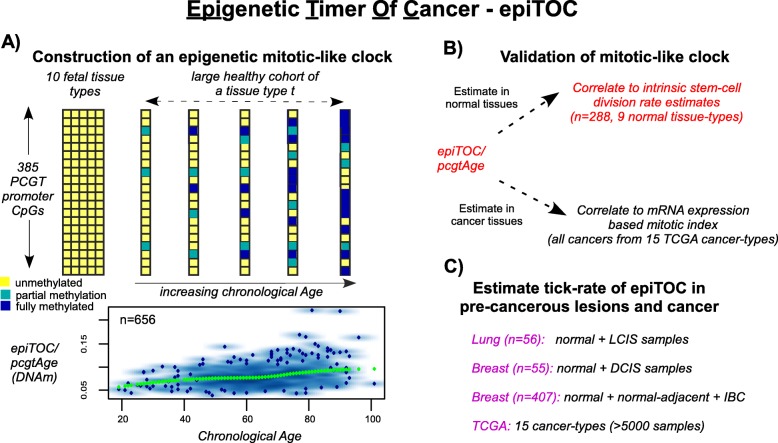
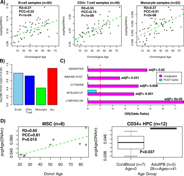
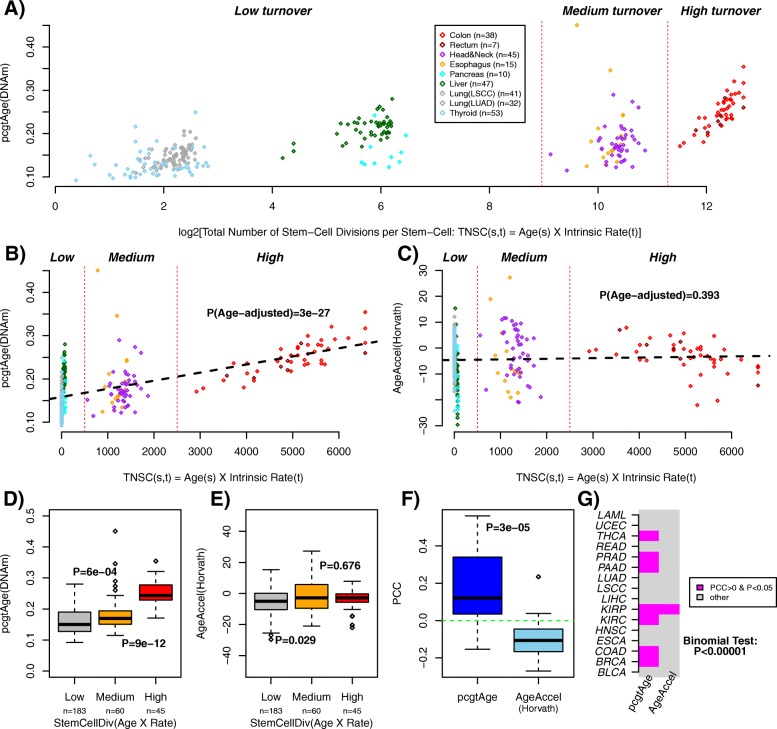
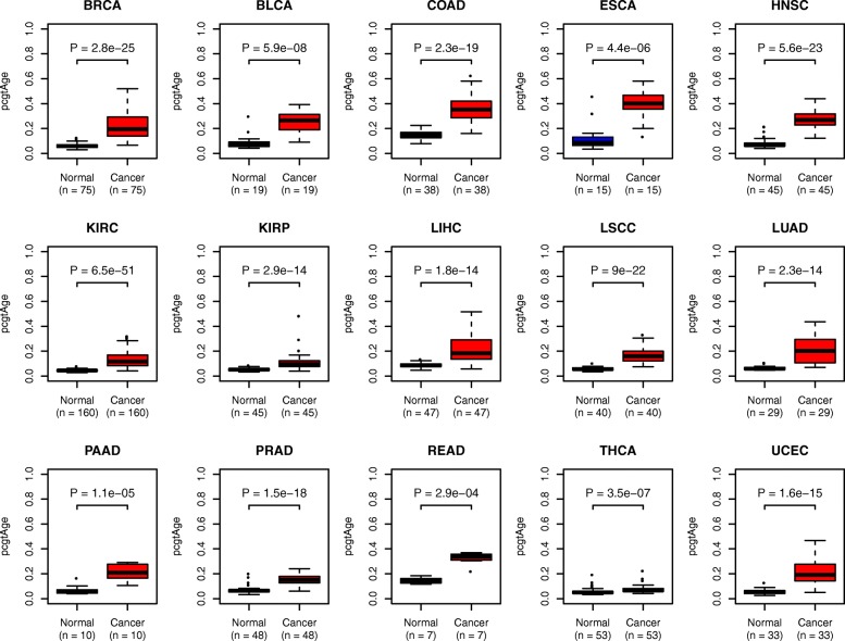
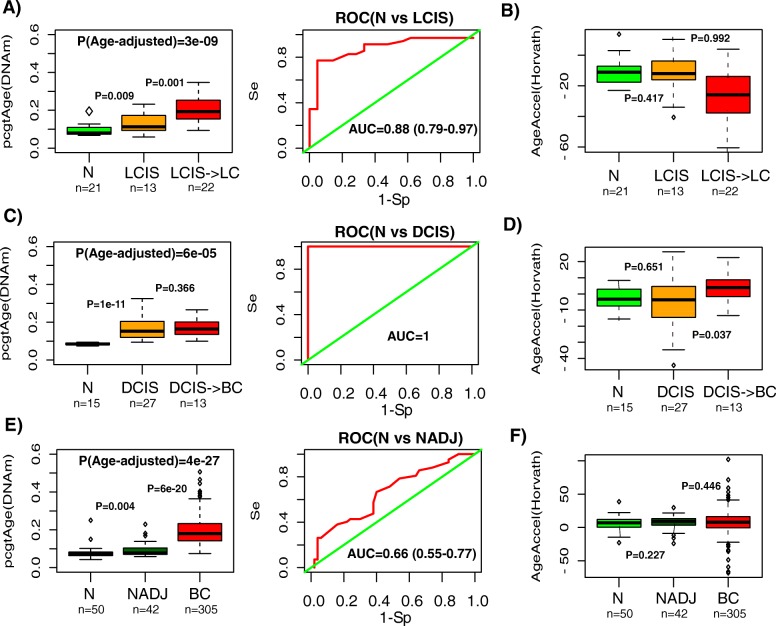
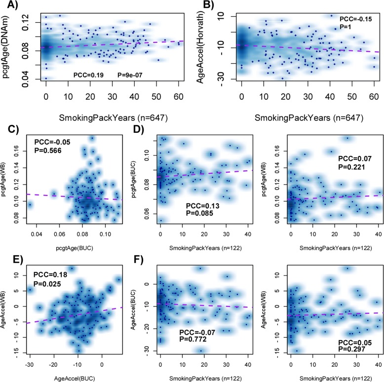
BACKGROUND: Variation in cancer risk among somatic tissues has been attributed to variations in the underlying rate of stem cell division. For a given tissue type, variable cancer risk between individuals is thought to be influenced by extrinsic factors which modulate this rate of stem cell division. To date, no molecular mitotic clock has been developed to approximate the number of stem cell divisions in a tissue of an individual and which is correlated with cancer risk. RESULTS: Here, we integrate mathematical modeling with prior biological knowledge to construct a DNA methylation-based age-correlative model which approximates a mitotic clock in both normal and cancer tissue. By focusing on promoter CpG sites that localize to Polycomb group target genes that are unmethylated in 11 different fetal tissue types, we show that increases in DNA methylation at these sites defines a tick rate which correlates with the estimated rate of stem cell division in normal tissues. Using matched DNA methylation and RNA-seq data, we further show that it correlates with an expression-based mitotic index in cancer tissue. We demonstrate that this mitotic-like clock is universally accelerated in cancer, including pre-cancerous lesions, and that it is also accelerated in normal epithelial cells exposed to a major carcinogen. CONCLUSIONS: Unlike other epigenetic and mutational clocks or the telomere clock, the epigenetic clock proposed here provides a concrete example of a mitotic-like clock which is universally accelerated in cancer and precancerous lesions.
Overview
The claims and statistics of this paper haven’t been checked yet.
Run the check — free, no account needed. It reads the open-access text and checks the sources it cites.
Integrity signals
Registry
Cites
Statistics
Figures
Verification
Claims the paper makes
The claims and statistics of this paper haven’t been checked yet.
Run the check — free, no account needed. It reads the open-access text and checks the sources it cites.
Claims other papers make about this one
Each is checked against this paper’s own text: does it say what the citing paper says it does?
In the citing paper
“In 2016, Yang et al.62 built an epigenetic clock associated with the number of cell divisions by pre-selecting CpG sites that had low levels of methylation in fetal tissue and increased methylation levels throughout adulthood and that mapped to Polycomb group target (PCGT) promoters, before training a sparse linear model.”
From Measuring biological age using omics data (2022) · cites this paper as [62]
In the citing paper
“Strikingly, this clock uniquely detected precancerous tissue samples to be age accelerated.”
From Measuring biological age using omics data (2022) · cites this paper as [62]
In the citing paper
“Yang et al.62 and Lu et al.167 took a different approach and trained clocks on cellular features — mitotic cell division and telomere length, respectively — to develop models with specific sensitivity for these aspects of cellular ageing.”
From Measuring biological age using omics data (2022) · cites this paper as [62]
Self-check
Percentages, sums and reported differences re-computed from the paper’s own numbers.
The self-check needs the paper’s full text.
Citations
Cites
The cancer genome atlas pan-cancer analysis project
The Cancer Genome Atlas Research Network, Weinstein, J. N., Collisson, E. A., Mills, G. B., Shaw, K. R. M., Ozenberger, B. A., Ellrott, K., Shmulevich, I., Sander, C., & Stuart, J. M. (2013) · Nature Genetics · doi:10.1038/ng.2764 · Cited by 9,092
DNA methylation age of human tissues and cell types
Horvath, S. (2013) · Genome Biology · doi:10.1186/gb-2013-14-10-r115 · Cited by 7,075
Age-related clonal hematopoiesis associated with adverse outcomes
Jaiswal, S., Fontanillas, P., Flannick, J., Manning, A., Grauman, P. V., Mar, B. G., Lindsley, R. C., Mermel, C. H., Burtt, N., Chavez, A., et al. (2014) · New England Journal of Medicine · doi:10.1056/nejmoa1408617 · Cited by 4,841
Genome-wide methylation profiles reveal quantitative views of human aging rates
Hannum, G., Guinney, J., Zhao, L., Zhang, L., Hughes, G., Sadda, S., Klotzle, B., Bibikova, M., Fan, J.-B., Gao, Y., et al. (2013) · Molecular Cell · doi:10.1016/j.molcel.2012.10.016 · Cited by 4,270
Clonal hematopoiesis and blood-cancer risk inferred from blood DNA sequence
Genovese, G., Kähler, A. K., Handsaker, R. E., Lindberg, J., Rose, S. A., Bakhoum, S. F., Chambert, K., Mick, E., Neale, B. M., Fromer, M., et al. (2014) · New England Journal of Medicine · doi:10.1056/nejmoa1409405 · Cited by 3,531
DNA methylation arrays as surrogate measures of cell mixture distribution
Houseman, E. A., Accomando, W. P., Koestler, D. C., Christensen, B. C., Marsit, C. J., Nelson, H. H., Wiencke, J. K., & Kelsey, K. T. (2012) · BMC Bioinformatics · doi:10.1186/1471-2105-13-86 · Cited by 3,175
Ben‐Porath, I., Thomson, M., Carey, V. J., Ge, R., Bell, G. W., Regev, A., & Weinberg, R. A. (2008) · Nature Genetics · doi:10.1038/ng.127 · Cited by 2,571
Control of Developmental Regulators by Polycomb in Human Embryonic Stem Cells
Lee, T. I., Jenner, R. G., Boyer, L. A., Guenther, M. G., Levine, S. S., Kumar, R., Chevalier, B. S., Johnstone, S. E., Cole, M. F., Isono, K., et al. (2006) · Cell · doi:10.1016/j.cell.2006.02.043 · Cited by 2,296
IDH1 mutation is sufficient to establish the glioma hypermethylator phenotype
Turcan, S., Rohle, D., Goenka, A., Walsh, L. A., Fang, F., Yilmaz, E., Campos, C., Fabius, A. W. M., Lu, C., Ward, P. S., et al. (2012) · Nature · doi:10.1038/nature10866 · Cited by 1,974
Variation in cancer risk among tissues can be explained by the number of stem cell divisions
Tomasetti, C., & Vogelstein, B. (2015) · Science · doi:10.1126/science.1260825 · Cited by 1,927
Cited by
Sorted by citations
DNA methylation-based biomarkers and the epigenetic clock theory of ageing
Horvath, S., & Raj, K. (2018) · Nature Reviews Genetics · doi:10.1038/s41576-018-0004-3 · Cited by 3,095
DNA methylation aging clocks: challenges and recommendations
Bell, C. G., Lowe, R., Adams, P. D., Baccarelli, A. A., Beck, S., Bell, J. T., Christensen, B. C., Gladyshev, V. N., Heijmans, B. T., Horvath, S., et al. (2019) · Genome Biology · doi:10.1186/s13059-019-1824-y · Cited by 1,025
Self-citationDNA Methylation Clocks in Aging: Categories, Causes, and Consequences
Field, A. E., Robertson, N., Wang, T., Havas, A. P., Ideker, T., & Adams, P. D. (2018) · Molecular Cell · doi:10.1016/j.molcel.2018.08.008 · Cited by 644
Oblak, L., van der Zaag, J., Higgins‐Chen, A., Levine, M. E., & Boks, M. P. (2021) · Ageing Research Reviews · doi:10.1016/j.arr.2021.101348 · Cited by 610
Measuring biological age using omics data
Rutledge, J., Oh, H., & Wyss-Coray, T. (2022) · Nature Reviews Genetics · doi:10.1038/s41576-022-00511-7 · Cited by 570
Metadata
- Authors
- Yang, Z., Wong, A., Kuh, D., Paul, D. S., Rakyan, V. K., Leslie, R. D., Zheng, S. C., Widschwendter, M., Beck, S., & Teschendorff, A. E.
- Affiliations
- Chinese Academy of Sciences (CN)
- MRC Unit for Lifelong Health and Ageing (GB)
- University College London (GB)
- Queen Mary University of London (GB)
- Corresponding author
- Teschendorff, Andrew E.
- ORCID
- Venue
- Genome Biology
- Published
- 2016-10-03
- Volume
- 17
- Issue
- 1
- Pages
- 205-205
- ISSN
- 1465-6906 (ISSN-L)
- 1474-760X
- PMC
- PMC5046977
- OpenAlex
- W2529317472
- Cited by
- 325
- References
- 71
- Citations by year
- 2026: 31 · 2025: 52 · 2024: 43 · 2023: 40 · 2022: 53 · 2021: 32 · 2020: 25 · 2019: 17 · 2018: 18 · 2017: 12 · 2016: 1
- FWCI
- 8.27
- Type
- journal article
- Language
- en
- Open access
- gold
- doi.org
- Licence
- cc-by
- Locations
- Genome biology (publishedVersion, cc-by)
- PubMed Central (publishedVersion, cc-by)
- PubMed (publishedVersion)
- UCL Discovery (University College London) (submittedVersion)
- Queen Mary Research Online (Queen Mary University of London) (submittedVersion, cc-by)
- Apollo (University of Cambridge) (submittedVersion, cc-by)
- Apollo (cc-by)
- Funders
- Royal Society (NAF 164914)
- National Science Foundation of China (NSFC 31571359)
- Topics
- Life Sciences › Biochemistry, Genetics and Molecular Biology › Molecular Biology › Epigenetics and DNA Methylation
- Life Sciences › Biochemistry, Genetics and Molecular Biology › Cancer Research › Cancer Genomics and Diagnostics
- Life Sciences › Biochemistry, Genetics and Molecular Biology › Cell Biology › Telomeres, Telomerase, and Senescence
- Keywords
- epigenetic clocks, DNA methylation, stem cell division, cancer risk, promoter CpG sites, fetal tissue, carcinogen exposure, precancerous lesions, cancer risk stratification
- MeSH terms
- Aging, Humans, Mitosis, Mitotic Index, Models, Theoretical, Neoplasms, Promoter Regions, Genetic, Neoplastic Stem Cells, Gene Expression Regulation, Neoplastic, CpG Islands, DNA Methylation, Epigenesis, Genetic, High-Throughput Nucleotide Sequencing, Polycomb-Group Proteins
About these checks
Registry, retraction, Cites and statistics results are computed, not judged, with one exception: a printed reference that no index lists is read once by a model before it counts as a citation error. Claims are extracted by a model and kept only when their quote appears verbatim in the text; each cited source is then checked, with a model’s help, for whether it says what the claim relies on. Those verdicts can be wrong — read the claim’s quote and the cited source before relying on one.
Statistics and arithmetic are recomputed, not judged.
Confidence bands are shown only for fields calibrated on our annotated test set. None is calibrated yet, so no band is shown.
What counts as a claim ›How we measure confidence ›How we check this ›
Are you an author, or do you see a problem with this page? Request a review
