

The DOI matches its Crossref record; 30 of the 30 references it cites were found in a registry or an index.
Gram-negative bacterial molecules associate with Alzheimer disease pathology
Open accessAbstract
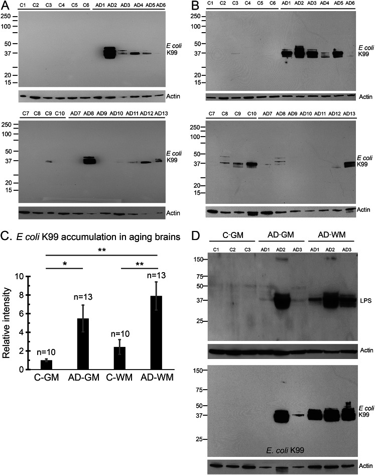
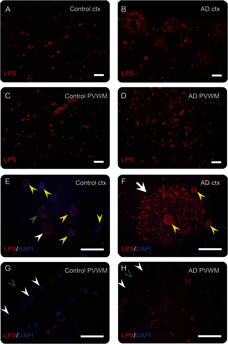
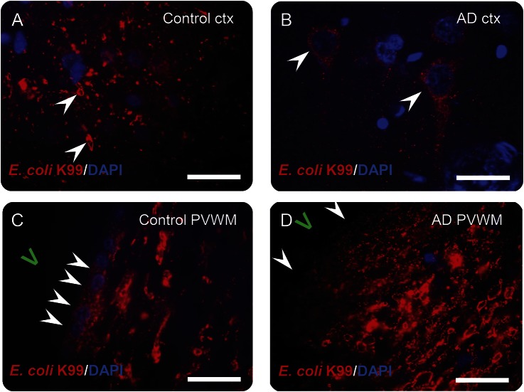
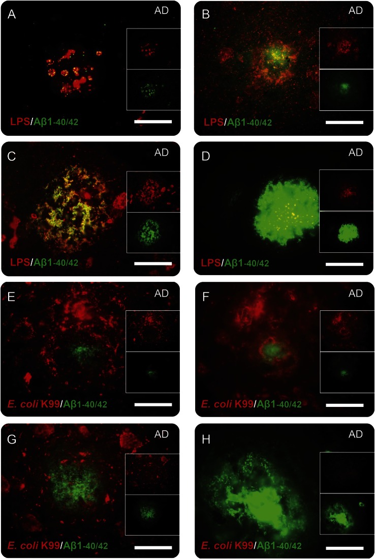
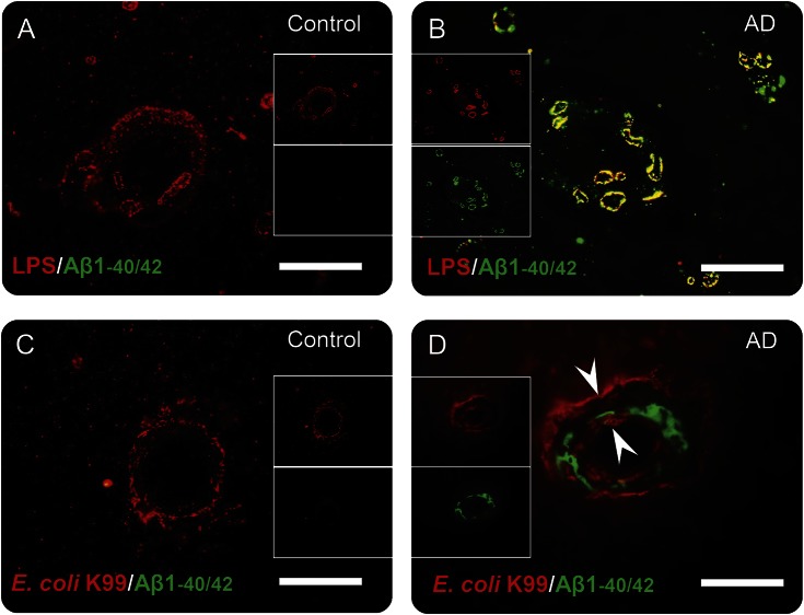
Objective: We determined whether Gram-negative bacterial molecules are associated with Alzheimer disease (AD) neuropathology given that previous studies demonstrate Gram-negative Escherichia coli bacteria can form extracellular amyloid and Gram-negative bacteria have been reported as the predominant bacteria found in normal human brains. Methods: Brain samples from gray and white matter were studied from patients with AD (n = 24) and age-matched controls (n = 18). Lipopolysaccharide (LPS) and E coli K99 pili protein were evaluated by Western blots and immunocytochemistry. Human brain samples were assessed for E coli DNA followed by DNA sequencing. Results: LPS and E coli K99 were detected immunocytochemically in brain parenchyma and vessels in all AD and control brains. K99 levels measured using Western blots were greater in AD compared to control brains (p < 0.01) and K99 was localized to neuron-like cells in AD but not control brains. LPS levels were also greater in AD compared to control brain. LPS colocalized with Aβ1-40/42 in amyloid plaques and with Aβ1-40/42 around vessels in AD brains. DNA sequencing confirmed E coli DNA in human control and AD brains. Conclusions: E coli K99 and LPS levels were greater in AD compared to control brains. LPS colocalized with Aβ1-40/42 in amyloid plaques and around vessels in AD brain. The data show that Gram-negative bacterial molecules are associated with AD neuropathology. They are consistent with our LPS-ischemia-hypoxia rat model that produces myelin aggregates that colocalize with Aβ and resemble amyloid-like plaques.
Overview
The claims and statistics of this paper haven’t been checked yet.
Run the check — free, no account needed. It reads the open-access text and checks the sources it cites.
Integrity signals
Registry
Cites
Statistics
Figures
Verification
Claims the paper makes
The claims and statistics of this paper haven’t been checked yet.
Run the check — free, no account needed. It reads the open-access text and checks the sources it cites.
Claims other papers make about this one
Each is checked against this paper’s own text: does it say what the citing paper says it does?
The source backs part of the claim, not all of it — or backs it for a different population, condition or comparison.
In the citing paper
“A variety of genetic and environmental factors have been implicated in AD etiopathogenesis, including contributions from gut microbiota [13–15].”
From The gut microbiota-derived metabolite trimethylamine N-oxide is elevated in Alzheimer’s disease (2018) · cites this paper as [14]
Experimental — accuracy not yet measuredThe source backs part of the claim, not all of it — or backs it for a different population, condition or comparison.
In the citing paper
“E. coli particles and LPS particles have been shown to associate directly with pathology in the brains of AD patients [41].”
From Alzheimer’s disease-associated protective variant Plcg2-P522R modulates peripheral macrophage function in a sex-dimorphic manner (2024) · cites this paper as [41]
Experimental — accuracy not yet measuredIn the citing paper
“Blood and brain endotoxin levels are elevated in Alzheimer’s disease”
From The endotoxin hypothesis of neurodegeneration (2019) · cites this paper as [79]
In the citing paper
“Brain endotoxin levels are increased two- or threefold in AD patients [78, 79], and endotoxin is also found in AD amyloid plaques [79].”
From The endotoxin hypothesis of neurodegeneration (2019) · cites this paper as [79]
In the citing paper
“There is a large increase in bacterial load and bacterial species in AD brains [79, 90], including P. gingivalis, which can live intracellularly in glia and neurons [91].”
From The endotoxin hypothesis of neurodegeneration (2019) · cites this paper as [79]
Self-check
Percentages, sums and reported differences re-computed from the paper’s own numbers.
The self-check needs the paper’s full text.
Citations
Cites
Frequency of Stages of Alzheimer-Related Lesions in Different Age Categories
Braak, H., & Braak, E. (1997) · Neurobiology of Aging · doi:10.1016/s0197-4580(97)00056-0 · Cited by 1,656
Role of Escherichia coli Curli Operons in Directing Amyloid Fiber Formation
Chapman, M. R., Robinson, L. S., Pinkner, J. S., Roth, R. A., Heuser, J., Hammar, M., Normark, S. J., & Hultgren, S. J. (2002) · Science · doi:10.1126/science.1067484 · Cited by 1,339
Functional amyloid – from bacteria to humans
Fowler, D. M., Koulov, A. V., Balch, W. E., & Kelly, J. W. (2007) · Trends in Biochemical Sciences · doi:10.1016/j.tibs.2007.03.003 · Cited by 1,052
Early inflammation and dementia: A 25‐year follow‐up of the Honolulu‐Asia aging study
Schmidt, R., Schmidt, H., Curb, J. D., Masaki, K., White, L. R., & Launer, L. J. (2002) · Annals of Neurology · doi:10.1002/ana.10265 · Cited by 710
Protective effects of NSAIDs on the development of Alzheimer disease
Vlad, S. C., Miller, D. R., Kowall, N. W., & Felson, D. T. (2008) · Neurology · doi:10.1212/01.wnl.0000311269.57716.63 · Cited by 535
Bacterial adhesins: function and structure
Klemm, P., & Schembri, M. A. (2000) · International Journal of Medical Microbiology · doi:10.1016/s1438-4221(00)80102-2 · Cited by 417
Enterotoxigenic Escherichia coli (ETEC) in farm animals.
Nagy, B., & Fekete, P. Z. (1999) · PubMed · Cited by 390
Bartzokis, G., Sultzer, D., Lu, P. H., Nuechterlein, K. H., Mintz, J., & Cummings, J. L. (2004) · Neurobiology of Aging · doi:10.1016/j.neurobiolaging.2003.09.005 · Cited by 363
Risk factors for Alzheimer's disease: a population-based, longitudinal study in Manitoba, Canada
Tyas, S. L., Manfreda, J., Strain, L. A., & Montgomery, P. R. (2001) · International Journal of Epidemiology · doi:10.1093/ije/30.3.590 · Cited by 272
Serum IgG Antibody Levels to Periodontal Microbiota Are Associated with Incident Alzheimer Disease
Noble, J. M., Scarmeas, N., Celenti, R. S., Elkind, M. S. V., Wright, C. B., Schupf, N., & Papapanou, P. N. (2014) · PLoS ONE · doi:10.1371/journal.pone.0114959 · Cited by 222
Association between Dementia and Infectious Disease
Dunn, N. R., Mullee, M., Perry, V. H., & Holmes, C. (2005) · Alzheimer Disease & Associated Disorders · doi:10.1097/01.wad.0000165511.52746.1f · Cited by 202
Porphyromonas gingivalis Periodontal Infection and Its Putative Links with Alzheimer’s Disease
Singhrao, S. K., Harding, A., Poole, S., Kesavalu, L., & Crean, S. (2015) · Mediators of Inflammation · doi:10.1155/2015/137357 · Cited by 195
Schmidt, C. (2015) · Nature · doi:10.1038/518s13a · Cited by 158
Branton, W. G., Ellestad, K. K., Maingat, F., Wheatley, B. M., Rud, E., Warren, R. L., Holt, R. A., Surette, M. G., & Power, C. (2013) · PLoS ONE · doi:10.1371/journal.pone.0054673 · Cited by 149 · cited 3×
“Our findings complement a recent RNAseq study that showed bacterially encoded 16s RNA sequences in all human brains with Gram-negativea-Proteobacteria representing over 70% of bacterial sequences.(16) The other 30% of bacterial classes varied widely, with g-Proteobacteria, including E coli, being present at ,5% of tot…”
BackgroundResultInfluentialEtiology of Diarrhea in Older Children, Adolescents and Adults: A Systematic Review
Walker, C. L. F., Sack, D. A., & Black, R. E. (2010) · PLoS neglected tropical diseases · doi:10.1371/journal.pntd.0000768 · Cited by 145
Erickson, M. A., Hartvigson, P. E., Morofuji, Y., Owen, J. B., Butterfield, D. A., & Banks, W. A. (2012) · Journal of Neuroinflammation · doi:10.1186/1742-2094-9-150 · Cited by 145
Bacterial peptidoglycan and immune reactivity in the central nervous system in multiple sclerosis
Schrijver, I. A. (2001) · Brain · doi:10.1093/brain/124.8.1544 · Cited by 139
Past exposure to vaccines and subsequent risk of Alzheimer's disease.
Verreault, R., Laurin, D., Lindsay, J., & Serres, G. D. (2001) · Europe PMC (PubMed Central) · Cited by 111
Zhan, X., Jickling, G. C., Ander, B. P., Stamova, B. S., Liu, D., Kao, P. F., Zelin, M. A., Jin, L., DeCarli, C. S., & Sharp, F. R. (2015) · Journal of Alzheimer s Disease · doi:10.3233/jad-142013 · Cited by 109
Self-citationAn update on the efficacy of non-steroidal anti-inflammatory drugs in Alzheimer's disease
Imbimbo, B. P. (2009) · Expert Opinion on Investigational Drugs · doi:10.1517/13543780903066780 · Cited by 109
Systemic inflammation and Alzheimer's disease
Holmes, C., & Butchart, J. (2011) · Biochemical Society Transactions · doi:10.1042/bst0390898 · Cited by 92
Peripheral Biomarkers of Alzheimer's Disease
Khan, T. K., & Alkon, D. L. (2015) · Journal of Alzheimer s Disease · doi:10.3233/jad-142262 · Cited by 80
Myelin Injury and Degraded Myelin Vesicles in Alzheimer’s Disease
Zhan, X., Jickling, G. C., Ander, B. P., Liu, D., Stamova, B. S., Cox, C. D., Jin, L. W., DeCarli, C. S., & Sharp, F. R. (2014) · Current Alzheimer Research · doi:10.2174/1567205011666140131120922 · Cited by 80
Self-citationDissecting the Contribution of Vascular Alterations and Aging to Alzheimer’s Disease
Janota, C. S., Lemere, C. A., & Brito, M. A. (2015) · Molecular Neurobiology · doi:10.1007/s12035-015-9319-7 · Cited by 70
Visser, L. H., Melief, M., van Riel, D. A. J., van Meurs, M., Sick, E. A., Inamura, S., Bajramovic, J. J., Amor, S., Hintzen, R. Q., Boven, L. A., et al. (2006) · American Journal Of Pathology · doi:10.2353/ajpath.2006.060143 · Cited by 65
Civitelli, L., Marcocci, M. E., Celestino, I., Piacentini, R., Garaci, E., Grassi, C., Chiara, G. D., & Palamara, A. T. (2015) · Journal of NeuroVirology · doi:10.1007/s13365-015-0344-0 · Cited by 50
Link Between Chronic Bacterial Inflammation and Alzheimer Disease
Bibi, F., Yasir, M., Sohrab, S. S., Azhar, E. I., Al-Qahtani, M., Abuzenadah, A. M., Kamal, M. A., & Naseer, M. I. (2014) · CNS & Neurological Disorders - Drug Targets · doi:10.2174/1871527313666140917115741 · Cited by 43
Zhan, X., Cox, C. D., Ander, B. P., Liu, D., Stamova, B. S., Jin, L., Jickling, G. C., & Sharp, F. R. (2015) · Journal of Alzheimer s Disease · doi:10.3233/jad-143072 · Cited by 41
Self-citationQuiñones, B., Swimley, M. S., Narm, K.-E., Patel, R. N., Cooley, M. B., & Mandrell, R. E. (2012) · Frontiers in Cellular and Infection Microbiology · doi:10.3389/fcimb.2012.00061 · Cited by 25
Inflammatory Proteins in Plasma and the Risk of Dementia: The Rotterdam Study
Engelhart, M. J., Geerlings, M. I., Meijer, J., Kiliaan, A., Ruitenberg, A., van Swieten, J. C., Stijnen, T., Hofman, A., Witteman, J. C. M., & Breteler, M. M. B. (2004) · Archives of Neurology · doi:10.1001/archneur.61.5.668 · Cited by 10
Cited by
Sorted by citations
Gut microbiome alterations in Alzheimer’s disease
Vogt, N. M., Kerby, R. L., Dill-McFarland, K. A., Harding, S. J., Merluzzi, A. P., Johnson, S. C., Carlsson, C. M., Asthana, S., Zetterberg, H., Blennow, K., et al. (2017) · Scientific Reports · doi:10.1038/s41598-017-13601-y · Cited by 2,137
A systemic view of Alzheimer disease — insights from amyloid-β metabolism beyond the brain
Wang, J., Gu, B. J., Masters, C. L., & Wang, Y.-J. (2017) · Nature Reviews Neurology · doi:10.1038/nrneurol.2017.111 · Cited by 808
Liu, P., Wu, L., Peng, G., Han, Y., Tang, R., Ge, J., Zhang, L., Jia, L., Yue, S., Zhou, K., et al. (2019) · Brain, Behavior, and Immunity · doi:10.1016/j.bbi.2019.05.008 · Cited by 669
Kim, M.-S., Kim, Y., Choi, H., Kim, W., Park, S., Lee, D., Kim, D. K., Kim, H. J., Choi, H., Hyun, D.-W., et al. (2020) · Gut · doi:10.1136/gutjnl-2018-317431 · Cited by 614
Inflammation: the link between comorbidities, genetics, and Alzheimer’s disease
Newcombe, E. A., Camats-Perna, J., Silva, M. L., Valmas, N., Huat, T. J., & Medeiros, R. (2018) · Journal of Neuroinflammation · doi:10.1186/s12974-018-1313-3 · Cited by 532
Metadata
- Authors
- Zhan, X., Stamova, B., Jin, L.-W., DeCarli, C., Phinney, B., & Sharp, F. R.
- Affiliations
- University of California Davis Medical Center (US)
- Corresponding author
- Zhan, Xinhua
- Venue
- Neurology
- Published
- 2016-11-29
- Volume
- 87
- Issue
- 22
- Pages
- 2324-2332
- ISSN
- 0028-3878 (ISSN-L)
- 1526-632X
- PMC
- PMC5135029
- OpenAlex
- W2545292312
- Cited by
- 521
- References
- 30
- Citations by year
- 2026: 38 · 2025: 41 · 2024: 69 · 2023: 49 · 2022: 69 · 2021: 86 · 2020: 75 · 2019: 40 · 2018: 30 · 2017: 24
- FWCI
- 25.23
- Type
- journal article
- Language
- en
- Open access
- hybrid
- doi.org
- Licence
- cc-by-nc-nd
- Locations
- Neurology (publishedVersion, cc-by-nc-nd)
- Europe PMC (PubMed Central) (publishedVersion, cc-by-nc-nd)
- PubMed Central (publishedVersion, cc-by-nc-nd)
- PubMed (publishedVersion)
- eScholarship (California Digital Library) (submittedVersion)
- eScholarship (California Digital Library) (submittedVersion, cc-by-nc-nd)
- Funders
- National Institute on Aging (R01AG042292)
- Topics
- Life Sciences › Neuroscience › Cellular and Molecular Neuroscience › Alzheimer's disease research and treatments
- Life Sciences › Neuroscience › Neurology › Neuroinflammation and Neurodegeneration Mechanisms
- Health Sciences › Medicine › Psychiatry and Mental health › Dementia and Cognitive Impairment Research
- Keywords
- Alzheimer's disease, Gram-negative bacteria, Escherichia coli, lipopolysaccharide, amyloid plaques, amyloid-beta, neuroinflammation
- MeSH terms
- Aged, Aged, 80 and over, Alzheimer Disease, Brain, DNA, Bacterial, Escherichia coli, Female, Fluorescent Antibody Technique, Humans, Lipopolysaccharides, Male, Peptide Fragments, Blotting, Western, Polymerase Chain Reaction, Amyloid beta-Peptides, Sequence Analysis, DNA, Escherichia coli Proteins, Plaque, Amyloid, White Matter, Gray Matter
About these checks
Registry, retraction, Cites and statistics results are computed, not judged, with one exception: a printed reference that no index lists is read once by a model before it counts as a citation error. Claims are extracted by a model and kept only when their quote appears verbatim in the text; each cited source is then checked, with a model’s help, for whether it says what the claim relies on. Those verdicts can be wrong — read the claim’s quote and the cited source before relying on one.
Statistics and arithmetic are recomputed, not judged.
Confidence bands are shown only for fields calibrated on our annotated test set. None is calibrated yet, so no band is shown.
What counts as a claim ›How we measure confidence ›How we check this ›
Are you an author, or do you see a problem with this page? Request a review
