

The DOI matches its Crossref record; 111 of the 114 references it cites were found in a registry or an index.
The pathophysiology of traumatic brain injury at a glance
Open accessAbstract
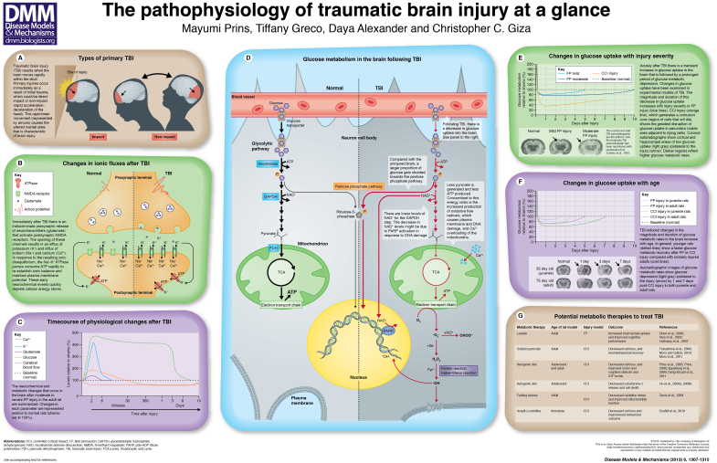
Summary: Traumatic brain injury (TBI) is defined as an impact, penetration or rapid movement of the brain within the skull that results in altered mental state. TBI occurs more than any other disease, including breast cancer, AIDS, Parkinson’s disease and multiple sclerosis, and affects all age groups and both genders. In the US and Europe, the magnitude of this epidemic has drawn national attention owing to the publicity received by injured athletes and military personnel. This increased public awareness has uncovered a number of unanswered questions concerning TBI, and we are increasingly aware of the lack of treatment options for a crisis that affects millions. Although each case of TBI is unique and affected individuals display different degrees of injury, different regional patterns of injury and different recovery profiles, this review and accompanying poster aim to illustrate some of the common underlying neurochemical and metabolic responses to TBI. Recognition of these recurrent features could allow elucidation of potential therapeutic targets for early intervention.
Overview
The claims and statistics of this paper haven’t been checked yet.
Run the check — free, no account needed. It reads the open-access text and checks the sources it cites.
Integrity signals
Registry
Cites
Statistics
Figures
Verification
Claims the paper makes
The claims and statistics of this paper haven’t been checked yet.
Run the check — free, no account needed. It reads the open-access text and checks the sources it cites.
Claims other papers make about this one
Each is checked against this paper’s own text: does it say what the citing paper says it does?
The check wasn’t confident either way, so it gives no verdict — never disputed.
In the citing paper
“We counted brainstem neurons in a blinded manner using the same stereological procedures as previously described.9”
From Damage to Arousal-Promoting Brainstem Neurons with Traumatic Brain Injury (2016) · cites this paper as [9]
Experimental — accuracy not yet measuredThe check wasn’t confident either way, so it gives no verdict — never disputed.
In the citing paper
“Additional factors that may protect the brainstem include lower metabolic requirements and a blood supply less likely to be damaged by direct traumatic forces.9”
From Damage to Arousal-Promoting Brainstem Neurons with Traumatic Brain Injury (2016) · cites this paper as [9]
Experimental — accuracy not yet measured
Self-check
Percentages, sums and reported differences re-computed from the paper’s own numbers.
The self-check needs the paper’s full text.
Citations
Cites
National Center for Injury Prevention and Control (U.S.). Division of Injury Response., Faul, M., Wald, M. M., Wu, L., & Coronado, V. G. (2010) · doi:10.15620/cdc.5571 · Cited by 3,494
Langlois, J., Rutland-Brown, W., & Thomas, K. (2006) · PsycEXTRA Dataset · doi:10.1037/e721222007-001 · Cited by 3,003
Chronic traumatic encephalopathy in athletes: Progressive tauopathy after repetitive head injury
McKee, A. C., Cantu, R. C., Nowinski, C. J., Hedley-Whyte, E. T., Gavett, B. E., Budson, A. E., Santini, V. E., Lee, H.-S., Kubilus, C. A., & Stern, R. A. (2009) · Journal of Neuropathology & Experimental Neurology · doi:10.1097/nen.0b013e3181a9d503 · Cited by 2,295
Brain Metabolism during Fasting*
Owen, O. E., Morgan, A. P., Kemp, H. G., Sullivan, J. M., Herrera, M. G., & Cahill, G. F. (1967) · Journal of Clinical Investigation · doi:10.1172/jci105650 · Cited by 1,662
A new model of diffuse brain injury in rats
Marmarou, A., Foda, M. A. A.-E., van den Brink, W., Campbell, J., Kita, H., & Demetriadou, K. (1994) · Journal of neurosurgery · doi:10.3171/jns.1994.80.2.0291 · Cited by 1,244
Katayama, Y., Becker, D. P., Tamura, T., & Hovda, D. A. (1990) · Journal of neurosurgery · doi:10.3171/jns.1990.73.6.0889 · Cited by 1,091
Veech, R. L. (2004) · Prostaglandins Leukotrienes and Essential Fatty Acids · doi:10.1016/j.plefa.2003.09.007 · Cited by 779
Free Radical Pathways in CNS Injury
LEWÉN, A., MATZ, P., & CHAN, P. H. (2000) · Journal of Neurotrauma · doi:10.1089/neu.2000.17.871 · Cited by 759
Bergsneider, M., Hovda, D. A., Shalmon, E., Kelly, D. F., Vespa, P. M., Martin, N. A., Phelps, M. E., McArthur, D. L., Caron, M., Kraus, J. F., et al. (1997) · Journal of neurosurgery · doi:10.3171/jns.1997.86.2.0241 · Cited by 639
Yoshino, A., Hovda, D. A., Kawamata, T., Katayama, Y., & Becker, D. P. (1991) · Brain Research · doi:10.1016/0006-8993(91)90755-k · Cited by 592
Research works printed but not linked: 1
- Hovda, 1994, Long-term changes in metabolic rates for glucose following mild, moderate and severe concussive head injuries in adult rats, J. Neurosci., 20, 845
Cited by
Sorted by citations
Recent Advances in Pathophysiology of Traumatic Brain Injury
Kaur, P., & Sharma, S. (2017) · Current Neuropharmacology · doi:10.2174/1570159x15666170613083606 · Cited by 471
“Phosphatidylserine translocation includes distinct however gradual membrane breakdown together with nuclear membranes disintegration, DNA-fragmentation, and chromatin condensation [117].”
InfluentialHenderson, T. A., & Morries, L. D. (2015) · Neuropsychiatric Disease and Treatment · doi:10.2147/ndt.s78182 · Cited by 432
“This energy crisis promotes the increased concentration of free radicals, due to increased pentose phosphate metabolism, reduced mitochondrial function, and impaired free radical scavenger mechanisms.(19) The consequences of increased free radicals can be far-reaching including propagation of additional free radicals,…”
Bidirectional Microglia–Neuron Communication in Health and Disease
Szepesi, Z., Manouchehrian, O., Bachiller, S., & Deierborg, T. (2018) · Frontiers in Cellular Neuroscience · doi:10.3389/fncel.2018.00323 · Cited by 459
“The primary injury of TBIs, caused by trauma to the head which for example might stretch, compress or tear blood vessels and axons, is not the only reason why patients die or are disabled (Prins et al., 2013).”
BackgroundThe synapse in traumatic brain injury
Jamjoom, A. A. B., Rhodes, J. K. J., Andrews, P., & Grant, S. G. N. (2020) · Brain · doi:10.1093/brain/awaa321 · Cited by 260
“The mechanical injury (primary insult) causes instantaneous tissue damage, which is followed by multiple secondary injury processes (Prins et al., 2013).”
BackgroundThe NLRP3 inflammasome in traumatic brain injury: potential as a biomarker and therapeutic target
O’Brien, W. T., Pham, L., Symons, G. F., Monif, M., Shultz, S. R., & McDonald, S. J. (2020) · Journal of Neuroinflammation · doi:10.1186/s12974-020-01778-5 · Cited by 233
Repeated Mild Traumatic Brain Injury
Fehily, B., & Fitzgerald, M. (2017) · Cell Transplantation · doi:10.1177/0963689717714092 · Cited by 240
In vivo characterization of chronic traumatic encephalopathy using [F-18]FDDNP PET brain imaging
Barrio, J. R., Small, G. W., Wong, K. P., Huang, S., Liu, J., Merrill, D. A., Giza, C. C., Fitzsimmons, R. P., Omalu, B. I., Bailes, J. E., et al. (2015) · Proceedings of the National Academy of Sciences · doi:10.1073/pnas.1409952112 · Cited by 211
“Focal types of brain injury such as contusions or lacerations that commonly occur in moderate and severe traumatic injuries in humans or primate models of traumatic brain injury (27, 31, 32) are only rarely reported in concussions (33).”
BackgroundSelf-citationEmery, C. A., Barlow, K. M., Brooks, B. L., Max, J. E., Villavicencio-Requis, A., Gnanakumar, V., Robertson, H., Schneider, K., & Yeates, K. O. (2016) · The Canadian Journal of Psychiatry · doi:10.1177/0706743716643741 · Cited by 164
“They are typically defined as injuries to the brain resulting from external physical forces and identified clinically by Glasgow Coma Scale scores of 13 to 15, disorientation or other mental status changes, loss of consciousness for no longer than 30 minutes, and/or posttraumatic amnesia lasting no longer than 24 hour…”
BackgroundThe role of the complement system in traumatic brain injury: a review
Hammad, A., Westacott, L. J., & Zaben, M. (2018) · Journal of Neuroinflammation · doi:10.1186/s12974-018-1066-z · Cited by 175
“TBI can also be classified on the basis of severity, according to the Glasgow Coma Scale (GCS), as mild, moderate or severe [3].”
BackgroundAcosta, S. A., Tajiri, N., de la Peña, I., Bastawrous, M., Sanberg, P. R., Kaneko, Y., & Borlongan, C. V. (2014) · Journal of Cellular Physiology · doi:10.1002/jcp.24830 · Cited by 160
“…at a higher risk of developing idiopathic Parkinsonism or PD- like syndrome, of which symptoms develop several years after the primary injury of TBI (Prins et al., 2013; Kokjohn et al., 2012; Karantzoulis and Randolph, 2013; Smith, 2013; Smith et al., 2013; Chauhan, 2014; Levin and Bhardwaj, 2014).”
BackgroundAltered Mitochondrial Dynamics and TBI Pathophysiology
Fischer, T. D., Hylin, M. J., Zhao, J., Moore, A. N., Waxham, M. N., & Dash, P. K. (2016) · Frontiers in Systems Neuroscience · doi:10.3389/fnsys.2016.00029 · Cited by 160
“Both clinical and experimental studies have shown there is an acute period of increased glucose utilization after TBI (Giza and Hovda, 2001; Prins et al., 2013; Gajavelli et al., 2015).”
BackgroundMesenchymal Stem Cells in the Treatment of Traumatic Brain Injury
Hasan, A., Deeb, G., Rahal, R., Atwi, K. M., Mondello, S., Marei, H. E. S., Gali, A., & Sleiman, E. (2017) · Frontiers in Neurology · doi:10.3389/fneur.2017.00028 · Cited by 143
Traumatic brain injury: a risk factor for neurodegenerative diseases
Gupta, R. K., & Sen, N. (2015) · Reviews in the Neurosciences · doi:10.1515/revneuro-2015-0017 · Cited by 143
“Brain injury leads to tissue deformation at the time of injury, damages blood vessels, shears axons, and produces cellular damage (Prins et al., 2013).”
BackgroundQian, F., Han, Y., Han, Z., Zhang, D., Zhang, L., Zhao, G., Li, S., Jin, G., Yu, R., & Liu, H. (2021) · Biomaterials · doi:10.1016/j.biomaterials.2021.120675 · Cited by 137
Eastman, C. L., D’Ambrosio, R., & Ganesh, T. (2019) · Neuropharmacology · doi:10.1016/j.neuropharm.2019.107907 · Cited by 140
Traumatic Brain Injury and NADPH Oxidase: A Deep Relationship
Angeloni, C., Prata, C., Sega, F. V. D., Piperno, R., & Hrelia, S. (2015) · Oxidative Medicine and Cellular Longevity · doi:10.1155/2015/370312 · Cited by 138
“naptic voltage-dependent Ca channels and in massive release of excitatory neurotransmitters like glutamate and aspartate into the extracellular space [46, 47].”
BackgroundKatzenberger, R. J., Chtarbanova, S., Rimkus, S. A., Fischer, J. A., Kaur, G., Seppala, J. M., Swanson, L. C., Zajac, J. E., Ganetzky, B., & Wassarman, D. A. (2015) · eLife · doi:10.7554/elife.04790 · Cited by 125
“…not only due to primary injuries, that is, mechanical injuries that occur at the moment of impact to the brain, but also to secondary injuries, that is, non-mechanical injuries that evolve over time in response to primary injuries (Masel and DeWitt, 2010; Blennow et al., 2012; Prins et al., 2013).”
BackgroundInfluentialLogan, S., Arzua, T., Canfield, S. G., Seminary, E. R., Sison, S. L., Ebert, A. D., & Bai, X. (2019) · Comprehensive physiology · doi:10.1002/cphy.c180025 · Cited by 143
Diffuse axonal injury in brain trauma: insights from alterations in neurofilaments
Siedler, D. G., Chuah, M. I., Kirkcaldie, M., Vickers, J. C., & King, A. E. (2014) · Frontiers in Cellular Neuroscience · doi:10.3389/fncel.2014.00429 · Cited by 121
“This places strain on the system, since mitochondria sequester calcium to counter the cytosolic excess (Buki et al., 2000), which generates reactive oxygen species and disrupts oxidative metabolism (Figure 1; Prins et al., 2013) through unknown mechanisms.”
Namjoshi, D., Good, C. A., Cheng, W. H., Panenka, W. J., Richards, D., Cripton, P. A., & Wellington, C. L. (2013) · Disease Models & Mechanisms · doi:10.1242/dmm.011320 · Cited by 118
“…calcium influx, elevated oxidative stress, free-radical- mediated damage, excitatory neurotransmitter release, inflammation and cell death (McIntosh et al., 1996; Blumbergs, 1997; Davis, 2000; Giza and Hovda, 2001; Werner and Engelhard, 2007; McAllister, 2011) (reviewed by Prins et al., 2013).”
NADPH oxidases in traumatic brain injury – Promising therapeutic targets?
Ma, M. W., Wang, J., Dhandapani, K. M., Wang, R., & Brann, D. W. (2018) · Redox Biology · doi:10.1016/j.redox.2018.03.005 · Cited by 116
The Role of NLRP3 Inflammasome in the Pathogenesis of Traumatic Brain Injury
Irrera, N., Russo, M., Pallio, G., Bitto, A., Mannino, F., Minutoli, L., Altavilla, D., & Squadrito, F. (2020) · International Journal of Molecular Sciences · doi:10.3390/ijms21176204 · Cited by 112
“The reason of the increase/decrease switch is still unknown, but it could be due to i) the reduction of blood availability, ii) the presence of glucose transporters defects, iii) the decrease of metabolic glucose request [34].”
BackgroundPhosphodiesterase-4 enzyme as a therapeutic target in neurological disorders
Bhat, A., Ray, B., Mahalakshmi, A. M., Tuladhar, S., Nandakumar, D., Srinivasan, M., Essa, M. M., Chidambaram, S. B., Guillemin, G. J., & Sakharkar, M. K. (2020) · Pharmacological Research · doi:10.1016/j.phrs.2020.105078 · Cited by 109
Donat, C. K., Lopez, M. Y., Sastre, M., Baxan, N., Goldfinger, M., Seeamber, R., Müller, F., Davies, P., Hellyer, P. J., Siegkas, P., et al. (2020) · Brain · doi:10.1093/brain/awaa336 · Cited by 103
Oxidized phospholipid signaling in traumatic brain injury
Anthonymuthu, T. S., Kenny, E., Lamade, A. M., Kagan, V. E., & Bayır, H. (2018) · Free Radical Biology and Medicine · doi:10.1016/j.freeradbiomed.2018.06.031 · Cited by 99
“Consequently, secondary injury is initiated at the time of the insult and develops in the postinjury period through signaling of multiple interconnected molecular pathways, including but not limited to: inflammation, excitotoxicity, ion/fluid imbalance, oxidative stress, and mitochondrial dysfunction [11,12].”
Traumatic brain injury: Mechanisms, manifestations, and visual sequelae
Rauchman, S. H., Zubair, A. S., Jacob, B., Rauchman, D., Pinkhasov, A., Placantonakis, D. G., & Reiss, A. B. (2023) · Frontiers in Neuroscience · doi:10.3389/fnins.2023.1090672 · Cited by 115
“A crucial feature of TBI is the acute phase, in which there is an immediate intractable excessive release of excitatory neurotransmitters that results from the stretching and tearing of brain tissue (Prins et al., 2013).”
BackgroundOzga, J. E., Povroznik, J. M., Engler-Chiurazzi, E. B., & Haar, C. V. (2018) · Behavioural Pharmacology · doi:10.1097/fbp.0000000000000430 · Cited by 96
“neurochemical and ionic disequilibrium in neurons, depletion of cellular energy reserves, mitochondrial dysfunction, lipid peroxidation and DNA damage, upregulated neuroinflammation, and compromised glial and endothelial support networks, that coalesce to contribute to enduring dysfunction after TBI (Prins et al., 201…”
BackgroundJanigro, D., Bailey, D. M., Lehmann, S., Badaut, J., O’flynn, R., Hirtz, C., & Marchi, N. (2021) · Frontiers in Neurology · doi:10.3389/fneur.2020.577312 · Cited by 98
Bhatti, J. A., Nascimento, B., Akhtar, U., Rhind, S. G., Tien, H. C. N., Nathens, A. B., & da Luz, L. T. (2018) · Frontiers in Neurology · doi:10.3389/fneur.2017.00744 · Cited by 86
“These processes consist of a cascade of metabolic, cellular, and molecular events related to extensive tissue destruction and repair (10).”
BackgroundZhou, J., Fang, C., Rong, C., Luo, T., Liu, J., & Zhang, K. (2023) · Smart Materials in Medicine · doi:10.1016/j.smaim.2023.01.004 · Cited by 87
Emili, M., Guidi, S., Uguagliati, B., Giacomini, A., Bartesaghi, R., & Stagni, F. (2020) · Critical Reviews in Food Science and Nutrition · doi:10.1080/10408398.2020.1810625 · Cited by 83
Traumatic Brain Injury: A Comprehensive Review of Biomechanics and Molecular Pathophysiology
Orr, T. J., Lesha, E., Kramer, A. H., Cecia, A. F., Dugan, J. E., Schwartz, B., & Einhaus, S. L. (2024) · World Neurosurgery · doi:10.1016/j.wneu.2024.01.084 · Cited by 96
The pericyte–glia interface at the blood–brain barrier
Giannoni, P., Badaut, J., Dargazanli, C., De Maudave, A. F., Klement, W., Costalat, V., & Marchi, N. (2018) · Clinical Science · doi:10.1042/cs20171634 · Cited by 81
Nanoparticle-Based Technology Approaches to the Management of Neurological Disorders
Sim, T. M., Dinesh, T., Dheen, S. T., Bay, B., & Srinivasan, D. K. (2020) · International Journal of Molecular Sciences · doi:10.3390/ijms21176070 · Cited by 84
“TBI is caused by a violent collision or rapid acceleration or deceleration of the brain which leads to altered mental function [83].”
BackgroundSahel, D. K., Kaira, M., Aran, K. R., Sharma, S., & Singh, S. (2019) · Neuroscience Letters · doi:10.1016/j.neulet.2019.134347 · Cited by 85
The Kynurenine Pathway in Traumatic Brain Injury: Implications for Psychiatric Outcomes
Meier, T. B., & Savitz, J. B. (2021) · Biological Psychiatry · doi:10.1016/j.biopsych.2021.05.021 · Cited by 80
Secretome of Mesenchymal Stem Cells and Its Potential Protective Effects on Brain Pathologies
Báez-Jurado, E., Hidalgo‐Lanussa, O., Barrera-Bailón, B., Sahebkar, A., Ashraf, G. M., Echeverrı́a, V., & Barreto, G. E. (2019) · Molecular Neurobiology · doi:10.1007/s12035-019-1570-x · Cited by 77
Mitochondrial damage & lipid signaling in traumatic brain injury
Lamade, A. M., Anthonymuthu, T. S., Hier, Z. E., Gao, Y., Kagan, V. E., & Bayır, H. (2020) · Experimental Neurology · doi:10.1016/j.expneurol.2020.113307 · Cited by 76
“Beginning in hours and continuing for weeks, the secondary phase of TBI is marked by excitotoxic, pro-death, and inflammatory responses (Prins et al., 2013).”
BackgroundProtection by Neuroglobin Expression in Brain Pathologies
Báez, E., Echeverría, V., Cabezas, R., Avila-Rodriguez, M. F., Garcia-Segura, L., & Barreto, G. (2016) · Frontiers in Neurology · doi:10.3389/fneur.2016.00146 · Cited by 70
“5 ml min−1), which leads to irreversible tissue damage by the small amount of glucose and oxygen available (8), and (ii) increased intracellular calcium that leads to damaging calcium levels in mitochondria (54).”
BackgroundMicroglia Receptors in Animal Models of Traumatic Brain Injury
Younger, D., Murugan, M., Rao, K. V. R., Wu, L., & Chandra, N. (2018) · Molecular Neurobiology · doi:10.1007/s12035-018-1428-7 · Cited by 69
Chen, X., Gao, C. H., Yan, Y., Cheng, Z., Chen, G., Rui, T., Luo, C., Gao, Y. Z., Wang, T., Chen, X., et al. (2021) · Experimental Neurology · doi:10.1016/j.expneurol.2021.113762 · Cited by 68
Hubbard, W. B., Joseph, B., Spry, M. L., Vekaria, H. J., Saatman, K. E., & Sullivan, P. G. (2018) · Journal of Neurotrauma · doi:10.1089/neu.2018.5990 · Cited by 68
“The importance that mitochondria play in cellular vulnerability after repeated mTBI has also been implied by assessing cerebral metabolic rate of glucose (CMRglc).(28,29) When there is a shorter interval between repeated injuries, the post-traumatic drop in CMRglc is exacerbated and prolonged.”
BackgroundDiscovery of Lipidome Alterations Following Traumatic Brain Injury via High-Resolution Metabolomics
Hogan, S., Phan, J. H., Alvarado‐Velez, M., Wang, M. D., Bellamkonda, R. V., Fernández, F. M., & LaPlaca, M. C. (2018) · Journal of Proteome Research · doi:10.1021/acs.jproteome.8b00068 · Cited by 68
Chen, X., Huang, X., Liu, C., Li, S., Yang, Z., Zhang, F., Chen, X., Shan, H., Tao, L., & Zhang, M. (2022) · Acta Biomaterialia · doi:10.1016/j.actbio.2022.11.021 · Cited by 67
Pathophysiology of Pediatric Traumatic Brain Injury
Serpa, R. O., Ferguson, L. A., Larson, C. P., Bailard, J., Cooke, S., Greco, T., & Prins, M. L. (2021) · Frontiers in Neurology · doi:10.3389/fneur.2021.696510 · Cited by 70
Self-citationJia, H., Liu, X., Cao, Y., Niu, H., Zhang, L., Li, R., Li, F., Sun, D., Shi, M., Wa, L., et al. (2023) · Brain Research · doi:10.1016/j.brainres.2023.148383 · Cited by 66
Green, T. R. F., Ortiz, J. B., Wonnacott, S., Williams, R. J., & Rowe, R. K. (2020) · Frontiers in Neuroscience · doi:10.3389/fnins.2020.00894 · Cited by 68
“More recent hypotheses suggest a cellular need for sleep in regulating brain plasticity (Puentes-Mestril and Aton, 2017), learning, and memory (Tononi and Cirelli, 2003; Huber et al., 2004; Tononi and Cirelli, 2014; Miyamoto et al., 2017; Raven et al., 2018).”
BackgroundKhalili, H., Derakhshan, N., Niakan, A., Ghaffarpasand, F., Salehi, M. S., Eshraghian, H., Shakibafard, A., & Zahabi, B. (2017) · World Neurosurgery · doi:10.1016/j.wneu.2017.01.103 · Cited by 66
The ketogenic diet as a treatment for traumatic brain injury: a scoping review
McDougall, A., Bayley, M. T., & Munce, S. (2018) · Brain Injury · doi:10.1080/02699052.2018.1429025 · Cited by 63
“Subsequently, intracranial pressure increases due to cerebral oedema and programmed celldeath and further diminishes cerebral perfusion, thus worsening the energy crisis (4,19,35).”
BackgroundInfluentialStem cells and combination therapy for the treatment of traumatic brain injury
Dekmak, A., Mantash, S., Shaito, A. A., Toutonji, A., Ramadan, N., Ghazale, H., Kassem, N. A., Darwish, H., & Zibara, K. (2016) · Behavioural Brain Research · doi:10.1016/j.bbr.2016.12.039 · Cited by 64
“7 million new head injury victims per year, ffecting all age groups from both genders, TBI has a higher incience rate than any other disease, including breast cancer, AIDS, arkinson’s disease and multiple sclerosis [4,5].”
BackgroundKhatri, N., Bommaraju, S., Kumar, S., Kaundal, R. K., Sharma, S., & Datusalia, A. K. (2021) · Current Neuropharmacology · doi:10.2174/1570159x19666210215123914 · Cited by 65
“This accumulation of lactic acid in extracellular space may lead to acidosis, membrane damage, disruption of BBB cerebral edema and ultimately neuronal dysfunction [9, 155].”
BackgroundPeng, W., Sun, J., Sheng, C., Wang, Z., Wang, Y., Zhang, C., & Fan, R. (2015) · Stem Cell Research & Therapy · doi:10.1186/s13287-015-0034-0 · Cited by 61
“In the USA alone, TBI leaves 80,000 individuals with permanent disabilities and costs more than US$77 billion on average per year [2].”
BackgroundBarlow, K. M., Esser, M. J., Veidt, M., & Boyd, R. N. (2018) · Journal of Neurotrauma · doi:10.1089/neu.2018.5752 · Cited by 59
“4 In humans, MEL achieves maximum concentrations at * 45 min when given orally (2h if given intravenously), except when slow-release preparations are used (2 h), and its half-life is * 2h. 57 Here, although earlier treatment was found to be beneficial in one larger study, 69 we did not find an effect…”
Cheng, Y., Pereira, M. G., Raukar, N. P., Reagan, J. L., Queseneberry, M., Goldberg, L. R., Borgovan, T., LaFrance, W. C., Jr., Dooner, M. S., Deregibus, M. C., et al. (2019) · Journal of Cellular Physiology · doi:10.1002/jcp.28139 · Cited by 62
“, 2013), and chronic traumatic encephalopathy (CTE; McKee et al., 2009; Mez et al., 2017; Prins et al., 2013), which is specifically described in patients that have a history of repeated head impacts (Gavett, Stern, Cantu, Nowinski, & McKee, 2010).”
BackgroundMethodInfluentialPannexin-1 Channels as Mediators of Neuroinflammation
Seo, J. H., Dalal, M. S., & Contreras, J. E. (2021) · International Journal of Molecular Sciences · doi:10.3390/ijms22105189 · Cited by 61
Glucose metabolism: A link between traumatic brain injury and Alzheimer’s disease
Xu, X., Yang, M.-S., Zhang, B., Niu, F., Dong, J., & Liu, B. (2020) · Chinese Journal of Traumatology · doi:10.1016/j.cjtee.2020.10.001 · Cited by 59
Muresanu, D. F., Florian, I. Ş., Hömberg, V., Matula, C., von Steinbüchel, N., Vos, P. E., Wild, K. V., Bîrle, C., Mureșanu, I. A., Slăvoacă, D., et al. (2020) · Neurological Sciences · doi:10.1007/s10072-019-04181-y · Cited by 56
Peng, W., Xing, Z., Yang, J., Wang, Y., Wang, W., & Huang, W. (2014) · Journal of neurosurgery · doi:10.3171/2014.6.jns132577 · Cited by 56
Gold, E. M., Vasilevko, V., Hasselmann, J., Tiefenthaler, C. M., Hoa, D., Ranawaka, K. H., Cribbs, D. H., & Cummings, B. J. (2018) · ASN NEURO · doi:10.1177/1759091418781921 · Cited by 56
“…of 5 m/s were 43.4 s for shams ( 1.9 SEM), 174.5 s for 1-hit ( 35.8), 242.7 s for 5-hit ( 19.0), and 186.5 s for 10-hit ( 10.3) animals, comparable with other rmTBI models of concussion/mTBI in rodents (Prins et al., 2010, 2013; Namjoshi et al., 2014; Acabchuk et al., 2016; Nichols et al., 2016).”
MethodInfluentialZibara, K., Ballout, N., Mondello, S., Karnib, N., Ramadan, N., Omais, S., Nabbouh, A. I., Caliz, D., Clavijo, A., Hu, Z., et al. (2018) · Neuropharmacology · doi:10.1016/j.neuropharm.2018.09.032 · Cited by 52
“TBI, unlike other defined disorders or diseases, is matched by the multimodal complexity of the brain pathophysiology (Prins et al., 2013).”
Huang, X., Ye, Y., Zhang, J., Zhang, X., Ma, H., Zhang, Y.-K., Fu, X., Tang, J., Jiang, N., Han, Y., et al. (2022) · ACS Applied Materials & Interfaces · doi:10.1021/acsami.2c04930 · Cited by 51
Lewandowski, S. A., Fredriksson, L., Lawrence, D. A., & Eriksson, U. (2016) · Pharmacology & Therapeutics · doi:10.1016/j.pharmthera.2016.07.016 · Cited by 54
“For example: traumatic brain injury (TBI) with its clinical spectrum (Jordan, 2013) and acute pathophysiology (DeKosky et al., 2013; Prins et al., 2013) can have a wide array of long-term consequences.”
Yang, L.-Y., Greig, N. H., Tweedie, D., Jung, Y. J., Chiang, Y. H., Hoffer, B. J., Miller, J. P., Chang, K.-H., & Wang, J. (2019) · Experimental Neurology · doi:10.1016/j.expneurol.2019.113135 · Cited by 51
“This is followed by an extended second phase involving cascades of biological processes initiated at the time of injury that persist over much longer times, leading to chronic ischemia, neuroinflammation, glutamate toxicity, astrocyte reactivity, neuronal dysfunction and, ultimately, apoptosis (Baratz et al., 2015; Ba…”
BackgroundEvidence to support mitochondrial neuroprotection, in severe traumatic brain injury
Gajavelli, S., Sinha, V. K., Mazzeo, A. T., Spurlock, M. S., Lee, S. W., Ahmed, A. I., Yokobori, S., & Bullock, R. M. (2014) · Journal of Bioenergetics and Biomembranes · doi:10.1007/s10863-014-9589-1 · Cited by 51
Glycolytic inhibitor 2-deoxyglucose prevents cortical hyperexcitability after traumatic brain injury
Koenig, J. B., Cantu, D., Low, C., Sommer, M., Noubary, F., Croker, D., Whalen, M. J., Kong, D., & Dulla, C. G. (2019) · JCI Insight · doi:10.1172/jci.insight.126506 · Cited by 50
Damage to Arousal-Promoting Brainstem Neurons with Traumatic Brain Injury
Valko, P. O., Gavrilov, Y. V., Yamamoto, M., Noaín, D., Reddy, H., Haybaeck, J., Weis, S., Baumann, C. R., & Scammell, T. E. (2016) · Sleep · doi:10.5665/sleep.5844 · Cited by 49
“Additional factors that may protect the brainstem include lower metabolic requirements and a blood supply less likely to be damaged by direct traumatic forces.(9) Within the brainstem, neuronal loss was most apparent in the serotonergic neurons of the DRN and the noradrenergic neurons of the LC.”
BackgroundMethodResultInfluentialBrain-on-a-chip Devices for Drug Screening and Disease Modeling Applications
Miccoli, B., Braeken, D., & Li, Y. E. (2019) · Current Pharmaceutical Design · doi:10.2174/1381612825666190220161254 · Cited by 49
Sparvero, L. J., Amoscato, A. A., Fink, A. B., Anthonymuthu, T. S., New, L. A., Kochanek, P. M., Watkins, S. C., Kagan, V. E., & Bayır, H. (2016) · Journal of Neurochemistry · doi:10.1111/jnc.13840 · Cited by 48
“A portion of these hippocampal mitochondria appeared to sustain damage, exhibiting swelling, ballooned membranes and a reduced mitochondrial permeability transition pore (mPTP) sensitivity to Ca 2+ (Prins et al. 2013; Werner et al. 2007; Lifshitz et al. 2004; Lifshitz et al. 2003).”
InfluentialFree radical biology in neurological manifestations: mechanisms to therapeutics interventions
Tripathi, R. S., Gupta, R. V., Sahu, M., Srivastava, D., Das, A., Ambasta, R. K., & Kumar, P. (2021) · Environmental Science and Pollution Research · doi:10.1007/s11356-021-16693-2 · Cited by 54
“This results in an excessive increase in FR species and inflammatory cytokines, causing damage to nucleic acids, proteins, and lipids, ultimately leading to neuronal death (Prins et al. 2013).”
BackgroundHsieh, T.-H., Kang, J.-W., Lai, J.-H., Huang, Y.-Z., Rotenberg, A., Chen, K.-Y., Wang, J.-Y., Chan, S.-Y., Chen, S.-C., Chiang, Y., et al. (2017) · PLoS ONE · doi:10.1371/journal.pone.0178186 · Cited by 43
“Traumatic brain injury (TBI) is one of the most common brain injuries caused by an external mechanical force, such as crushing, rapid acceleration or deceleration impact, and projectile penetration [1].”
BackgroundAstrocytes Mediate Protective Actions of Estrogenic Compounds after Traumatic Brain Injury
Martín-Jiménez, C., Gaitán-Vaca, D. M., Mazo, N. A., Echeverrı́a, V., Ashraf, G. M., González, J., Sahebkar, A., García‐Segura, L. M., & Barreto, G. E. (2018) · Neuroendocrinology · doi:10.1159/000495078 · Cited by 45
George, A. K., Behera, J., Homme, R. P., Tyagi, N., Tyagi, S. C., & Singh, M. (2021) · Molecular Neurobiology · doi:10.1007/s12035-021-02357-2 · Cited by 44
Assessing Drug Administration Techniques in Zebrafish Models of Neurological Disease
Chaoul, V., Dib, E., Bedran, J., Khoury, C., Shmoury, O., Harb, F., & Soueid, J. (2023) · International Journal of Molecular Sciences · doi:10.3390/ijms241914898 · Cited by 47
“Traumatic brain injury (TBI) is a sudden and brutal insult [236] to the brain caused by an external mechanical force that results in temporary or permanent impairment [237].”
BackgroundLi, Q., Wang, P., Huang, C., Chen, B., Liu, J., Zhao, M., & Zhao, J. (2019) · Journal of Molecular Neuroscience · doi:10.1007/s12031-019-01263-6 · Cited by 44
Beard, K., Meaney, D. F., & Issadore, D. (2020) · Journal of Neurotrauma · doi:10.1089/neu.2020.6990 · Cited by 44
Pioglitazone Therapy and Fractures: Systematic Review and Meta- Analysis
Pavlova, V., Filipova, E. P., Uzunova, K. H., Kalinov, K., & Vekov, T. Y. (2018) · Endocrine Metabolic & Immune Disorders - Drug Targets · doi:10.2174/1871530318666180423121833 · Cited by 42
Rodriguez‐Grande, B., Obenaus, A., Ichkova, A., Aussudre, J., Bessy, T., Barse, E., Hiba, B., Catheline, G., Barrière, G., & Badaut, J. (2018) · Glia · doi:10.1002/glia.23336 · Cited by 44
Pandya, J. D., Musyaju, S., Modi, H. R., Cao, Y. J., Flerlage, W. J., Huynh, L., Kociuba, B., Visavadiya, N. P., Kobeissy, F. H., Wang, K., et al. (2023) · Free Radical Biology and Medicine · doi:10.1016/j.freeradbiomed.2023.02.001 · Cited by 42
Urban, K. J., Riggs, L., Wells, G. D., Keightley, M., Chen, J., Ptito, A., Fait, P. E., Taha, T., & Sinopoli, K. J. (2016) · Journal of Neurotrauma · doi:10.1089/neu.2016.4502 · Cited by 42
“8 Development of newer analysis techniques that evaluate microstructural damage has provided new knowledge on the subtle effects of mTBIs.”
Circular Ribonucleic Acid Expression Profile in Mouse Cortex after Traumatic Brain Injury
Jiang, Y., Cao, S., Gao, L., Wang, Y., Zhou, B., Hu, X., Pu, Y., Li, Z.-L., Wang, Q., Xiao, X., et al. (2018) · Journal of Neurotrauma · doi:10.1089/neu.2018.5647 · Cited by 44
“3 T cells are recruited rapidly and provide assistant recovery after TBI.”
Neurodegeneration and Sensorimotor Deficits in the Mouse Model of Traumatic Brain Injury
Bhowmick, S., D’Mello, V., Ponery, N., & Muneer, P. M. A. (2018) · Brain Sciences · doi:10.3390/brainsci8010011 · Cited by 42
“Although the effects of the primary injury are immediate upon impact, secondary neurodegeneration is initiated by delayed, long-term cellular and biochemical changes at the site of impact that progressively lead to psychomotor dysfunction [7].”
Role of Astrocytes in Post-traumatic Epilepsy
Xu, S., Sun, Q., Fan, J., Jiang, Y., Yang, W., Cui, Y., Yu, Z., Jiang, H., & Li, B. (2019) · Frontiers in Neurology · doi:10.3389/fneur.2019.01149 · Cited by 44
“Outcome from traumatic brain injury is divided into two substantially different stages (27).”
BackgroundWelcome, M. O. (2020) · PharmaNutrition · doi:10.1016/j.phanu.2020.100176 · Cited by 41
Cell therapy attempted as a novel approach for chronic traumatic brain injury – a pilot study
Sharma, A. K., Sane, H. M., Kulkarni, P., Yadav, J., Gokulchandran, N., Biju, H., & Badhe, P. B. (2015) · SpringerPlus · doi:10.1186/s40064-015-0794-0 · Cited by 40
“TBI leads to alterations in cerebral blood flow and oxygenation, cell death and generalized atrophy (Prins et al. 2013).”
BackgroundBáez-Jurado, E., Guio-Vega, G. P., Aliev, G. M., Tarasov, V. V., Esquinas, P., Echeverrı́a, V., & Barreto, G. E. (2017) · Molecular Neurobiology · doi:10.1007/s12035-017-0481-y · Cited by 40
Mitochondria-Targeted Antioxidant Therapeutics for Traumatic Brain Injury
Modi, H. R., Musyaju, S., Ratcliffe, M., Shear, D. A., Scultetus, A. H., & Pandya, J. D. (2024) · Antioxidants · doi:10.3390/antiox13030303 · Cited by 44
Pearson, A., Koprivica, M., Eisenbaum, M., Ortiz, C., Browning, M., Vincennie, T., Tinsley, C., MULLAN, M. J., Crawford, F., & Ojo, J. O. (2024) · Journal of Neuroinflammation · doi:10.1186/s12974-024-03173-w · Cited by 40
“Secondary injury is more prolonged and widespread, involving patho-physiological mechanisms such as neuroinflammation, metabolic dysfunction, oxidative stress, and changes in synaptic plasticity and integrity [103, 104, 110].”
BackgroundBonds, B. W., Dhanda, A., Wade, C., Diaz, C., Massetti, J. M., & Stein, D. M. (2015) · Journal of Neurotrauma · doi:10.1089/neu.2014.3742 · Cited by 39
“3 Trauma care for moderate and severe TBI focuses on the ultimate goal of providing for the most favorable functional outcomes and to inform the patient or family when that seems unlikely to be achieved.”
Acetazolamide Mitigates Astrocyte Cellular Edema Following Mild Traumatic Brain Injury
Sturdivant, N. M., Smith, S. G., Ali, S. F., Wolchok, J. C., & Balachandran, K. (2016) · Scientific Reports · doi:10.1038/srep33330 · Cited by 39
Aiming for the target: Mitochondrial drug delivery in traumatic brain injury
Lamade, A. M., Kenny, E., Anthonymuthu, T. S., Soysal, E., Clark, R. S. B., Kagan, V. E., & Bayır, H. (2018) · Neuropharmacology · doi:10.1016/j.neuropharm.2018.07.014 · Cited by 41
“The first phase centers on the direct physical force on brain tissue, but the second phase includes all the complex reactions of the sterile inflammatory response and discoordinated homeostasis that result from the initial mechanical injury (Prins et al., 2013; Werner and Engelhard, 2007).”
Janatpour, Z. C., Korotcov, A. V., Bosomtwi, A., Dardzinski, B. J., & Symes, A. J. (2019) · Journal of Neurotrauma · doi:10.1089/neu.2019.6376 · Cited by 40
“4,5 The ability to pharmacologically mitigate these cascades has the potential to reduce secondary injury, and ultimately improve functional outcomes.”
InfluentialIn Vivo Detection of Extracellular Adenosine Triphosphate in a Mouse Model of Traumatic Brain Injury
Faroqi, A. H., Lim, M. J., Kee, E. C., Lee, J. H., Burgess, J. D., Chen, R., Virgilio, F. D., Delenclos, M., & McLean, P. J. (2020) · Journal of Neurotrauma · doi:10.1089/neu.2020.7226 · Cited by 39
Wu, X., Wang, C., Wang, J., Zhu, M., Yao, Y., & Liu, J. (2020) · Neurosurgical Review · doi:10.1007/s10143-019-01228-8 · Cited by 44
Estrogen Formation and Inactivation Following TBI: What we Know and Where we Could go
Duncan, K. A. (2020) · Frontiers in Endocrinology · doi:10.3389/fendo.2020.00345 · Cited by 39
Metabolic aspects of neuronal degeneration: From a NAD+ point of view
Sasaki, Y. (2018) · Neuroscience Research · doi:10.1016/j.neures.2018.07.001 · Cited by 39
Zhuang, Z., Shen, Z., Chen, Y., Dai, Z., Zhang, X., Mao, Y., Zhang, B., Zeng, H., Chen, P., & Wu, R. (2018) · ACS Chemical Neuroscience · doi:10.1021/acschemneuro.8b00482 · Cited by 37
Greco, T., Vespa, P. M., & Prins, M. L. (2020) · Experimental Neurology · doi:10.1016/j.expneurol.2020.113289 · Cited by 38
Self-citationArneson, D. V., Zhang, G., Ahn, I. S., Ying, Z., Diamante, G., Cely, I., Palafox-Sánchez, V., Gómez‐Pinilla, F., & Yang, X. (2022) · Cellular and Molecular Life Sciences · doi:10.1007/s00018-022-04495-9 · Cited by 40
Yang, H., Hong, Y., Gong, M., Cai, S., Yuan, Z., Feng, S., Chen, Q., Liu, X., & Mei, Z. (2024) · Frontiers in Pharmacology · doi:10.3389/fphar.2024.1480345 · Cited by 40
“The interplay of a cascade of complex neurochemical and metabolic processes makes the treatment of TBI dif fi cult (Prins et al., 2013).”
Brain energetics, mitochondria, and traumatic brain injury
Benaroya, H. (2020) · Reviews in the Neurosciences · doi:10.1515/revneuro-2019-0086 · Cited by 37
Metadata
- Authors
- Prins, M., Greco, T., Alexander, D., & Giza, C. C.
- Affiliations
- University of California, Los Angeles (US)
- Corresponding author
- Prins, Mayumi
- Venue
- Disease Models & Mechanisms
- Publisher
- The Company of Biologists
- Published
- 2013-01-01
- Volume
- 6
- Issue
- 6
- Pages
- 1307-15
- ISSN
- 1754-8403 (ISSN-L)
- 1754-8411
- PMC
- PMC3820255
- OpenAlex
- W2141923055
- Cited by
- 426
- References
- 114
- Citations by year
- 2026: 7 · 2025: 29 · 2024: 37 · 2023: 40 · 2022: 41 · 2021: 55 · 2020: 55 · 2019: 40 · 2018: 46 · 2017: 27 · 2016: 14 · 2015: 24 · 2014: 8 · 2013: 3
- FWCI
- 10.37
- Type
- journal article
- Language
- en
- Open access
- gold
- doi.org
- Locations
- Disease Models & Mechanisms (publishedVersion, cc-by)
- Europe PMC (PubMed Central) (publishedVersion, other-oa)
- PubMed Central (publishedVersion, cc-by)
- PubMed (publishedVersion)
- DOAJ (DOAJ: Directory of Open Access Journals) (submittedVersion, cc-by-sa)
- Funders
- National Institute of Neurological Disorders and Stroke (NS-058489, P01NS058489)
- Topics
- Health Sciences › Medicine › Clinical Neurology › Traumatic Brain Injury and Neurovascular Disturbances
- Health Sciences › Medicine › Clinical Neurology › Traumatic Brain Injury Research
- Physical Sciences › Engineering › Biomedical Engineering › Automotive and Human Injury Biomechanics
- Keywords
- traumatic brain injury, pathophysiology, brain metabolism, altered mental status, injury patterns, recovery profiles, therapeutic target, TBI epidemiology
- MeSH terms
- Brain, Brain Injuries, Free Radicals, Glucose, Humans, Mitochondria
About these checks
Registry, retraction, Cites and statistics results are computed, not judged, with one exception: a printed reference that no index lists is read once by a model before it counts as a citation error. Claims are extracted by a model and kept only when their quote appears verbatim in the text; each cited source is then checked, with a model’s help, for whether it says what the claim relies on. Those verdicts can be wrong — read the claim’s quote and the cited source before relying on one.
Statistics and arithmetic are recomputed, not judged.
Confidence bands are shown only for fields calibrated on our annotated test set. None is calibrated yet, so no band is shown.
What counts as a claim ›How we measure confidence ›How we check this ›
Are you an author, or do you see a problem with this page? Request a review
