

The DOI matches its Crossref record; 46 of the 46 references it cites were found in a registry or an index.
Neuroinflammation and α-Synuclein Dysfunction Potentiate Each Other, Driving Chronic Progression of Neurodegeneration in a Mouse Model of Parkinson’s Disease
Open accessAbstract
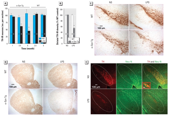
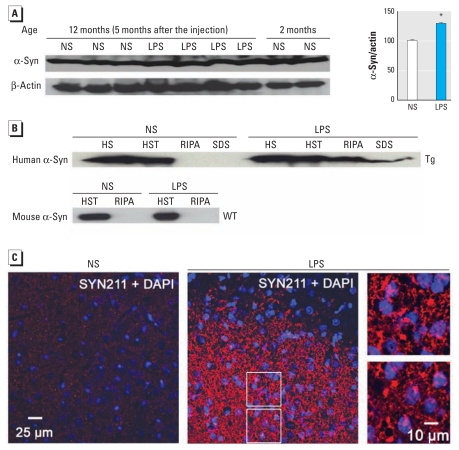
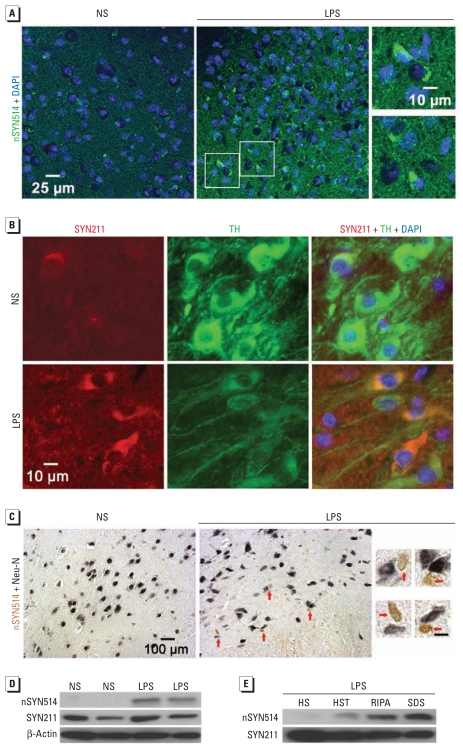
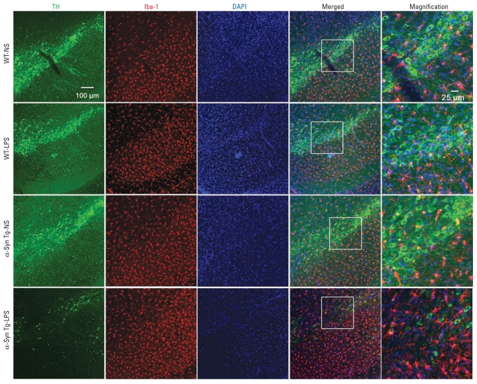
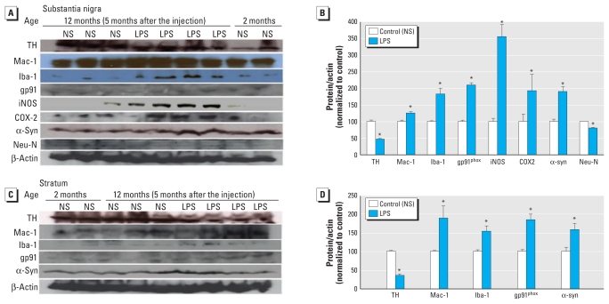
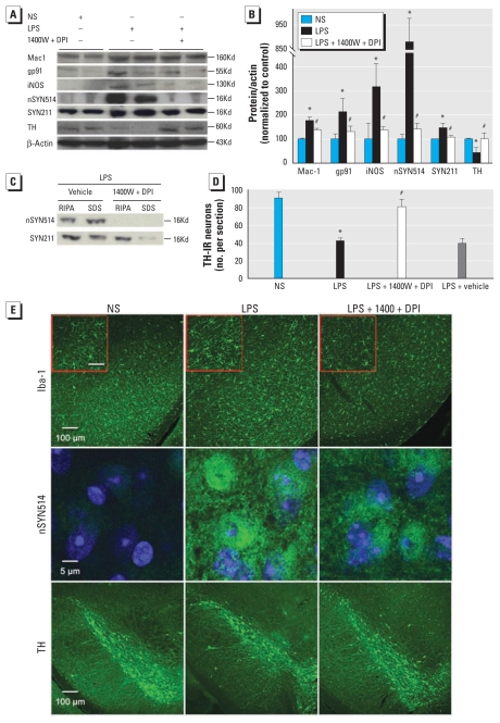
Background: Mechanisms whereby gene–environment interactions mediate chronic, progressive neurodegenerative processes in Parkinson’s disease (PD)—the second most common neurodegenerative disease—remain elusive. Objective: We created a two-hit [neuroinflammation and mutant α-synuclein (α-syn) overexpression] animal model to investigate mechanisms through which mutant α-syn and inflammation work in concert to mediate chronic PD neurodegeneration. Methods: We used an intraperitoneal injection of the inflammogen lipopolysaccharide (LPS; 3 × 10⁶ EU/kg) to initiate systemic and brain inflammation in wild-type (WT) mice and transgenic (Tg) mice overexpressing human A53T mutant α-syn. We then evaluated nigral dopaminergic neurodegeneration, α-syn pathology, and neuroinflammation. Results: After LPS injection, both WT and Tg mice initially displayed indistinguishable acute neuroinflammation; however, only Tg mice developed persistent neuroinflammation, chronic progressive degeneration of the nigrostriatal dopamine pathway, accumulation of aggregated, nitrated α-syn, and formation of Lewy body-like inclusions in nigral neurons. Further mechanistic studies indicated that 4-week infusion of two inhibitors of inducible nitric oxide synthase and NADPH oxidase, major free radical–generating enzymes in activated microglia, blocked nigral α-syn pathology and neurodegeneration in LPS-injected Tg mice. Conclusions: Microglia-derived oxidative stress bridged neuroinflammation and α-syn pathogenic alteration in mediating chronic PD progression. Our two-hit animal model involving both a genetic lesion and an environmental trigger reproduced key features of PD and demonstrated synergistic effects of genetic predisposition and environmental exposures in the development of PD. The chronic progressive nature of dopaminergic neurodegeneration, which is absent in most existing PD models, makes this new model invaluable for the study of mechanisms of PD progression.
Overview
The claims and statistics of this paper haven’t been checked yet.
Run the check — free, no account needed. It reads the open-access text and checks the sources it cites.
Integrity signals
Registry
Cites
Statistics
Figures
Verification
Claims the paper makes
The claims and statistics of this paper haven’t been checked yet.
Run the check — free, no account needed. It reads the open-access text and checks the sources it cites.
Claims other papers make about this one
Each is checked against this paper’s own text: does it say what the citing paper says it does?
The cited source says what the claim attributes to it.
In the citing paper
“Not only has chronic neuroinflammation been shown to increase α-synuclein deposition in various synucleinopathy animal models (51–56), but when these animals were treated with drugs to reduce neuroinflammation, α-synuclein neuropathology also decreased (57, 58).”
From LRRK2 mediates microglial neurotoxicity via NFATc2 in rodent models of synucleinopathies (2020) · cites this paper as [53]
In the citing paper
“This supports a dual-hit hypothesis for PD: elevated endotoxin plus aggregable α-synuclein results in neurodegeneration.”
From The endotoxin hypothesis of neurodegeneration (2019) · cites this paper as [101]
In the citing paper
“A single injection of peripheral endotoxin into wild-type mice and transgenic mice (expressing human A53T mutant α-synuclein) resulted in indistinguishable acute neuroinflammation, but only the transgenic mice treated with endotoxin developed persistent neuroinflammation, aggregated α-synuclein, progressive degeneration of dopamine neurons and Lewy body-like inclusions in nigral neurons [101].”
From The endotoxin hypothesis of neurodegeneration (2019) · cites this paper as [101]
Self-check
Percentages, sums and reported differences re-computed from the paper’s own numbers.
The self-check needs the paper’s full text.
Citations
Cites
Spillantini, M. G., Schmidt, M. L., Lee, V. M.-Y., Trojanowski, J. Q., Jakes, R., & Goedert, M. (1997) · Nature · doi:10.1038/42166 · Cited by 8,382 · cited 1×
“The low rate of concordance for PD in mono zygotic and dizygotic twins (Marttila et al. 1988; Tanner 2003) strongly suggests the involvement of environmental factors in PD. Epidemiologic and case–control studies have implicated rural living, well water consumption, and pesticide exposure as…”
BackgroundMutation in the α-Synuclein Gene Identified in Families with Parkinson's Disease
Polymeropoulos, M. H., Lavedan, C., Leroy, E., Ide, S. E., Dehejia, A., Dutra, A., Pike, B., Root, H., Rubenstein, J., Boyer, R., et al. (1997) · Science · doi:10.1126/science.276.5321.2045 · Cited by 8,240
α-Synuclein Locus Triplication Causes Parkinson's Disease
Singleton, A. B., Farrer, M., Johnson, J., Singleton, A., Hague, S., Kachergus, J., Hulihan, M., Peuralinna, T., Dutra, A., Nussbaum, R., et al. (2003) · Science · doi:10.1126/science.1090278 · Cited by 4,383 · cited 1×
“Furthermore, α-syn aggregates are the primary component of Lewy bodies in both familial and sporadic PD (Spillantini et al. 1997).”
BackgroundMicroglia-mediated neurotoxicity: uncovering the molecular mechanisms
Block, M. L., Zecca, L., & Hong, J.-S. (2007) · Nature Reviews Neuroscience · doi:10.1038/nrn2038 · Cited by 4,110 · cited 1×
“Emerging evidence has indicated that neuroinflammation contributes to PD (Block et al. 2007; Gao and Hong 2008; Hirsch and Hunot 2009).”
BackgroundSelf-citationAlaSOPro mutation in the gene encoding α-synuclein in Parkinson's disease
Krüger, R., Kuhn, W., Müller, T., Woitalla, D., Graeber, M., Kösel, S., Przuntek, H., Epplen, J. T., Schols, L., & Riess, O. (1998) · Nature Genetics · doi:10.1038/ng0298-106 · Cited by 3,836 · cited 1×
“…genes have been identified in familial PD. α-Synuclein (α-syn) is closely associated with both familial and sporadic PD. Missense mutations (A53T, A30P, and E46K) and multiplications of the α-syn gene (SNCA) cause autosomal-dominant PD (Farrer 2006; Krüger et al. 1998; Polymeropoulos et al. 1997).”
BackgroundMcGeer, P. L., Itagaki, S., Boyes, B. E., & McGeer, E. G. (1988) · Neurology · doi:10.1212/wnl.38.8.1285 · Cited by 2,990
Guide for the care and use of laboratory animals
(1997) · Choice Reviews Online · doi:10.5860/choice.34-3310 · Cited by 2,930
Systemic LPS causes chronic neuroinflammation and progressive neurodegeneration
Qin, L., Wu, X., Block, M. L., Liu, Y., Breese, G. R., Hong, J., Knapp, D. J., & Crews, F. T. (2007) · Glia · doi:10.1002/glia.20467 · Cited by 2,144 · cited 1×
“Morphologic changes of microglia (the resident brain immune cells) and accumulation of pro inflammatory factors are associated with degenerating DA neurons in the substantia nigra (SN) of PD patients and various PD animal models (Liberatore et al. 1999; McGeer et al. 1988; Sherer et al. 2003).”
BackgroundSelf-citationα-synuclein locus duplication as a cause of familial Parkinson's disease
Chartier-Harlin, M.-C., Kachergus, J., Roumier, C., Mouroux, V., Douay, X., Lincoln, S., Levecque, C., Larvor, L., Andrieux, J., Hulihan, M., et al. (2004) · The Lancet · doi:10.1016/s0140-6736(04)17103-1 · Cited by 2,029 · cited 1×
“Disease severity in PD patients with SNCA multiplications positively correlates with gene copy number (Chartier-Harlin et al. 2004; Ibanez et al. 2004; Singleton et al. 2003).”
BackgroundNeuroinflammation in Parkinson’s disease: A target for neuroprotection?
Hirsch, E. C., & Hunot, S. (2009) · The Lancet Neurology · doi:10.1016/s1474-4422(09)70062-6 · Cited by 1,984 · cited 1×
“Emerging evidence has indicated that neuroinflammation contributes to PD (Block et al. 2007; Gao and Hong 2008; Hirsch and Hunot 2009).”
BackgroundLi, J.-Y., Englund, E., Holton, J. L., Soulet, D., Hagell, P., Lees, A. J., Lashley, T., Quinn, N. P., Rehncrona, S., Björklund, A., et al. (2008) · Nature Medicine · doi:10.1038/nm1746 · Cited by 1,845 · cited 2×
“…inclusions in neurons grafted into the brains of PD patients and a transgenic (Tg) PD model (Desplats et al. 2009; Kordower et al. 2008; Li et al. 2008; Mendez et al. 2008) further emphasizes a pivotal role of α-syn in ongoing disease progression and host-to-graft disease propagation.”
BackgroundMasliah, E., Rockenstein, E. M., Veinbergs, I., Mallory, M. E., Hashimoto, M., Takeda, A., Sagara, Y., Sisk, A., & Mucke, L. (2000) · Science · doi:10.1126/science.287.5456.1265 · Cited by 1,779
Lewy body–like pathology in long-term embryonic nigral transplants in Parkinson's disease
Kordower, J. H., Chu, Y., Hauser, R. A., Freeman, T. B., & Olanow, C. W. (2008) · Nature Medicine · doi:10.1038/nm1747 · Cited by 1,722
Inclusion formation and neuronal cell death through neuron-to-neuron transmission of α-synuclein
Desplats, P., Lee, H.-J., Bae, E.-J., Patrick, C., Rockenstein, E., Crews, L., Spencer, B., Masliah, E., & Lee, S.-J. (2009) · Proceedings of the National Academy of Sciences · doi:10.1073/pnas.0903691106 · Cited by 1,485 · cited 2×
“The occurrence of α-syn–positive inclusions in neurons grafted into the brains of PD patients and a transgenic (Tg) PD model (Desplats et al. 2009; Kordower et al. 2008; Li et al. 2008; Mendez et al. 2008) further emphasizes a pivotal role of α-syn in ongoing disease progression and host-to-graft disease propagation.”
Backgroundα-Synuclein Blocks ER-Golgi Traffic and Rab1 Rescues Neuron Loss in Parkinson's Models
Cooper, A. A., Gitler, A. D., Cashikar, A. G., Haynes, C. M., Hill, K. J., Bhullar, B., Liu, K., Xu, K., Strathearn, K. E., Liu, F., et al. (2006) · Science · doi:10.1126/science.1129462 · Cited by 1,411 · cited 1×
“Aggregated α-syn may over whelm the ubiquitin–proteasome system and accumulate over time, leading to endoplasmic reticulum stress and mitochondrial impairment (Cooper et al. 2006).”
BackgroundNeuronal α-Synucleinopathy with Severe Movement Disorder in Mice Expressing A53T Human α-Synuclein
Giasson, B. I., Duda, J. E., Quinn, S. M., Zhang, B., Trojanowski, J. Q., & Lee, V. M.-Y. (2002) · Neuron · doi:10.1016/s0896-6273(02)00682-7 · Cited by 1,266
Zhang, W., Wang, T., Pei, Z., Miller, D. S., Wu, X., Block, M. L., Wilson, B., Zhang, W., Zhou, Y., Hong, J.-S., et al. (2005) · The FASEB Journal · doi:10.1096/fj.04-2751com · Cited by 1,265 · cited 1×
“Exogenous application of aggregated α-syn activates cultured microglia (Zhang et al. 2005).”
BackgroundSelf-citationCausal relation between α-synuclein locus duplication as a cause of familial Parkinson's disease
Ibáñez, P., Bonnet, A. M., Débarges, B., Lohmann, E., Tison, F., Agid, Y., Dürr, A., Brice, A., & Pollak, P. (2004) · The Lancet · doi:10.1016/s0140-6736(04)17104-3 · Cited by 1,063 · cited 1×
“Disease severity in PD patients with SNCA multiplications positively correlates with gene copy number (Chartier-Harlin et al. 2004; Ibanez et al. 2004; Singleton et al. 2003).”
BackgroundLiberatore, G. T., Jackson-Lewis, V., Vukosavic, S., Mandir, A. S., Vila, M., McAuliffe, W. G., Dawson, V. L., Dawson, T. M., & Przedborski, S. (1999) · Nature Medicine · doi:10.1038/70978 · Cited by 1,046 · cited 1×
“Morphologic changes of microglia (the resident brain immune cells) and accumulation of pro inflammatory factors are associated with degenerating DA neurons in the substantia nigra (SN) of PD patients and various PD animal models (Liberatore et al. 1999; McGeer et al. 1988; Sherer et al. 2003).”
BackgroundKim, W., Mohney, R. P., Wilson, B. C., Jeohn, G., Liu, B., & Hong, J. (2000) · Journal of Neuroscience · doi:10.1523/jneurosci.20-16-06309.2000 · Cited by 881
Self-citationGenetic Animal Models of Parkinson's Disease
Dawson, T. M., Ko, H. S., & Dawson, V. L. (2010) · Neuron · doi:10.1016/j.neuron.2010.04.034 · Cited by 866 · cited 1×
“Among different α-syn Tg mouse models, only a few exhibit overt degeneration of nigro striatal DA fibers and even fewer show loss of nigral DA neurons (Dawson et al. 2010; Lee et al. 2002; Masliah et al. 2000).”
BackgroundWhy neurodegenerative diseases are progressive: uncontrolled inflammation drives disease progression
Gao, H., & Hong, J. (2008) · Trends in Immunology · doi:10.1016/j.it.2008.05.002 · Cited by 852 · cited 2×
“Emerging evidence has indicated that neuroinflammation contributes to PD (Block et al. 2007; Gao and Hong 2008; Hirsch and Hunot 2009).”
BackgroundSelf-citationLee, M. K., Stirling, W., Xu, Y., Xu, X., Qui, D., Mandir, A. S., Dawson, T. M., Copeland, N. G., Jenkins, N. A., & Price, D. L. (2002) · Proceedings of the National Academy of Sciences · doi:10.1073/pnas.132197599 · Cited by 824
Hamza, T. H., Zabetian, C. P., Tenesa, A., Laederach, A., Montimurro, J. S., Yearout, D., Kay, D. M., Doheny, K. F., Paschall, J. E., Pugh, E., et al. (2010) · Nature Genetics · doi:10.1038/ng.642 · Cited by 808 · cited 1×
“DNA polymorphisms of several inflammatory cytokines and genetic variation in the HLA (human leukocyte antigen) region that contains numerous immune-related genes might increase the risk of PD (Hamza et al. 2010; Wahner et al. 2007).”
BackgroundGenetics of Parkinson disease: paradigm shifts and future prospects
Farrer, M. J. (2006) · Nature Reviews Genetics · doi:10.1038/nrg1831 · Cited by 724 · cited 1×
“Farrer MJ. 2006.”
Gao, H., Jiang, J., Wilson, B. C., Zhang, W., Hong, J., & Liu, B. (2002) · Journal of Neurochemistry · doi:10.1046/j.1471-4159.2002.00928.x · Cited by 690 · cited 1×
“We and others previously reported that an intra peritoneal injection or chronic nigral infusion of relatively high doses of lipo polysaccharide (LPS; a common inflammogen) led to time-dependent degeneration of nigral DA neurons in rodents (Gao et al. 2002; Ling et al. 2006; Qin et al. 2007).”
BackgroundSelf-citationUnderstanding cell death in parkinson's disease
Jenner, P., & Olanow, C. W. (1998) · Annals of Neurology · doi:10.1002/ana.410440712 · Cited by 686
Wu, D., Teismann, P., Tieu, K., Vila, M., Jackson-Lewis, V. R., Ischiropoulos, H., & Przedborski, S. (2003) · Proceedings of the National Academy of Sciences · doi:10.1073/pnas.0937239100 · Cited by 634 · cited 1×
“Exogenous application of aggregated α-syn activates cultured microglia (Zhang et al. 2005).”
BackgroundNeuroinflammation and Oxidation/Nitration of α-Synuclein Linked to Dopaminergic Neurodegeneration
Gao, H., Kotzbauer, P. T., Uryu, K., Leight, S., Trojanowski, J. Q., & Lee, V. M. ‐. (2008) · Journal of Neuroscience · doi:10.1523/jneurosci.0143-07.2008 · Cited by 453 · cited 6×
“For all other biochemical studies, we used the 2% SDS buffer to extract both soluble and aggregated proteins from stratum, SN, or whole brains (Gao et al. 2008; Giasson et al. 2002).”
BackgroundMethodInfluentialSelf-citationMendez, I. M., Viñuela, Á., Astradsson, A., Mukhida, K., Hallett, P. J., Robertson, H. A., Tierney, T. S., Holness, R. O., Dagher, A., Trojanowski, J. Q., et al. (2008) · Nature Medicine · doi:10.1038/nm1752 · Cited by 442
Chu, Y., & Kordower, J. H. (2006) · Neurobiology of Disease · doi:10.1016/j.nbd.2006.08.021 · Cited by 423 · cited 1×
“Additionally, age-related increases of α-syn in monkeys and humans are linked to nigrostriatal DA depletion (Chu and Kordower 2007).”
BackgroundNovel anti-inflammatory therapy for Parkinson's disease
Gao, H., Liu, B., Zhang, W., & Hong, J. (2003) · Trends in Pharmacological Sciences · doi:10.1016/s0165-6147(03)00176-7 · Cited by 325 · cited 1×
“Moreover, inhibition of inflammation is associated with reduced neuronal impairment in various PD models (Gao et al. 2003; Wu et al. 2003).”
BackgroundSelf-citationProfessional exposure to pesticides and Parkinson disease
Elbaz, A., Clavel, J., Rathouz, P. J., Moisan, F., Galanaud, J. ‐., Delemotte, B., Alpérovitch, A., & Tzourio, C. (2009) · Annals of Neurology · doi:10.1002/ana.21717 · Cited by 317 · cited 2×
“…twins (Marttila et al. 1988; Tanner 2003) strongly suggests the involvement of environmental factors in PD. Epidemiologic and case–control studies have implicated rural living, well water consumption, and pesticide exposure as potential environ mental risk factors for PD (Elbaz et al. 2009).”
BackgroundSelective microglial activation in the rat rotenone model of Parkinson's disease
Sherer, T., Betarbet, R. S., Kim, J.-H., & Greenamyre, J. T. (2003) · Neuroscience Letters · doi:10.1016/s0304-3940(03)00172-1 · Cited by 317 · cited 1×
“Disease severity in PD patients with SNCA multiplications positively correlates with gene copy number (Chartier-Harlin et al. 2004; Ibanez et al. 2004; Singleton et al. 2003).”
BackgroundParkin Deficiency Increases Vulnerability to Inflammation-Related Nigral Degeneration
Frank-Cannon, T. C., Tran, T. A. T., Ruhn, K. A., Martinez, T. N., Hong, J. J., Marvin, M. A., Hartley, M., Treviño, I., O’Brien, D. E., Casey, B. H., et al. (2008) · Journal of Neuroscience · doi:10.1523/jneurosci.3001-08.2008 · Cited by 268 · cited 1×
“Another newly developed two-hit mouse model combining a genetic lesion (Parkin–/–) and neuro inflammation has provided additional experimental evidence supporting a crucial role of gene–environment interactions in PD (Frank-Cannon et al. 2008).”
BackgroundSelf-citationα-Synuclein Overexpression Protects against Paraquat-Induced Neurodegeneration
Manning-Bog, A., McCormack, A. L., Purisai, M. G., Bolin, L. M., & Monte, D. A. D. (2003) · Journal of Neuroscience · doi:10.1523/jneurosci.23-08-03095.2003 · Cited by 242 · cited 1×
“Interestingly, it has been reported that over expression of mutant α-syn in mice protects against (Manning-Bog et al. 2003) or fails to affect paraquat neuro toxicity (Norris et al. 2007), augments neuro degeneration elicited by 1-methyl-4-phenyl-1,2,3,6-tetrahydropyridine and LPS (Gao et al. 2008;…”
Backgroundα‐Synuclein promoter confers susceptibility to Parkinson's disease
Pals, P., Lincoln, S., Manning, J. R., Heckman, M. G., Skipper, L. M., Hulihan, M., den Broeck, M. V., Pooter, T. D., Cras, P. P., Crook, J. E., et al. (2004) · Annals of Neurology · doi:10.1002/ana.20268 · Cited by 211
Zhang, W., Wang, T., Qin, L., Gao, H., Wilson, B. C., Ali, S. F., Zhang, W., Hong, J., & Liu, B. (2004) · The FASEB Journal · doi:10.1096/fj.03-0983fje · Cited by 206 · cited 1×
“To monitor DA neuro degeneration, two individuals blind to the treatment counted the number of TH-immunoreactive (TH-IR) neurons in the SN pars compacta (SNpc) of six evenly spaced brain sections from a series of 24 sections that covered the entire SN (Zhang et al. 2004).”
MethodSelf-citationLing, Z., Zhu, Y., Tong, C. W., Snyder, J. A., Lipton, J. W., & Carvey, P. M. (2006) · Experimental Neurology · doi:10.1016/j.expneurol.2006.01.010 · Cited by 160
Inflammatory Cytokine Gene Polymorphisms and Increased Risk of Parkinson Disease
Wahner, A. D., Sinsheimer, J. S., Bronstein, J. M., & Ritz, B. R. (2007) · Archives of Neurology · doi:10.1001/archneur.64.6.836 · Cited by 143 · cited 1×
“Moreover, inhibition of inflammation is associated with reduced neuronal impairment in various PD models (Gao et al. 2003; Wu et al. 2003).”
BackgroundRab11a and HSP90 Regulate Recycling of Extracellular α-Synuclein
Liu, J., Zhang, J., Shi, M. Y., Quinn, T. J., Bradner, J. M., Beyer, R., Chen, S., & Zhang, J. (2009) · Journal of Neuroscience · doi:10.1523/jneurosci.6202-08.2009 · Cited by 136
Pesticide Exposure Exacerbates α-Synucleinopathy in an A53T Transgenic Mouse Model
Norris, E. H., Uryu, K., Leight, S., Giasson, B. I., Trojanowski, J. Q., & Lee, V. M. ‐. (2007) · American Journal Of Pathology · doi:10.2353/ajpath.2007.060359 · Cited by 134 · cited 1×
“…has been reported that over expression of mutant α-syn in mice protects against (Manning-Bog et al. 2003) or fails to affect paraquat neuro toxicity (Norris et al. 2007), augments neuro degeneration elicited by 1-methyl-4-phenyl-1,2,3,6-tetrahydropyridine and LPS (Gao et al. 2008; Nieto et al.…”
BackgroundParkinson's disease in a nationwide twin cohort
Marttila, R. J., Kaprio, J. A., Koskenvuo, M., & Rinne, U. K. (1988) · Neurology · doi:10.1212/wnl.38.8.1217 · Cited by 132
Is the cause of Parkinson's disease environmental or hereditary? Evidence from twin studies.
Tanner, C. M. (2003) · PubMed · Cited by 130
Increased sensitivity to MPTP in human α-synuclein A30P transgenic mice
Nieto, M., Gil-Bea, F. J., Dalfó, E., Cuadrado‐Tejedor, M., Cabodevilla, F., Sánchez, B., Catena, S., Sesma, T., Ribé, E. M., Ferrer, I., et al. (2005) · Neurobiology of Aging · doi:10.1016/j.neurobiolaging.2005.04.010 · Cited by 100
Causal relation between α synuclein gene duplication and familial Parkinson’s disease
Inserm, U. (2005) · Dkgest of the World Latest Medical Information · Cited by 81
Cited by
Sorted by citations
Gut microbiota regulate motor deficits and neuroinflammation in a model of Parkinson's disease
Sampson, T. R., Debelius, J. W., Thron, T., Janssen, S., Shastri, G. G., Ilhan, Z. E., Challis, C., Schretter, C. E., Rocha, S., Gradinaru, V., et al. (2016) · Cell · doi:10.1016/j.cell.2016.11.018 · Cited by 3,622
“Intriguingly, an inflammatory environment is known to enhance aSyn aggregation, which may further activate microglia upon contact and promote a feed-forward cascade that leads to additional aSyn aggregation and propagation and progression of disease (Gao et al., 2011).”
The Role of Oxidative Stress in Parkinson's Disease
Dias, V., Junn, E., & Mouradian, M. M. (2013) · Journal of Parkinson’s Disease · doi:10.3233/jpd-130230 · Cited by 1,845
“synuclein and dopamine neuron degeneration [284].”
Forsyth, C. B., Shannon, K. M., Kordower, J. H., Voigt, R. M., Shaikh, M., Jaglin, J. A., Estes, J. D., Dodiya, H. B., & Keshavarzian, A. (2011) · PLoS ONE · doi:10.1371/journal.pone.0028032 · Cited by 974
“Only the transgenic mice, and not controls, developed sustained neuroinflammation including Lewy bodies staining of nigral neurons [40].”
BackgroundSystemic inflammation and microglial activation: systematic review of animal experiments
Hoogland, I. C. M., Houbolt, C., van Westerloo, D. J., van Gool, W. A., & van de Beek, D. (2015) · Journal of Neuroinflammation · doi:10.1186/s12974-015-0332-6 · Cited by 957
“Gao [59] B6C3F1/J U 7 m M E.”
Oxidative stress, mitochondrial dysfunction and neurodegenerative diseases; a mechanistic insight
Bhat, A. H., Dar, K. B., Anees, S., Zargar, M. A., Masood, A., Sofi, M. A., & Ganie, S. A. (2015) · Biomedicine & Pharmacotherapy · doi:10.1016/j.biopha.2015.07.025 · Cited by 962
Metadata
- Authors
- Gao, H.-M., Zhang, F., Zhou, H., Kam, W., Wilson, B., & Hong, J.-S.
- Affiliations
- National Institutes of Health (US)
- Corresponding author
- Gao, Hui-Ming
- Venue
- Environmental Health Perspectives
- Publisher
- American Chemical Society (ACS)
- Published
- 2011-01-18
- Volume
- 119
- Issue
- 6
- Pages
- 807-814
- ISSN
- 0091-6765 (ISSN-L)
- 1552-9924
- PMC
- PMC3114815
- OpenAlex
- W1971599063
- Cited by
- 355
- References
- 46
- Citations by year
- 2026: 16 · 2025: 13 · 2024: 36 · 2023: 23 · 2022: 28 · 2021: 37 · 2020: 39 · 2019: 17 · 2018: 28 · 2017: 20 · 2016: 18 · 2015: 16 · 2014: 19 · 2013: 18 · 2012: 19
- FWCI
- 13.00
- Type
- journal article
- Language
- en
- Open access
- gold
- doi.org
- Licence
- public-domain
- creativecommons.org/licenses/by/4.0/ (vor, 2537 d)
- Locations
- Environmental Health Perspectives (publishedVersion, public-domain)
- PubMed Central (publishedVersion, public-domain)
- PubMed (publishedVersion)
- Funders
- National Institutes of Health
- National Institute of Environmental Health Sciences
- Topics
- Health Sciences › Medicine › Clinical Neurology › Parkinson's Disease Mechanisms and Treatments
- Life Sciences › Neuroscience › Neurology › Neuroinflammation and Neurodegeneration Mechanisms
- Life Sciences › Biochemistry, Genetics and Molecular Biology › Molecular Biology › Nuclear Receptors and Signaling
- Keywords
- Parkinson's disease, neuroinflammation, alpha-synuclein, lipopolysaccharide, two-hit model, gene-environment interaction, microglia, oxidative stress, inducible nitric oxide synthase, NADPH oxidase, dopaminergic neuron loss, nigrostriatal pathway, Lewy bodies, chronic neurodegeneration
- MeSH terms
- Amidines, Animals, Benzylamines, Disease Models, Animal, Injections, Intraperitoneal, Lipopolysaccharides, Nerve Degeneration, Onium Compounds, Parkinson Disease, Substantia Nigra, Lewy Bodies, Oxidative Stress, NADPH Oxidases, Neurodegenerative Diseases, Animals, Genetically Modified, Mice, alpha-Synuclein, Nitric Oxide Synthase Type II
About these checks
Registry, retraction, Cites and statistics results are computed, not judged, with one exception: a printed reference that no index lists is read once by a model before it counts as a citation error. Claims are extracted by a model and kept only when their quote appears verbatim in the text; each cited source is then checked, with a model’s help, for whether it says what the claim relies on. Those verdicts can be wrong — read the claim’s quote and the cited source before relying on one.
Statistics and arithmetic are recomputed, not judged.
Confidence bands are shown only for fields calibrated on our annotated test set. None is calibrated yet, so no band is shown.
What counts as a claim ›How we measure confidence ›How we check this ›
Are you an author, or do you see a problem with this page? Request a review
