

Primary Visual Cortex Activity along the Apparent-Motion Trace Reflects Illusory Perception
Open accessThe DOI matches its Crossref record; 64 of the 64 references it cites were found in a registry or an index.
Abstract
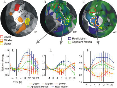
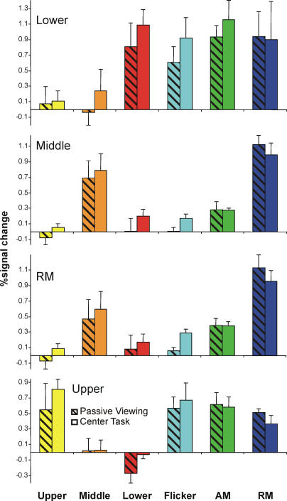
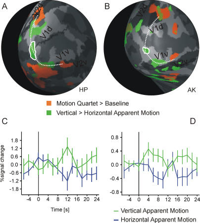
The illusion of apparent motion can be induced when visual stimuli are successively presented at different locations. It has been shown in previous studies that motion-sensitive regions in extrastriate cortex are relevant for the processing of apparent motion, but it is unclear whether primary visual cortex (V1) is also involved in the representation of the illusory motion path. We investigated, in human subjects, apparent-motion-related activity in patches of V1 representing locations along the path of illusory stimulus motion using functional magnetic resonance imaging. Here we show that apparent motion caused a blood-oxygenation-level-dependent response along the V1 representations of the apparent-motion path, including regions that were not directly activated by the apparent-motion-inducing stimuli. This response was unaltered when participants had to perform an attention-demanding task that diverted their attention away from the stimulus. With a bistable motion quartet, we confirmed that the activity was related to the conscious perception of movement. Our data suggest that V1 is part of the network that represents the illusory path of apparent motion. The activation in V1 can be explained either by lateral interactions within V1 or by feedback mechanisms from higher visual areas, especially the motion-sensitive human MT/V5 complex.
Overview
The claims and statistics of this paper haven’t been checked yet.
Free: it reads the open-access text and checks the first 5 claims that cite a source we can check. Create a free account to check every claim.
Integrity signals
Registry
Cites
Statistics
Figures



Verification
Claims the paper makes
The claims and statistics of this paper haven’t been checked yet.
Free: it reads the open-access text and checks the first 5 claims that cite a source we can check. Create a free account to check every claim.
Claims other papers make about this one
Each is checked against this paper’s own text: does it say what the citing paper says it does?
The source backs part of the claim, not all of it — or backs it for a different population, condition or comparison.
In the citing paper
“V5 feedback signals spread out to a large region in primary visual cortex (de-Wit et al. 2012; Muckli et al. 2005)”
From Whatever next? Predictive brains, situated agents, and the future of cognitive science (2013) · cites this paper as Muckli et al. 2005
Experimental — accuracy not yet measuredIn the citing paper
“Moreover, higher visual areas project back to V1 the associated color of grayscale objects [36] or the predicted motion path of an apparent motion illusion [37, 38].”
From Decoding sound and imagery content in early visual cortex (2014) · cites this paper as [37]
Self-check
Percentages, sums and reported differences re-computed from the paper’s own numbers.
The self-check needs the paper’s full text.
Citations
Cites
Neurophysiological investigation of the basis of the fMRI signal
Logothetis, N. K., Pauls, J., Augath, M., Trinath, T., & Oeltermann, A. (2001) · Nature · doi:10.1038/35084005 · Cited by 6,551 · cited 1×
“Current findings from simultaneous fMRI and electrophysiological recordings in monkeys have suggested that the BOLD signal might be especially sensitive to changes in local field potentials [ 53 ].”
BackgroundBorders of Multiple Visual Areas in Humans Revealed by Functional Magnetic Resonance Imaging
Sereno, M. I., Dale, A. M., Reppas, J. B., Kwong, K. K., Belliveau, J. W., Brady, T. J., Rosen, B. R., & Tootell, R. B. H. (1995) · Science · doi:10.1126/science.7754376 · Cited by 2,658
Linear Systems Analysis of Functional Magnetic Resonance Imaging in Human V1
Boynton, G. M., Engel, S. A., Glover, G. H., & Heeger, D. J. (1996) · The Journal of Neuroscience · doi:10.1523/jneurosci.16-13-04207.1996 · Cited by 2,314
Kastner, S., Pinsk, M. A., De Weerd, P., Desimone, R., & Ungerleider, L. G. (1999) · Neuron · doi:10.1016/s0896-6273(00)80734-5 · Cited by 1,622
Why visual attention and awareness are different
Lamme, V. A. F. (2003) · Trends in Cognitive Sciences · doi:10.1016/s1364-6613(02)00013-x · Cited by 1,172 · cited 1×
“In these studies, different imaging and electrophysiological methods have been used to investigate the functional properties of V1 during binocular rivalry [40– 43], perception of moving phosphenes [15,17], figure–ground segregation [34,44–46], the ‘‘line-motion’’ illusion [28], and color filling-in [47].”
MethodThe Underpinnings of the BOLD Functional Magnetic Resonance Imaging Signal
Logothetis, N. K. (2003) · The Journal of Neuroscience · doi:10.1523/jneurosci.23-10-03963.2003 · Cited by 1,039
Engel, S. A., Rumelhart, D. E., Wandell, B. A., Lee, A. T., Glover, G. H., Chichilnisky, E.-J., & Shadlen, M. N. (1994) · Nature · doi:10.1038/369525a0 · Cited by 990
Experimentelle Studien uber das Sehen von Bewegung
Wertheimer, M. (1912) · Cited by 975
Mechanisms of Directed Attention in the Human Extrastriate Cortex as Revealed by Functional MRI
Kastner, S., De Weerd, P., Desimone, R., & Ungerleider, L. G. (1998) · Science · doi:10.1126/science.282.5386.108 · Cited by 953
Are we aware of neural activity in primary visual cortex?
Crick, F., & Koch, C. (1995) · Nature · doi:10.1038/375121a0 · Cited by 932
Research works printed but not linked: 4
- Wertheimer, 1912, Z Psychol, 61, 161
- Albright, 1993, Rev Oculomot Res, 5, 177
- Neuhaus, 1930, Arch Gesamte Psychol, 75, 315
- Lee, 2002, J Vis, 2, 618
Cited by
Whatever next? Predictive brains, situated agents, and the future of cognitive science
Clark, A. (2013) · Behavioral and Brain Sciences · doi:10.1017/s0140525x12000477 · Cited by 5,924
How Do Expectations Shape Perception?
de Lange, F. P., Heilbron, M., & Kok, P. (2018) · Trends in Cognitive Sciences · doi:10.1016/j.tics.2018.06.002 · Cited by 975
Expectation in perceptual decision making: neural and computational mechanisms
Summerfield, C., & de Lange, F. P. (2014) · Nature Reviews Neuroscience · doi:10.1038/nrn3838 · Cited by 832
Predictions not commands: Active inference in the motor system
Adams, R. A., Shipp, S., & Friston, K. J. (2013) · Brain Structure and Function · doi:10.1007/s00429-012-0475-5 · Cited by 765
Stimulus Predictability Reduces Responses in Primary Visual Cortex
Alink, A., Schwiedrzik, C. M., Kohler, A., Singer, W., & Muckli, L. (2010) · The Journal of Neuroscience · doi:10.1523/jneurosci.3730-10.2010 · Cited by 435
Self-citationDecoding sound and imagery content in early visual cortex
Vetter, P., Smith, F. W., & Muckli, L. (2014) · Current Biology · doi:10.1016/j.cub.2014.04.020 · Cited by 323
Self-citationPressnitzer, D., & Hupé, J.-M. (2006) · Current Biology · doi:10.1016/j.cub.2006.05.054 · Cited by 302
Varieties of cognitive penetration in visual perception
Vetter, P., & Newen, A. (2014) · Consciousness and Cognition · doi:10.1016/j.concog.2014.04.007 · Cited by 263
Nonstimulated early visual areas carry information about surrounding context
Smith, F. W., & Muckli, L. (2010) · Proceedings of the National Academy of Sciences · doi:10.1073/pnas.1000233107 · Cited by 238
“For instance, the size of the activated region in retinotopic cortex corresponds to perceived size, not absolute size, in the context of a size illusion (2); moreover, the subjectively perceived apparent-motion illusion activates nonstimulated V1 on the motion trace (3), and object classification triggers discriminabl…”
BackgroundSelf-citationTop-down effects on early visual processing in humans: A predictive coding framework
Rauss, K., Schwartz, S., & Pourtois, G. (2011) · Neuroscience & Biobehavioral Reviews · doi:10.1016/j.neubiorev.2010.12.011 · Cited by 219
Network interactions: non-geniculate input to V1
Muckli, L., & Petro, L. S. (2013) · Current Opinion in Neurobiology · doi:10.1016/j.conb.2013.01.020 · Cited by 202
Self-citationTime-compressed preplay of anticipated events in human primary visual cortex
Ekman, M., Kok, P., & de Lange, F. P. (2017) · Nature Communications · doi:10.1038/ncomms15276 · Cited by 191
Wibral, M., Rahm, B., Rieder, M., Lindner, M., Vicente, R., & Kaiser, J. (2011) · Progress in Biophysics and Molecular Biology · doi:10.1016/j.pbiomolbio.2010.11.006 · Cited by 190
Laminar fMRI: applications for cognitive neuroscience
Lawrence, S. J. D., Formisano, E., Muckli, L., & de Lange, F. P. (2019) · NeuroImage · doi:10.1016/j.neuroimage.2017.07.004 · Cited by 186
Self-citationSmaller Primary Visual Cortex Is Associated with Stronger, but Less Precise Mental Imagery
Bergmann, J., Genç, E., Kohler, A., Singer, W., & Pearson, J. (2016) · Cerebral Cortex · doi:10.1093/cercor/bhv186 · Cited by 177
Self-citationPounder, Z., Jacob, J., Evans, S., Loveday, C., Eardley, A. F., & Silvanto, J. (2022) · Cortex · doi:10.1016/j.cortex.2021.12.010 · Cited by 168
Shape Perception Simultaneously Up- and Downregulates Neural Activity in the Primary Visual Cortex
Kok, P., & de Lange, F. P. (2014) · Current Biology · doi:10.1016/j.cub.2014.05.042 · Cited by 163
Neural correlates of the contents of visual awareness in humans
Rees, G. (2007) · Philosophical Transactions of the Royal Society B: Biological Sciences · doi:10.1098/rstb.2007.2094 · Cited by 167
Phantom perception: voluntary and involuntary nonretinal vision
Pearson, J., & Westbrook, F. (2015) · Trends in Cognitive Sciences · doi:10.1016/j.tics.2015.03.004 · Cited by 161
Primary visual cortex activation on the path of apparent motion is mediated by feedback from hMT+/V5
Sterzer, P., Haynes, J.-D., & Rees, G. (2006) · NeuroImage · doi:10.1016/j.neuroimage.2006.05.029 · Cited by 156
“Our finding of activity related to the perception of an AM path in V1 is in accordance with recent fMRI findings (Muckli et al., 2005), though one previous study did not find such AM-related activity differences in early visual cortical areas (Liu et al., 2004).”
BackgroundMethodResultInfluentialVisual Adaptation of the Perception of Causality
Rolfs, M., Dambacher, M., & Patrick Cavanagh, B. (2013) · Current Biology · doi:10.1016/j.cub.2012.12.017 · Cited by 156
Laycock, R., Crewther, S. G., & Crewther, D. P. (2006) · Neuroscience & Biobehavioral Reviews · doi:10.1016/j.neubiorev.2006.10.003 · Cited by 177
What is Bottom-Up and What is Top-Down in Predictive Coding?
Rauss, K. S., & Pourtois, G. (2013) · Frontiers in Psychology · doi:10.3389/fpsyg.2013.00276 · Cited by 196
Gender-Specific Left–Right Asymmetries in Human Visual Cortex
Amunts, K., Armstrong, E., Maliković, A., Hömke, L., Mohlberg, H., Schleicher, A., & Zilles, K. (2007) · Journal of Neuroscience · doi:10.1523/jneurosci.4753-06.2007 · Cited by 157
Functional neuroimaging findings on the human perception of illusory contours
Seghier, M. L., & Vuilleumier, P. (2006) · Neuroscience & Biobehavioral Reviews · doi:10.1016/j.neubiorev.2005.11.002 · Cited by 128
Imagery of a moving object: The role of occipital cortex and human MT/V5+
Kaas, A. L., Weigelt, S., Roebroeck, A. F., Kohler, A., & Muckli, L. F. (2009) · NeuroImage · doi:10.1016/j.neuroimage.2009.07.055 · Cited by 134
Self-citationMuckli, L. F., Kieß, S., Tonhausen, N., Singer, W., Goebel, R., & Sireteanu, R. (2005) · Vision Research · doi:10.1016/j.visres.2005.10.014 · Cited by 112
Self-citationInterhemispheric Connections Shape Subjective Experience of Bistable Motion
Genç, E., Bergmann, J., Singer, W., & Kohler, A. (2011) · Current Biology · doi:10.1016/j.cub.2011.08.003 · Cited by 107
Self-citationPredictive Coding in Sensory Cortex
Kok, P., & de Lange, F. P. (2015) · An Introduction to Model-Based Cognitive Neuroscience · doi:10.1007/978-1-4939-2236-9_11 · Cited by 98
Contextual modulation of primary visual cortex by auditory signals
Petro, L. S., Paton, A. T., & Muckli, L. (2017) · Philosophical Transactions of the Royal Society B: Biological Sciences · doi:10.1098/rstb.2016.0104 · Cited by 101
Self-citationCortical excitability controls the strength of mental imagery
Keogh, R., Bergmann, J., & Pearson, J. (2020) · eLife · doi:10.7554/elife.50232 · Cited by 94
“displays was used for presentation of the stimuli (MR Vision 2000; Resonance Technology Northridge, CA), which were generated with a custom-made program based on the Microsoft DirectX library (Muckli et al., 2005).”
MethodMuckli, L. (2010) · International Journal of Imaging Systems and Technology · doi:10.1002/ima.20236 · Cited by 94
“However, we, and others, were able to demonstrate that the apparent motion path can be stimulated by the visual motion illusion (Muckli et al., 2005; Larsen et al., 2006; Sterzer et al., 2006).”
BackgroundSelf-citationBilateral visual field maps in a patient with only one hemisphere
Muckli, L. F., Naumer, M. J., & Singer, W. (2009) · Proceedings of the National Academy of Sciences · doi:10.1073/pnas.0809688106 · Cited by 95
Self-citationAuditory Attraction: Activation of Visual Cortex by Music and Sound in Williams Syndrome
Thornton‐Wells, T. A., Cannistraci, C. J., Anderson, A. W., Kim, C.-Y., Eapen, M., Gore, J. C., Blake, R., & Dykens, E. M. (2010) · American Journal on Intellectual and Developmental Disabilities · doi:10.1352/1944-7588-115.172 · Cited by 79
Neural Anatomy of Primary Visual Cortex Limits Visual Working Memory
Bergmann, J., Genç, E., Kohler, A., Singer, W., & Pearson, J. (2014) · Cerebral Cortex · doi:10.1093/cercor/bhu168 · Cited by 78
Self-citationVisual cortical networks: of mice and men
Katzner, S., & Weigelt, S. (2013) · Current Opinion in Neurobiology · doi:10.1016/j.conb.2013.01.019 · Cited by 73
Variability of perceptual multistability: from brain state to individual trait
Kleinschmidt, A. K., Sterzer, P., & Rees, G. E. (2012) · Philosophical Transactions of the Royal Society B Biological Sciences · doi:10.1098/rstb.2011.0367 · Cited by 78
Mur, M., Ruff, D. A., Bodurka, J., Bandettini, P. A., & Kriegeskorte, N. (2010) · Cerebral Cortex · doi:10.1093/cercor/bhp272 · Cited by 79
Self-citationTMS Over V5 Disrupts Motion Prediction
Vetter, P., Grosbras, M.-H., & Muckli, L. (2015) · Cerebral Cortex · doi:10.1093/cercor/bht297 · Cited by 66
Self-citationMorgan, A., Petro, L. S., & Muckli, L. F. (2019) · Journal of Neuroscience · doi:10.1523/jneurosci.0852-19.2019 · Cited by 72
Self-citationThe Timing of Feedback to Early Visual Cortex in the Perception of Long-Range Apparent Motion
Wibral, M., Bledowski, C., Kohler, A., Singer, W., & Muckli, L. (2009) · Cerebral Cortex · doi:10.1093/cercor/bhn192 · Cited by 64
Self-citationContributions of cortical feedback to sensory processing in primary visual cortex
Petro, L. S., Vizioli, L., & Muckli, L. (2014) · Frontiers in Psychology · doi:10.3389/fpsyg.2014.01223 · Cited by 61
Self-citationSeparate cortical stages in amodal completion revealed by functional magnetic resonance adaptation
Weigelt, S., Singer, W., & Muckli, L. F. (2007) · BMC Neuroscience · doi:10.1186/1471-2202-8-70 · Cited by 63
Self-citationGori, S., Molteni, M., & Facoetti, A. (2016) · Frontiers in Human Neuroscience · doi:10.3389/fnhum.2016.00175 · Cited by 80
Chemla, S., Reynaud, A., di Volo, M., Zerlaut, Y., Perrinet, L., Destexhe, A., & Chavane, F. (2019) · Journal of Neuroscience · doi:10.1523/jneurosci.2792-18.2019 · Cited by 75
The neural correlates of hierarchical predictions for perceptual decisions
Weilnhammer, V. A., Stuke, H., Sterzer, P., & Schmack, K. (2018) · The Journal of Neuroscience · doi:10.1523/jneurosci.2901-17.2018 · Cited by 57
Microsaccade Rate Varies with Subjective Visibility during Motion-Induced Blindness
Hsieh, P., & Tse, P. U. (2009) · PLoS ONE · doi:10.1371/journal.pone.0005163 · Cited by 64
Brain activity dynamics in human parietal regions during spontaneous switches in bistable perception
Megumi, F., Bahrami, B., Kanai, R., & Rees, G. E. (2014) · NeuroImage · doi:10.1016/j.neuroimage.2014.12.018 · Cited by 63
Reconstructing representations of dynamic visual objects in early visual cortex
Chong, E., Familiar, A., & Shim, W. M. (2015) · Proceedings of the National Academy of Sciences · doi:10.1073/pnas.1512144113 · Cited by 59
Predictive feedback to V1 dynamically updates with sensory input
Edwards, G., Vetter, P., McGruer, F., Petro, L. S., & Muckli, L. (2017) · Scientific Reports · doi:10.1038/s41598-017-16093-y · Cited by 54
Self-citationThe Role of Dendritic Signaling in the Anesthetic Suppression of Consciousness
Meyer, K. (2015) · Anesthesiology · doi:10.1097/aln.0000000000000673 · Cited by 55
Primary sensory cortices, top-down projections and conscious experience
Meyer, K. (2011) · Progress in Neurobiology · doi:10.1016/j.pneurobio.2011.05.010 · Cited by 59
Deciding what to see: The role of intention and attention in the perception of apparent motion
Kohler, A., Haddad, L., Singer, W., & Muckli, L. F. (2008) · Vision Research · doi:10.1016/j.visres.2007.11.020 · Cited by 60
Self-citationGenç, E., Bergmann, J., Tong, F., Blake, R., Singer, W., & Kohler, A. (2011) · Frontiers in Human Neuroscience · doi:10.3389/fnhum.2011.00161 · Cited by 54
Identifying neural correlates of visual consciousness with ALE meta-analyses
Bisenius, S., Trapp, S., Neumann, J., & Schroeter, M. L. (2015) · NeuroImage · doi:10.1016/j.neuroimage.2015.07.070 · Cited by 54
Ragni, F., Tucciarelli, R., Andersson, P. U., & Lingnau, A. (2020) · Cortex · doi:10.1016/j.cortex.2020.02.020 · Cited by 57
Retinotopic effects during spatial audio-visual integration
Meienbrock, A., Naumer, M. J., Doehrmann, O., Singer, W., & Muckli, L. F. (2006) · Neuropsychologia · doi:10.1016/j.neuropsychologia.2006.05.018 · Cited by 54
Self-citationPhenomenal Variability and Introspective Reliability
HOHWY, J. (2011) · Mind & Language · doi:10.1111/j.1468-0017.2011.01418.x · Cited by 42
Maffei, V., Macaluso, E., Indovina, I., Orban, G. A., & Lacquaniti, F. (2009) · Journal of Neurophysiology · doi:10.1152/jn.00892.2009 · Cited by 47
Sireteanu, R., Oertel, V., Mohr, H. M., Linden, D. E. J., & Singer, W. (2008) · Perception · doi:10.1068/p6034 · Cited by 49
Self-citationModulation of Perception and Brain Activity by Predictable Trajectories of Facial Expressions
Furl, N., van Rijsbergen, N. J., Kiebel, S. J., Friston, K. J., Treves, A., & Dolan, R. J. (2009) · Cerebral Cortex · doi:10.1093/cercor/bhp140 · Cited by 51
Viera, G., & Nánay, B. (2020) · Cambridge University Press eBooks · doi:10.1017/9781108580298.015 · Cited by 41
Fundamental differences in change detection between vision and audition
Demany, L., Semal, C., Cazalets, J., & Pressnitzer, D. (2010) · Experimental Brain Research · doi:10.1007/s00221-010-2226-2 · Cited by 41
Images of Illusory Motion in Primary Visual Cortex
Larsen, A., Madsen, K. H., Lund, T. E., & Bundesen, C. (2006) · Journal of Cognitive Neuroscience · doi:10.1162/jocn.2006.18.7.1174 · Cited by 50
Genç, E., Bergmann, J., Singer, W., & Kohler, A. (2013) · Cerebral Cortex · doi:10.1093/cercor/bht342 · Cited by 47
Self-citationFunctional Connectivity Patterns of Visual Cortex Reflect its Anatomical Organization
Genç, E., Schölvinck, M. L., Bergmann, J., Singer, W., & Kohler, A. (2015) · Cerebral Cortex · doi:10.1093/cercor/bhv175 · Cited by 49
Self-citationContributions of magno- and parvocellular channels to conscious and non-conscious vision
Breitmeyer, B. G. (2014) · Philosophical Transactions of the Royal Society B Biological Sciences · doi:10.1098/rstb.2013.0213 · Cited by 51
Clark, A. (2014) · Oxford University Press eBooks · doi:10.1093/acprof:oso/9780199832798.003.0002 · Cited by 51
Higher brain functions served by the lowly rodent primary visual cortex
Gavornik, J. P., & Bear, M. F. (2014) · Learning & Memory · doi:10.1101/lm.034355.114 · Cited by 46
Arstila, V. (2015) · Phenomenology and the Cognitive Sciences · doi:10.1007/s11097-015-9418-y · Cited by 44
Rekauzke, S., Nortmann, N., Staadt, R., Hock, H. S., Schöner, G., & Jancke, D. (2016) · Journal of Neuroscience · doi:10.1523/jneurosci.3235-15.2016 · Cited by 40
Decoding face categories in diagnostic subregions of primary visual cortex
Petro, L. S., Smith, F. W., Schyns, P. G., & Muckli, L. F. (2013) · European Journal of Neuroscience · doi:10.1111/ejn.12129 · Cited by 46
Self-citationFeature integration across space, time, and orientation.
Otto, T. U., Öğmen, H., & Herzog, M. H. (2009) · Journal of Experimental Psychology Human Perception & Performance · doi:10.1037/a0015798 · Cited by 41
Decoding information about dynamically occluded objects in visual cortex
Erlikhman, G., & Caplovitz, G. P. (2016) · NeuroImage · doi:10.1016/j.neuroimage.2016.09.024 · Cited by 42
A spatio-temporal interaction on the apparent motion trace
Schwiedrzik, C. M., Alink, A., Kohler, A., Singer, W., & Muckli, L. F. (2007) · Vision Research · doi:10.1016/j.visres.2007.10.004 · Cited by 41
Self-citationThe Time of Experience and the Experience of Time
Arstila, V. (2015) · Springer eBooks · doi:10.1007/978-3-319-22195-3_9 · Cited by 40
Visual awareness suppression by pre-stimulus brain stimulation; a neural effect
Jacobs, C., Goebel, R., & Sack, A. T. (2011) · NeuroImage · doi:10.1016/j.neuroimage.2011.07.090 · Cited by 33
Rajaei, N., Aoki, N., Takahashi, H. K., Miyaoka, T., Kochiyama, T., Ohka, M., Sadato, N., & Kitada, R. (2018) · Human Brain Mapping · doi:10.1002/hbm.24323 · Cited by 32
Arstila, V. (2015) · Consciousness and Cognition · doi:10.1016/j.concog.2015.10.001 · Cited by 31
Neural and behavioral signatures of the multidimensionality of manipulable object processing
Almeida, J., Fracasso, A., Kristensen, S., Valério, D., Bergström, F., Chakravarthi, R., Tal, Z., & Walbrin, J. (2023) · Communications Biology · doi:10.1038/s42003-023-05323-x · Cited by 30
Seeing Motion and Apparent Motion
Hoerl, C. (2012) · European Journal of Philosophy · doi:10.1111/j.1468-0378.2012.00565.x · Cited by 25
Adaptation to feedback representation of illusory orientation produced from flash grab effect
Ge, Y., Zhou, H., Qian, C., Zhang, P., Wang, L., & He, S. (2020) · Nature Communications · doi:10.1038/s41467-020-17786-1 · Cited by 27
Face processing regions are sensitive to distinct aspects of temporal sequence in facial dynamics
Reinl, M., & Bartels, A. (2014) · NeuroImage · doi:10.1016/j.neuroimage.2014.08.011 · Cited by 27
Does Area V3A Predict Positions of Moving Objects?
Maus, G. W., Weigelt, S., Nijhawan, R., & Muckli, L. (2010) · Frontiers in Psychology · doi:10.3389/fpsyg.2010.00186 · Cited by 25
Self-citationInterpolation and extrapolation on the path of apparent motion
Hogendoorn, H., Carlson, T. A., & Verstraten, F. A. J. (2008) · Vision Research · doi:10.1016/j.visres.2007.12.019 · Cited by 29
Transfer of Predictive Signals Across Saccades
Vetter, P., Edwards, G., & Muckli, L. (2012) · Frontiers in Psychology · doi:10.3389/fpsyg.2012.00176 · Cited by 24
“Using an apparent motion illusion, we induced an internal motion model that is known to produce a spatio-temporal prediction signal along the apparent motion trace in V1 (Muckli et al., 2005; Alink et al., 2010).”
BackgroundMethodSelf-citationSound Induces Change in Orientation Preference of V1 Neurons: Audio-Visual Cross-Influence
Chanauria, N., Bharmauria, V., Bachatene, L., Cattan, S., Rouat, J., & Molotchnikoff, S. (2019) · Neuroscience · doi:10.1016/j.neuroscience.2019.01.039 · Cited by 25
Stimulus Motion Propels Traveling Waves in Binocular Rivalry
Knapen, T. H. J., van Ee, R., & Blake, R. (2007) · PLoS ONE · doi:10.1371/journal.pone.0000739 · Cited by 24
The Brain Structural Hub of Interhemispheric Information Integration for Visual Motion Perception
Shimono, M., Mano, H., & Niki, K. (2011) · Cerebral Cortex · doi:10.1093/cercor/bhr108 · Cited by 28
Real-motion signals in human early visual cortex
Nau, M., Schindler, A., & Bartels, A. (2018) · NeuroImage · doi:10.1016/j.neuroimage.2018.04.012 · Cited by 25
Perceptual teleology: expectations of action efficiency bias social perception
Hudson, M., McDonough, K. L., Edwards, R., & Bach, P. (2018) · Proceedings of the Royal Society B Biological Sciences · doi:10.1098/rspb.2018.0638 · Cited by 30
The time-course of prediction formation and revision in human visual motion processing
Blom, T., Bode, S., & Hogendoorn, H. (2021) · Cortex · doi:10.1016/j.cortex.2021.02.008 · Cited by 28
Affordance matching predictively shapes the perceptual representation of others’ ongoing actions.
McDonough, K. L., Costantini, M., Hudson, M., Ward, E., & Bach, P. (2020) · Journal of Experimental Psychology Human Perception & Performance · doi:10.1037/xhp0000745 · Cited by 28
Orgs, G., Kirsch, L. P., & Haggard, P. (2013) · Experimental Brain Research · doi:10.1007/s00221-013-3502-8 · Cited by 23
Orientation-specific adaptation to mentally generated lines in human visual cortex
Mohr, H. M., Linder, N. S., Linden, D. E. J., Kaiser, J., & Sireteanu, R. (2009) · NeuroImage · doi:10.1016/j.neuroimage.2009.03.045 · Cited by 23
Illusory Contours Do Not Pass through the “Blind Spot”
Maertens, M., & Pollmann, S. (2007) · Journal of Cognitive Neuroscience · doi:10.1162/jocn.2007.19.1.91 · Cited by 24
Apparent Motion Is Computed in Perceptual Coordinates
Hui, J., Wang, Y., Zhang, P., Tse, P. U., & Cavanagh, P. (2020) · i-Perception · doi:10.1177/2041669520933309 · Cited by 20
A Generic Mechanism for Perceptual Organization in the Parietal Cortex
Grassi, P. R., Zaretskaya, N., & Bartels, A. (2018) · Journal of Neuroscience · doi:10.1523/jneurosci.0436-18.2018 · Cited by 26
Ajina, S., Jünemann, K., Sahraie, A., & Bridge, H. (2021) · Journal of Neuroscience · doi:10.1523/jneurosci.2790-20.2021 · Cited by 26
Priming with real motion biases visual cortical response to bistable apparent motion
Zhang, Q., Wen, Y., Zhang, D., She, L., Wu, J., Dan, Y., & Poo, M. (2012) · Proceedings of the National Academy of Sciences · doi:10.1073/pnas.1218654109 · Cited by 22
Metadata
- Authors
- Muckli, L., Kohler, A., Kriegeskorte, N., & Singer, W.
- Affiliations
- Max Planck Institute for Brain Research (DE)
- Corresponding author
- Muckli, Lars
- Venue
- PLoS Biology
- Publisher
- Public Library of Science (PLoS)
- Published
- 2005-07-19
- Volume
- 3
- Issue
- 8
- Pages
- e265
- ISSN
- 1544-9173 (ISSN-L)
- 1545-7885
- PMC
- PMC1175820
- OpenAlex
- W2033471768
- Cited by
- 225
- References
- 64
- Citations by year
- 2026: 4 · 2025: 5 · 2024: 4 · 2023: 9 · 2022: 6 · 2021: 8 · 2020: 13 · 2019: 10 · 2018: 11 · 2017: 13 · 2016: 12 · 2015: 17 · 2014: 19 · 2013: 15 · 2012: 12
- FWCI
- 4.45
- Type
- journal article
- Language
- en
- Open access
- gold
- doi.org
- Locations
- PLoS Biology (publishedVersion, cc-by)
- Europe PMC (PubMed Central) (publishedVersion, cc-by)
- PubMed Central (publishedVersion, cc-by)
- PubMed (publishedVersion)
- ENLIGHTEN (Jurnal Bimbingan dan Konseling Islam) (acceptedVersion)
- DOAJ (DOAJ: Directory of Open Access Journals) (submittedVersion)
- DOAJ (DOAJ: Directory of Open Access Journals) (submittedVersion, cc-by-sa)
- Max Planck Institute for Plasma Physics (submittedVersion)
- Figshare (submittedVersion, cc-by)
- Publication Server of Goethe University Frankfurt am Main (Goethe University Frankfurt) (submittedVersion, cc-by)
- Funders
- Deutsche Forschungsgemeinschaft
- Bundesministerium für Bildung und Forschung
- Max-Planck-Gesellschaft
- Topics
- Life Sciences › Neuroscience › Cognitive Neuroscience › Visual perception and processing mechanisms
- Social Sciences › Psychology › Experimental and Cognitive Psychology › Multisensory perception and integration
- Life Sciences › Neuroscience › Cognitive Neuroscience › Motor Control and Adaptation
- Keywords
- apparent motion, primary visual cortex, illusory motion, motion perception, visual attention
- MeSH terms
- Adult, Brain Mapping, Female, Humans, Magnetic Resonance Imaging, Male, Motion Perception, Optical Illusions, Photic Stimulation, Visual Cortex, Vision Screening
About these checks
Registry, retraction, Cites and statistics results are computed, not judged, with one exception: a printed reference that no index lists is read once by a model before it counts as a citation error. Claims are extracted by a model and kept only when their quote appears verbatim in the text; each cited source is then checked, with a model’s help, for whether it says what the claim relies on. Those verdicts can be wrong — read the claim’s quote and the cited source before relying on one.
Statistics and arithmetic are recomputed, not judged.
Confidence bands are shown only for fields calibrated on our annotated test set. None is calibrated yet, so no band is shown.
What counts as a claim ›How we measure confidence ›How we check this ›
Are you an author, or do you see a problem with this page? Request a review
Data sources
Bibliographic data: Semantic Scholar, OpenAlex, Crossref.
Contains information from the Semantic Scholar Academic Graph Datasets, made available under the ODC Attribution License (ODC-BY 1.0).
Semantic Scholar’s data is described in: Kinney, R., et al. (2023). The Semantic Scholar Open Data Platform. arXiv:2301.10140.
