

Primary Visual Cortex Activity along the Apparent-Motion Trace Reflects Illusory Perception
Open accessThe DOI matches its Crossref record; 64 of the 64 references it cites were found in a registry or an index.
Abstract
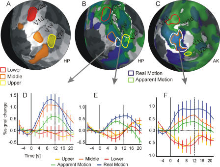
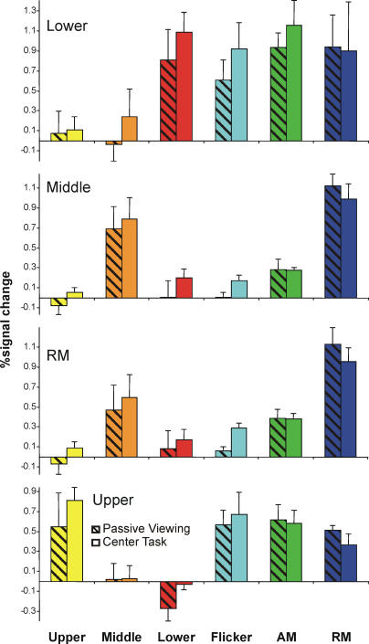
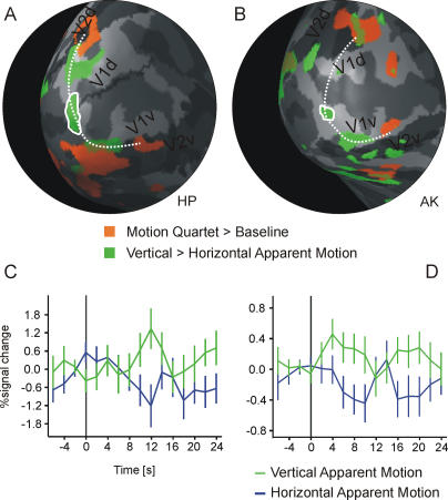
The illusion of apparent motion can be induced when visual stimuli are successively presented at different locations. It has been shown in previous studies that motion-sensitive regions in extrastriate cortex are relevant for the processing of apparent motion, but it is unclear whether primary visual cortex (V1) is also involved in the representation of the illusory motion path. We investigated, in human subjects, apparent-motion-related activity in patches of V1 representing locations along the path of illusory stimulus motion using functional magnetic resonance imaging. Here we show that apparent motion caused a blood-oxygenation-level-dependent response along the V1 representations of the apparent-motion path, including regions that were not directly activated by the apparent-motion-inducing stimuli. This response was unaltered when participants had to perform an attention-demanding task that diverted their attention away from the stimulus. With a bistable motion quartet, we confirmed that the activity was related to the conscious perception of movement. Our data suggest that V1 is part of the network that represents the illusory path of apparent motion. The activation in V1 can be explained either by lateral interactions within V1 or by feedback mechanisms from higher visual areas, especially the motion-sensitive human MT/V5 complex.
Overview
The claims and statistics of this paper haven’t been checked yet.
Free: it reads the open-access text and checks the first 5 claims that cite a source we can check. Create a free account to check every claim.
Integrity signals
Registry
Cites
Statistics
Figures



Verification
Claims the paper makes
The claims and statistics of this paper haven’t been checked yet.
Free: it reads the open-access text and checks the first 5 claims that cite a source we can check. Create a free account to check every claim.
Claims other papers make about this one
Each is checked against this paper’s own text: does it say what the citing paper says it does?
The source backs part of the claim, not all of it — or backs it for a different population, condition or comparison.
In the citing paper
“V5 feedback signals spread out to a large region in primary visual cortex (de-Wit et al. 2012; Muckli et al. 2005)”
From Whatever next? Predictive brains, situated agents, and the future of cognitive science (2013) · cites this paper as Muckli et al. 2005
Experimental — accuracy not yet measuredIn the citing paper
“Moreover, higher visual areas project back to V1 the associated color of grayscale objects [36] or the predicted motion path of an apparent motion illusion [37, 38].”
From Decoding sound and imagery content in early visual cortex (2014) · cites this paper as [37]
Self-check
Percentages, sums and reported differences re-computed from the paper’s own numbers.
The self-check needs the paper’s full text.
Citations
Cites
Neurophysiological investigation of the basis of the fMRI signal
Logothetis, N. K., Pauls, J., Augath, M., Trinath, T., & Oeltermann, A. (2001) · Nature · doi:10.1038/35084005 · Cited by 6,551 · cited 1×
“Current findings from simultaneous fMRI and electrophysiological recordings in monkeys have suggested that the BOLD signal might be especially sensitive to changes in local field potentials [ 53 ].”
BackgroundBorders of Multiple Visual Areas in Humans Revealed by Functional Magnetic Resonance Imaging
Sereno, M. I., Dale, A. M., Reppas, J. B., Kwong, K. K., Belliveau, J. W., Brady, T. J., Rosen, B. R., & Tootell, R. B. H. (1995) · Science · doi:10.1126/science.7754376 · Cited by 2,658
Linear Systems Analysis of Functional Magnetic Resonance Imaging in Human V1
Boynton, G. M., Engel, S. A., Glover, G. H., & Heeger, D. J. (1996) · The Journal of Neuroscience · doi:10.1523/jneurosci.16-13-04207.1996 · Cited by 2,314
Kastner, S., Pinsk, M. A., De Weerd, P., Desimone, R., & Ungerleider, L. G. (1999) · Neuron · doi:10.1016/s0896-6273(00)80734-5 · Cited by 1,622
Why visual attention and awareness are different
Lamme, V. A. F. (2003) · Trends in Cognitive Sciences · doi:10.1016/s1364-6613(02)00013-x · Cited by 1,172 · cited 1×
“In these studies, different imaging and electrophysiological methods have been used to investigate the functional properties of V1 during binocular rivalry [40– 43], perception of moving phosphenes [15,17], figure–ground segregation [34,44–46], the ‘‘line-motion’’ illusion [28], and color filling-in [47].”
MethodThe Underpinnings of the BOLD Functional Magnetic Resonance Imaging Signal
Logothetis, N. K. (2003) · The Journal of Neuroscience · doi:10.1523/jneurosci.23-10-03963.2003 · Cited by 1,039
Engel, S. A., Rumelhart, D. E., Wandell, B. A., Lee, A. T., Glover, G. H., Chichilnisky, E.-J., & Shadlen, M. N. (1994) · Nature · doi:10.1038/369525a0 · Cited by 990
Experimentelle Studien uber das Sehen von Bewegung
Wertheimer, M. (1912) · Cited by 975
Mechanisms of Directed Attention in the Human Extrastriate Cortex as Revealed by Functional MRI
Kastner, S., De Weerd, P., Desimone, R., & Ungerleider, L. G. (1998) · Science · doi:10.1126/science.282.5386.108 · Cited by 953
Are we aware of neural activity in primary visual cortex?
Crick, F., & Koch, C. (1995) · Nature · doi:10.1038/375121a0 · Cited by 932
Cortical feedback improves discrimination between figure and background by V1, V2 and V3 neurons
Hupé, J. M., James, A. C., Payne, B. R., Lomber, S. G., Girard, P., & Bullier, J. (1998) · Nature · doi:10.1038/29537 · Cited by 864
Hemodynamic Signals Correlate Tightly with Synchronized Gamma Oscillations
Niessing, J., Ebisch, B., Schmidt, K. E., Niessing, M., Singer, W., & Galuske, R. A. W. (2005) · Science · doi:10.1126/science.1110948 · Cited by 806 · cited 1×
“A more recent study established a tight link between BOLD signal and neuronal synchronization [54].”
BackgroundSelf-citationCircuits for local and global signal integration in primary visual cortex
Angelucci, A., Levitt, J. B., Walton, E. J. S., Hupé, J.-M., Bullier, J., & Lund, J. S. (2002) · The Journal of Neuroscience · doi:10.1523/jneurosci.22-19-08633.2002 · Cited by 789
Fast Backprojections from the Motion to the Primary Visual Area Necessary for Visual Awareness
Pascual-Leone, A., & Walsh, V. (2001) · Science · doi:10.1126/science.1057099 · Cited by 761 · cited 1×
“In these studies, different imaging and electrophysiological methods have been used to investigate the functional properties of V1 during binocular rivalry [40– 43], perception of moving phosphenes [15,17], figure–ground segregation [34,44–46], the ‘‘line-motion’’ illusion [28], and color filling-in [47].”
MethodA physiological correlate of the 'spotlight' of visual attention
Brefczynski, J. A., & DeYoe, E. A. (1999) · Nature Neuroscience · doi:10.1038/7280 · Cited by 723 · cited 1×
“It has been demonstrated in previous fMRI studies that spatial attention strongly modulates activation in visual cortical areas down to V1 [22–27].”
BackgroundThe Retinotopy of Visual Spatial Attention
Tootell, R. B. H., Hadjikhani, N., Hall, E. K., Marrett, S., Vanduffel, W., Vaughan, J. T., & Dale, A. M. (1998) · Neuron · doi:10.1016/s0896-6273(00)80659-5 · Cited by 712
Spatial attention affects brain activity in human primary visual cortex
Gandhi, S. P., Heeger, D. J., & Boynton, G. M. (1999) · Proceedings of the National Academy of Sciences · doi:10.1073/pnas.96.6.3314 · Cited by 682
Functional MRI reveals spatially specific attentional modulation in human primary visual cortex
Somers, D. C., Dale, A. M., Seiffert, A. E., & Tootell, R. B. H. (1999) · Proceedings of the National Academy of Sciences · doi:10.1073/pnas.96.4.1663 · Cited by 653
Corticocortical connections in the visual system: structure and function
Salin, P. A., & Bullier, J. (1995) · Physiological Reviews · doi:10.1152/physrev.1995.75.1.107 · Cited by 632
Dubner, R., & Zeki, S. M. (1971) · Brain Research · doi:10.1016/0006-8993(71)90494-x · Cited by 624
Neuronal activity in human primary visual cortex correlates with perception during binocular rivalry
Polonsky, A., Blake, R., Braun, J., & Heeger, D. J. (2000) · Nature Neuroscience · doi:10.1038/80676 · Cited by 587
Primary visual cortex and visual awareness
Tong, F. (2003) · Nature Reviews Neuroscience · doi:10.1038/nrn1055 · Cited by 535
Lateral Connectivity and Contextual Interactions in Macaque Primary Visual Cortex
Stettler, D. D., Das, A., Bennett, J., & Gilbert, C. D. (2002) · Neuron · doi:10.1016/s0896-6273(02)01029-2 · Cited by 504
Visual topography of V2 in the macaque
Gattass, R., Gross, C. G., & Sandell, J. H. (1981) · Journal of Comparative Neurology · doi:10.1002/cne.902010405 · Cited by 488
Goebel, R., Khorram‐Sefat, D., Muckli, L., Hacker, H., & Singer, W. (1998) · European Journal of Neuroscience · doi:10.1046/j.1460-9568.1998.00181.x · Cited by 474
Self-citationTwo distinct modes of sensory processing observed in monkey primary visual cortex (V1)
Supèr, H., Spekreijse, H., & Lamme, V. A. F. (2001) · Nature Neuroscience · doi:10.1038/85170 · Cited by 467
Low-level and high-level processes in apparent motion
Braddick, O. J. (1980) · Philosophical Transactions of the Royal Society of London. B, Biological Sciences · doi:10.1098/rstb.1980.0087 · Cited by 456
Interocular rivalry revealed in the human cortical blind-spot representation
Tong, F., & Engel, S. A. (2001) · Nature · doi:10.1038/35075583 · Cited by 453
Estimating Receptive Field Size from fMRI Data in Human Striate and Extrastriate Visual Cortex
Smith, A. T. (2001) · Cerebral Cortex · doi:10.1093/cercor/11.12.1182 · Cited by 357
Mikami, A., Newsome, W. T., & Wurtz, R. H. (1986) · Journal of Neurophysiology · doi:10.1152/jn.1986.55.6.1308 · Cited by 350
Mikami, A., Newsome, W. T., & Wurtz, R. H. (1986) · Journal of Neurophysiology · doi:10.1152/jn.1986.55.6.1328 · Cited by 299
Traveling waves of activity in primary visual cortex during binocular rivalry
Lee, S.-H., Blake, R., & Heeger, D. J. (2005) · Nature Neuroscience · doi:10.1038/nn1365 · Cited by 256
Striate cortex (V1) activity gates awareness of motion
Silvanto, J., Cowey, A., Lavie, N., & Walsh, V. (2005) · Nature Neuroscience · doi:10.1038/nn1379 · Cited by 246 · cited 1×
“In these studies, different imaging and electrophysiological methods have been used to investigate the functional properties of V1 during binocular rivalry [40– 43], perception of moving phosphenes [15,17], figure–ground segregation [34,44–46], the ‘‘line-motion’’ illusion [28], and color filling-in [47].”
MethodThe representation of the ipsilateral visual field in human cerebral cortex
Tootell, R. B. H., Mendola, J. D., Hadjikhani, N. K., Liu, A. K., & Dale, A. M. (1998) · Proceedings of the National Academy of Sciences · doi:10.1073/pnas.95.3.818 · Cited by 243
Imaging cortical correlates of illusion in early visual cortex
Jancke, D., Chavane, F., Naaman, S., & Grinvald, A. (2004) · Nature · doi:10.1038/nature02396 · Cited by 243
Kriegeskorte, N., & Goebel, R. (2001) · NeuroImage · doi:10.1006/nimg.2001.0831 · Cited by 242
Self-citationModulation of the motion aftereffect by selective attention
Chaudhuri, A. (1990) · Nature · doi:10.1038/344060a0 · Cited by 241
Motion selectivity in macaque visual cortex. III. Psychophysics and physiology of apparent motion
Newsome, W. T., Mikami, A., & Wurtz, R. H. (1986) · Journal of Neurophysiology · doi:10.1152/jn.1986.55.6.1340 · Cited by 210
Chapter 13 The role of feedback connections in shaping the responses of visual cortical neurons
Bullier, J., Hupé, J.-M., James, A. C., & Girard, P. (2001) · Progress in Brain Research · doi:10.1016/s0079-6123(01)34014-1 · Cited by 204
Optical Imaging of a Tactile Illusion in Area 3b of the Primary Somatosensory Cortex
Chen, L. M., Friedman, R. M., & Roe, A. W. (2003) · Science · doi:10.1126/science.1087846 · Cited by 180
Internal State of Monkey Primary Visual Cortex (V1) Predicts Figure–Ground Perception
Supèr, H., van der Togt, C., Spekreijse, H., & Lamme, V. A. F. (2003) · Journal of Neuroscience · doi:10.1523/jneurosci.23-08-03407.2003 · Cited by 171
Task-dependent influences of attention on the activation of human primary visual cortex
Watanabe, T., Harner, A. M., Miyauchi, S., Sasaki, Y., Nielsen, M., Palomo, D., & Mukai, I. (1998) · Proceedings of the National Academy of Sciences · doi:10.1073/pnas.95.19.11489 · Cited by 159
Attentional inhibition of visual processing in human striate and extrastriate cortex
Slotnick, S. D., Schwarzbach, J., & Yantis, S. (2003) · NeuroImage · doi:10.1016/s1053-8119(03)00187-3 · Cited by 159
Modulation of moving phosphene thresholds by transcranial direct current stimulation of V1 in human
Antal, A., Kincses, Z. T., Nitsche, M. A., & Paulus, W. (2003) · Neuropsychologia · doi:10.1016/s0028-3932(03)00181-7 · Cited by 149
Representation of motion boundaries in retinotopic human visual cortical areas
Reppas, J. B., Niyogi, S. A., Dale, A. M., Sereno, M. I., & Tootell, R. B. H. (1997) · Nature · doi:10.1038/40633 · Cited by 130
Muckli, L., Kriegeskorte, N., Lanfermann, H., Zanella, F. E., Singer, W., & Goebel, R. (2002) · The Journal of Neuroscience · doi:10.1523/jneurosci.22-09-j0003.2002 · Cited by 128
Self-citationSeiffert, A. E., Somers, D. C., Dale, A. M., & Tootell, R. B. H. (2003) · Cerebral Cortex · doi:10.1093/cercor/13.4.340 · Cited by 126
The primary visual cortex fills in color
Sasaki, Y., & Watanabe, T. (2004) · Proceedings of the National Academy of Sciences · doi:10.1073/pnas.0406293102 · Cited by 124
Perceptual Organization in Multistable Apparent Motion
Ramachandran, V. S., & Anstis, S. (1985) · Perception · doi:10.1068/p140135 · Cited by 120
Moving Illusory Contours Activate Primary Visual Cortex: an fMRI Study
Seghier, M., Dojat, M., Delon-Martin, C., Rubin, C., Warnking, J., Segebarth, C., & Bullier, J. (2000) · Cerebral Cortex · doi:10.1093/cercor/10.7.663 · Cited by 118
Cortical processing of visual motion.
Albright, T. D. (1993) · PubMed · Cited by 109
The role of feedback in shaping neural representations in cat visual cortex
Galuske, R. A. W., Schmidt, K. E., Goebel, R., Lomber, S. G., & Payne, B. R. (2002) · Proceedings of the National Academy of Sciences · doi:10.1073/pnas.242399199 · Cited by 106
Visual interactions in the path of apparent motion
Yantis, S., & Nakama, T. (1998) · Nature Neuroscience · doi:10.1038/2226 · Cited by 103
V1 activity is reduced during binocular rivalry
Lee, S.-H., & Blake, R. (2002) · Journal of Vision · doi:10.1167/2.9.4 · Cited by 100
Linden, D. E. J., Kallenbach, U., Heinecke, A., Singer, W., & Goebel, R. (1999) · Perception · doi:10.1068/p2820 · Cited by 99
Self-citationHuman MT+ mediates perceptual filling-in during apparent motion
Liu, T.-S., Slotnick, S., & Yantis, S. (2004) · NeuroImage · doi:10.1016/j.neuroimage.2003.12.025 · Cited by 88
Visuotopic organization of corticocortical connections in the visual system of the cat
Salin, P. A., Girard, P., Kennedy, H., & Bullier, J. (1992) · The Journal of Comparative Neurology · doi:10.1002/cne.903200402 · Cited by 87
Neuroimaging of Direction-Selective Mechanisms for Second-Order Motion
Nishida, S., Sasaki, Y., Murakami, I., Watanabe, T., & Tootell, R. B. H. (2003) · Journal of Neurophysiology · doi:10.1152/jn.00693.2003 · Cited by 78
Correlates of figure-ground segregation in fMRI
Skiera, G., Petersen, D., Skalej, M., & Fahle, M. W. (2000) · Vision Research · doi:10.1016/s0042-6989(00)00038-9 · Cited by 38
Responses of extrastriate cortex to switching perception of ambiguous visual motion stimuli
Sterzer, P., Eger, E., & Kleinschmidt, A. K. (2003) · Neuroreport · doi:10.1097/00001756-200312190-00010 · Cited by 32
Mikami, A. (1991) · International Journal of Neuroscience · doi:10.3109/00207459108986278 · Cited by 32
Some experiments on the breakdown effect in apparent motion
Finlay, D., & von Grünau, M. W. (1987) · Perception & Psychophysics · doi:10.3758/bf03207984 · Cited by 31
Time-till-breakdown and scalp electrical potential maps of long-range apparent motion
Selmes, C. M., Fulham, W. R., Finlay, D. C., Chorlton, M. C., & Manning, M. L. (1997) · Perception & Psychophysics · doi:10.3758/bf03211858 · Cited by 9
Givre, S. J. (2003) · Journal of Neuro-Ophthalmology · doi:10.1097/00041327-200306000-00022 · Cited by 5
Research works printed but not linked: 4
- Wertheimer, 1912, Z Psychol, 61, 161
- Albright, 1993, Rev Oculomot Res, 5, 177
- Neuhaus, 1930, Arch Gesamte Psychol, 75, 315
- Lee, 2002, J Vis, 2, 618
Cited by
Whatever next? Predictive brains, situated agents, and the future of cognitive science
Clark, A. (2013) · Behavioral and Brain Sciences · doi:10.1017/s0140525x12000477 · Cited by 5,924
How Do Expectations Shape Perception?
de Lange, F. P., Heilbron, M., & Kok, P. (2018) · Trends in Cognitive Sciences · doi:10.1016/j.tics.2018.06.002 · Cited by 975
Expectation in perceptual decision making: neural and computational mechanisms
Summerfield, C., & de Lange, F. P. (2014) · Nature Reviews Neuroscience · doi:10.1038/nrn3838 · Cited by 832
Predictions not commands: Active inference in the motor system
Adams, R. A., Shipp, S., & Friston, K. J. (2013) · Brain Structure and Function · doi:10.1007/s00429-012-0475-5 · Cited by 765
Stimulus Predictability Reduces Responses in Primary Visual Cortex
Alink, A., Schwiedrzik, C. M., Kohler, A., Singer, W., & Muckli, L. (2010) · The Journal of Neuroscience · doi:10.1523/jneurosci.3730-10.2010 · Cited by 435
Self-citation
Metadata
- Authors
- Muckli, L., Kohler, A., Kriegeskorte, N., & Singer, W.
- Affiliations
- Max Planck Institute for Brain Research (DE)
- Corresponding author
- Muckli, Lars
- Venue
- PLoS Biology
- Publisher
- Public Library of Science (PLoS)
- Published
- 2005-07-19
- Volume
- 3
- Issue
- 8
- Pages
- e265
- ISSN
- 1544-9173 (ISSN-L)
- 1545-7885
- PMC
- PMC1175820
- OpenAlex
- W2033471768
- Cited by
- 225
- References
- 64
- Citations by year
- 2026: 4 · 2025: 5 · 2024: 4 · 2023: 9 · 2022: 6 · 2021: 8 · 2020: 13 · 2019: 10 · 2018: 11 · 2017: 13 · 2016: 12 · 2015: 17 · 2014: 19 · 2013: 15 · 2012: 12
- FWCI
- 4.45
- Type
- journal article
- Language
- en
- Open access
- gold
- doi.org
- Locations
- PLoS Biology (publishedVersion, cc-by)
- Europe PMC (PubMed Central) (publishedVersion, cc-by)
- PubMed Central (publishedVersion, cc-by)
- PubMed (publishedVersion)
- ENLIGHTEN (Jurnal Bimbingan dan Konseling Islam) (acceptedVersion)
- DOAJ (DOAJ: Directory of Open Access Journals) (submittedVersion)
- DOAJ (DOAJ: Directory of Open Access Journals) (submittedVersion, cc-by-sa)
- Max Planck Institute for Plasma Physics (submittedVersion)
- Figshare (submittedVersion, cc-by)
- Publication Server of Goethe University Frankfurt am Main (Goethe University Frankfurt) (submittedVersion, cc-by)
- Funders
- Deutsche Forschungsgemeinschaft
- Bundesministerium für Bildung und Forschung
- Max-Planck-Gesellschaft
- Topics
- Life Sciences › Neuroscience › Cognitive Neuroscience › Visual perception and processing mechanisms
- Social Sciences › Psychology › Experimental and Cognitive Psychology › Multisensory perception and integration
- Life Sciences › Neuroscience › Cognitive Neuroscience › Motor Control and Adaptation
- Keywords
- apparent motion, primary visual cortex, illusory motion, motion perception, visual attention
- MeSH terms
- Adult, Brain Mapping, Female, Humans, Magnetic Resonance Imaging, Male, Motion Perception, Optical Illusions, Photic Stimulation, Visual Cortex, Vision Screening
About these checks
Registry, retraction, Cites and statistics results are computed, not judged, with one exception: a printed reference that no index lists is read once by a model before it counts as a citation error. Claims are extracted by a model and kept only when their quote appears verbatim in the text; each cited source is then checked, with a model’s help, for whether it says what the claim relies on. Those verdicts can be wrong — read the claim’s quote and the cited source before relying on one.
Statistics and arithmetic are recomputed, not judged.
Confidence bands are shown only for fields calibrated on our annotated test set. None is calibrated yet, so no band is shown.
What counts as a claim ›How we measure confidence ›How we check this ›
Are you an author, or do you see a problem with this page? Request a review
Data sources
Bibliographic data: Semantic Scholar, OpenAlex, Crossref.
Contains information from the Semantic Scholar Academic Graph Datasets, made available under the ODC Attribution License (ODC-BY 1.0).
Semantic Scholar’s data is described in: Kinney, R., et al. (2023). The Semantic Scholar Open Data Platform. arXiv:2301.10140.
