Cited by 82

The DOI matches its Crossref record; 3 of the 3 references it cites were found in a registry or an index.

Limiting the Persistence of a Chromosome Break Diminishes Its Mutagenic Potential

Bennardo, Nicole, Gunn, Amanda, Cheng, Anita, Hasty, Paul, Stark, Jeremy M.·2009·PLoS Genetics

Overview

The claims and statistics of this paper haven’t been checked yet.

Run the check — free, no account needed. It reads the open-access text and checks the sources it cites.

A few free checks a day · papers already verified open instantly · results are public

Integrity signals

Registry

Crossref · 10.1371/journal.pgen.1000683

Cites

3 of 3 references verified0 citation errors · 0 not checkable

Statistics

Figures

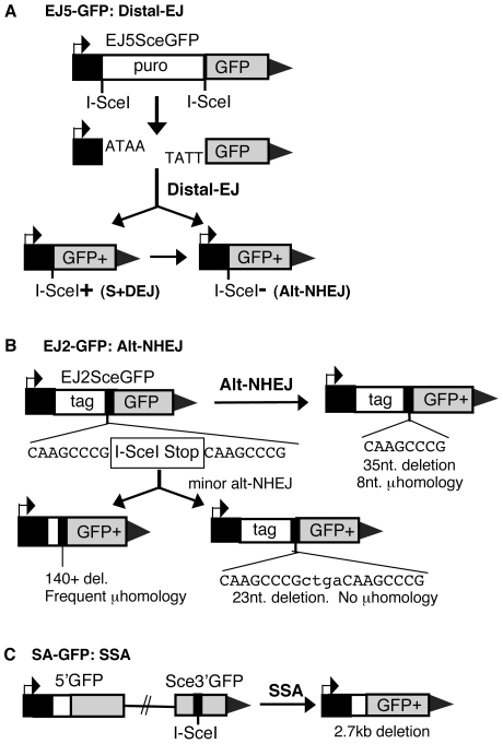
Figure 1 Reporters for EJ and SSA repair pathways. (A) EJ5-GFP is shown along with products of EJ between distal DSB ends (Distal-EJ) that restores a GFP expression cassette, including Ku-dependent I-SceI site restoration (S+DEJ), and Ku-independent I-SceI-resistant products (Alt-NHEJ). (B) EJ2-GFP is shown along with products of Alt-NHEJ that restore the reading frame of the GFP expression cassette. The most abundant Alt-NHEJ product involves 8 nt of microhomology at the repair junction and a 35 nt deletion. (C) SA-GFP is shown along with the SSA repair product that utilizes 266 nt of homology between the tandem GFP segments, thereby restoring a GFP expression cassette.
Figure 1 Reporters for EJ and SSA repair pathways. (A) EJ5-GFP is shown along with products of EJ between distal DSB ends (Distal-EJ) that restores a GFP expression cassette, including Ku-dependent I-SceI site restoration (S+DEJ), and Ku-independent I-SceI-resistant products (Alt-NHEJ). (B) EJ2-GFP is shown along with products of Alt-NHEJ that restore the reading frame of the GFP expression cassette. The most abundant Alt-NHEJ product involves 8 nt of microhomology at the repair junction and a 35 nt deletion. (C) SA-GFP is shown along with the SSA repair product that utilizes 266 nt of homology between the tandem GFP segments, thereby restoring a GFP expression cassette.CC BY · PubMed Central
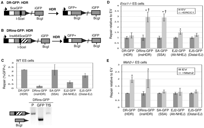
Figure 2 Ercc1 promotes repair pathways that require removal of a nonhomologous segment, whereas Msh2 promotes HDR. (A) DR-GFP is shown along with the HDR product that uses iGFP as the template for nascent DNA synthesis, which results in restoration of a GFP expression cassette. (B) Shown is a distinct reporter for HDR (DRins-GFP) that is similar to DR-GFP, except it contains an insertion of 464 nt downstream of the I-SceI site that needs to be removed during HDR to restore a GFP expression cassette. (C) Requiring removal of a nonhomologous insertion during HDR decreases the efficiency of repair. DR-GFP and DRins-GFP were integrated at the identical locus (Pim1) of wild-type (J1 strain) mouse ES cells, and the efficiency of HDR (%GFP+ cells) was determined following transient expression of I-SceI. Shown is the efficiency of repair (%GFP+ cells) from these reporters, along with those from Figure 1 that are also in WT ES cells. Also shown is confirmation of the structure of the GFP+ repair product for DRins-GFP via PCR/sequencing analysis of sorted GFP+ cells. The labels P, GFP, and TfS denote the parental cell line, GFP+ sorted cells, and total I-SceI-transfected cells, respectively. (D) Ercc1 specifically promotes DSB repair pathways that require removal of an extended nonhomologous segment. Each of the reporters depicted in Figure 1 and Figure 2A and 2B were integrated into mouse ES cells deficient in Ercc1. These individual cell lines were transfected with an expression vector for I-SceI, along with either a complementation vector for Ercc1, or the empty expression vector (EV). Repair is measured as percent GFP+ cells, which is normalized to the EV samples transfected in parallel. Asterisks denote a statistical difference in repair efficiency from EV (p<0.0001). The dagger denotes a statistical difference in fold-complementation compared to HDR of the DR-GFP reporter (p<0.0001). (E) Msh2 specifically promotes HDR. Analysis of repair in Msh2−/− cells was performed as described above for Ercc1, where asterisks denote the same statistical differences as described above.
Figure 2 Ercc1 promotes repair pathways that require removal of a nonhomologous segment, whereas Msh2 promotes HDR. (A) DR-GFP is shown along with the HDR product that uses iGFP as the template for nascent DNA synthesis, which results in restoration of a GFP expression cassette. (B) Shown is a distinct reporter for HDR (DRins-GFP) that is similar to DR-GFP, except it contains an insertion of 464 nt downstream of the I-SceI site that needs to be removed during HDR to restore a GFP expression cassette. (C) Requiring removal of a nonhomologous insertion during HDR decreases the efficiency of repair. DR-GFP and DRins-GFP were integrated at the identical locus (Pim1) of wild-type (J1 strain) mouse ES cells, and the efficiency of HDR (%GFP+ cells) was determined following transient expression of I-SceI. Shown is the efficiency of repair (%GFP+ cells) from these reporters, along with those from Figure 1 that are also in WT ES cells. Also shown is confirmation of the structure of the GFP+ repair product for DRins-GFP via PCR/sequencing analysis of sorted GFP+ cells. The labels P, GFP, and TfS denote the parental cell line, GFP+ sorted cells, and total I-SceI-transfected cells, respectively. (D) Ercc1 specifically promotes DSB repair pathways that require removal of an extended nonhomologous segment. Each of the reporters depicted in Figure 1 and Figure 2A and 2B were integrated into mouse ES cells deficient in Ercc1. These individual cell lines were transfected with an expression vector for I-SceI, along with either a complementation vector for Ercc1, or the empty expression vector (EV). Repair is measured as percent GFP+ cells, which is normalized to the EV samples transfected in parallel. Asterisks denote a statistical difference in repair efficiency from EV (p<0.0001). The dagger denotes a statistical difference in fold-complementation compared to HDR of the DR-GFP reporter (p<0.0001). (E) Msh2 specifically promotes HDR. Analysis of repair in Msh2−/− cells was performed as described above for Ercc1, where asterisks denote the same statistical differences as described above.CC BY · PubMed Central
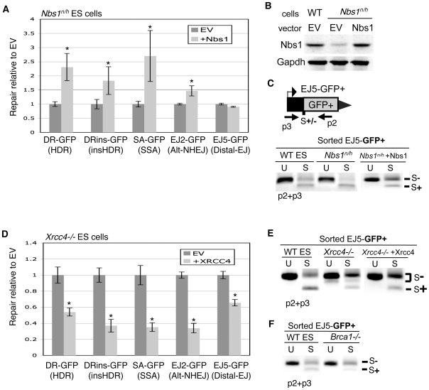
Figure 3 Nbs1, Xrcc4, and Brca1 play distinct roles during individual repair events. (A) Nbs1 promotes HDR, SSA, and Alt-NHEJ, but is dispensable for Distal-EJ. Reporters from Figure 1 and Figure 2 were integrated into Nbs1n/h cells, and the effect of Nbs1 complementation on repair was determined as for Ercc1 in Figure 2D. Asterisks denote a statistical difference in repair efficiency from EV (p<0.0001). (B) Nbs1n/h cells show a reduced level of Nbs1 that is restored to wild-type levels with transient expression. Shown are immunoblot signals for Nbs1 from transfections with Nbs1n/h, and from WT cells. (C) The repair event S+DEJ is increased in Nbs1-deficient cells. The diagram depicts the primers used for amplification (p2, p3). Shown are amplification products from sorted GFP+ cells derived from I-SceI transfection of EJ5-GFP-containing WT and Nbs1n/h cells, as well as Nbs1n/h cells transiently complemented with Nbs1. The products have been left uncut (U) and cut with I-SceI (S). (D) Xrcc4 suppresses HDR, SSA, Alt-NHEJ, and Distal-EJ. Shown are repair levels of reporters integrated into Xrcc4−/− ES cells that were assayed with/without transient complementation of Xrcc4 as described for Ercc1 in Figure 2D. Asterisks denote a statistical difference in repair efficiency from EV (p<0.0001). (E) Xrcc4-deficient cells show a decrease in S+DEJ. Shown are amplification products using the same primers and annotation as shown in C, from sorted GFP+ cells derived from I-SceI transfection of EJ5-GFP-containing WT cells, Xrcc4−/− cells, and Xrcc4−/− cells transiently complemented with Xrcc4 as in D. (F) Brca1-deficient cells show a decrease in S+DEJ. As in E, amplification products are shown from sorted GFP+ cells derived from I-SceI transfection of EJ5-GFP for WT and Brca1−/− cell lines.
Figure 3 Nbs1, Xrcc4, and Brca1 play distinct roles during individual repair events. (A) Nbs1 promotes HDR, SSA, and Alt-NHEJ, but is dispensable for Distal-EJ. Reporters from Figure 1 and Figure 2 were integrated into Nbs1n/h cells, and the effect of Nbs1 complementation on repair was determined as for Ercc1 in Figure 2D. Asterisks denote a statistical difference in repair efficiency from EV (p<0.0001). (B) Nbs1n/h cells show a reduced level of Nbs1 that is restored to wild-type levels with transient expression. Shown are immunoblot signals for Nbs1 from transfections with Nbs1n/h, and from WT cells. (C) The repair event S+DEJ is increased in Nbs1-deficient cells. The diagram depicts the primers used for amplification (p2, p3). Shown are amplification products from sorted GFP+ cells derived from I-SceI transfection of EJ5-GFP-containing WT and Nbs1n/h cells, as well as Nbs1n/h cells transiently complemented with Nbs1. The products have been left uncut (U) and cut with I-SceI (S). (D) Xrcc4 suppresses HDR, SSA, Alt-NHEJ, and Distal-EJ. Shown are repair levels of reporters integrated into Xrcc4−/− ES cells that were assayed with/without transient complementation of Xrcc4 as described for Ercc1 in Figure 2D. Asterisks denote a statistical difference in repair efficiency from EV (p<0.0001). (E) Xrcc4-deficient cells show a decrease in S+DEJ. Shown are amplification products using the same primers and annotation as shown in C, from sorted GFP+ cells derived from I-SceI transfection of EJ5-GFP-containing WT cells, Xrcc4−/− cells, and Xrcc4−/− cells transiently complemented with Xrcc4 as in D. (F) Brca1-deficient cells show a decrease in S+DEJ. As in E, amplification products are shown from sorted GFP+ cells derived from I-SceI transfection of EJ5-GFP for WT and Brca1−/− cell lines.CC BY · PubMed Central
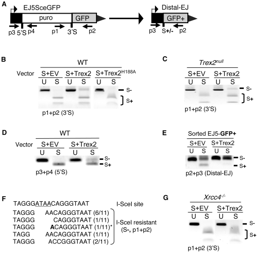
Figure 4 Expression of the Trex2 exonuclease promotes the formation of Xrcc4-dependent I-SceI-resistant EJ products. (A) Shown are primers for EJ5-GFP that are used to analyze proximal-EJ at the 5′ and 3′ I-SceI sites (shown as 5′S and 3′S, respectively), as well as Distal-EJ. (B) Expression of the Trex2 exonuclease promotes the formation of I-SceI-resistant EJ products between proximal DSB ends at the 3′ I-SceI site in EJ5-GFP. WT ES cells with EJ5-GFP were transfected with an I-SceI expression vector (S) along with a Trex2 expression vector (Trex2), an exonuclease-deficient mutant of Trex2 (Trex2-H188A), or EV. Shown are amplification products from these transfections using the primers p1 and p2, which were either left uncut (U) or were cut with I-SceI (S). (C) Trex2 expression also promotes I-SceI-resistant EJ products in Trex2null cells. Analysis was performed on Trex2null cells with the EJ5-GFP reporter as described in B. (D) Trex2 expression promotes I-SceI-resistant EJ products at the 5′ I-SceI site of EJ5-GFP. Shown are amplification products using the primers p3 and p4, using the same transfection conditions and annotation as described in B. (E) Trex2 expression promotes I-SceI-resistant Distal-EJ products. Shown are amplification products from sorted GFP+ cells derived from the transfections shown in C, using primers p3 and p2, with the same annotation shown in B. (F) EJ products via Trex2 show deletion of segments of the I-SceI 3′ overhang (underlined). The I-SceI-resistant products shown in C were cloned, and 11 individual clones were sequenced. Shown are the sequences of these clones, where the numerator in parenthesis depicts the number of times a given sequence was identified. An asterisk denotes the one clone with evidence of microhomology (1 nt., A in bold). (G) I-SceI-resistant proximal EJ products via Trex2 are dependent on Xrcc4. Analysis of the effect on Trex2 expression on the 3′ I-SceI site of the EJ5-GFP reporter was performed on Xrcc4−/− cells as described in B.
Figure 4 Expression of the Trex2 exonuclease promotes the formation of Xrcc4-dependent I-SceI-resistant EJ products. (A) Shown are primers for EJ5-GFP that are used to analyze proximal-EJ at the 5′ and 3′ I-SceI sites (shown as 5′S and 3′S, respectively), as well as Distal-EJ. (B) Expression of the Trex2 exonuclease promotes the formation of I-SceI-resistant EJ products between proximal DSB ends at the 3′ I-SceI site in EJ5-GFP. WT ES cells with EJ5-GFP were transfected with an I-SceI expression vector (S) along with a Trex2 expression vector (Trex2), an exonuclease-deficient mutant of Trex2 (Trex2-H188A), or EV. Shown are amplification products from these transfections using the primers p1 and p2, which were either left uncut (U) or were cut with I-SceI (S). (C) Trex2 expression also promotes I-SceI-resistant EJ products in Trex2null cells. Analysis was performed on Trex2null cells with the EJ5-GFP reporter as described in B. (D) Trex2 expression promotes I-SceI-resistant EJ products at the 5′ I-SceI site of EJ5-GFP. Shown are amplification products using the primers p3 and p4, using the same transfection conditions and annotation as described in B. (E) Trex2 expression promotes I-SceI-resistant Distal-EJ products. Shown are amplification products from sorted GFP+ cells derived from the transfections shown in C, using primers p3 and p2, with the same annotation shown in B. (F) EJ products via Trex2 show deletion of segments of the I-SceI 3′ overhang (underlined). The I-SceI-resistant products shown in C were cloned, and 11 individual clones were sequenced. Shown are the sequences of these clones, where the numerator in parenthesis depicts the number of times a given sequence was identified. An asterisk denotes the one clone with evidence of microhomology (1 nt., A in bold). (G) I-SceI-resistant proximal EJ products via Trex2 are dependent on Xrcc4. Analysis of the effect on Trex2 expression on the 3′ I-SceI site of the EJ5-GFP reporter was performed on Xrcc4−/− cells as described in B.CC BY · PubMed Central
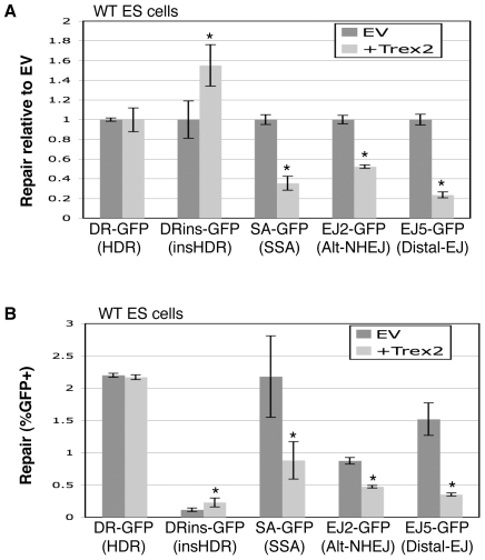
Figure 5 Trex2 expression causes a significant decrease in Distal-EJ, Alt-NHEJ, and SSA, but not HDR. WT ES cell lines with individual reporters were co-transfected with expression vectors for I-SceI and Trex2. (A) Shown are repair values normalized to parallel co-transfections with I-SceI and EV. Asterisks denote a statistical difference from EV (p<0.0001, DRins-GFP p = 0.0008). (B) Shown are primary repair values (%GFP+) from the experiment shown in A, to allow a direct comparison of the frequencies of different repair events. The error bars are somewhat larger in this panel as compared to A, since the primary repair levels show greater experimental variation versus the relatively consistent fold-effect of Trex2 expression. Asterisks denote a statistical difference from EV (p<0.0001 for EJ2-GFP and EJ5-GFP, p = 0.001 for SA-GFP, and p = 0.005 for DRins-GFP).
Figure 5 Trex2 expression causes a significant decrease in Distal-EJ, Alt-NHEJ, and SSA, but not HDR. WT ES cell lines with individual reporters were co-transfected with expression vectors for I-SceI and Trex2. (A) Shown are repair values normalized to parallel co-transfections with I-SceI and EV. Asterisks denote a statistical difference from EV (p<0.0001, DRins-GFP p = 0.0008). (B) Shown are primary repair values (%GFP+) from the experiment shown in A, to allow a direct comparison of the frequencies of different repair events. The error bars are somewhat larger in this panel as compared to A, since the primary repair levels show greater experimental variation versus the relatively consistent fold-effect of Trex2 expression. Asterisks denote a statistical difference from EV (p<0.0001 for EJ2-GFP and EJ5-GFP, p = 0.001 for SA-GFP, and p = 0.005 for DRins-GFP).CC BY · PubMed Central
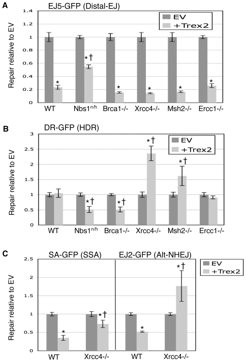
Figure 6 Roles of individual genetic factors during repair of DSBs with non-cohesive ends generated by co-expression of I-SceI with Trex2. Cell lines with individual reporters were co-transfected with expression vectors for I-SceI and Trex2. Repair values are quantified and normalized to parallel co-transfections with I-SceI and EV. (A) Nbs1-deficient cells show a diminished suppression of Distal-EJ from Trex2 expression. Shown are the effects of Trex2 expression on the EJ5-GFP reporter integrated into DNA repair-deficient mouse ES cell lines described in Figure 2 and Figure 3. Asterisks denote a statistical difference from EV (p<0.0001), and the dagger denotes a statistical difference from WT (p<0.0001). (B) Trex2 expression causes a decrease in HDR in cells deficient in Nbs1 and Brca1, but an increase in HDR in cells deficient in Xrcc4 and Msh2. Shown are the effects of Trex2 expression on the DR-GFP reporter in the cell lines shown in A. Asterisks denote a statistical difference from EV (p<0.0001, Msh2 p = 0.001), and the dagger denotes a statistical difference from WT (p<0.0001, Msh2 p = 0.0027). (C) Trex2 expression in Xrcc4−/− cells shows an increase in Alt-NHEJ, and a relative increase in SSA compared to WT. Shown are the effects of Trex2 expression on the EJ2-GFP and SA-GFP reporters in Xrcc4−/− cells, along with WT. Asterisks and daggers are as in A.
Figure 6 Roles of individual genetic factors during repair of DSBs with non-cohesive ends generated by co-expression of I-SceI with Trex2. Cell lines with individual reporters were co-transfected with expression vectors for I-SceI and Trex2. Repair values are quantified and normalized to parallel co-transfections with I-SceI and EV. (A) Nbs1-deficient cells show a diminished suppression of Distal-EJ from Trex2 expression. Shown are the effects of Trex2 expression on the EJ5-GFP reporter integrated into DNA repair-deficient mouse ES cell lines described in Figure 2 and Figure 3. Asterisks denote a statistical difference from EV (p<0.0001), and the dagger denotes a statistical difference from WT (p<0.0001). (B) Trex2 expression causes a decrease in HDR in cells deficient in Nbs1 and Brca1, but an increase in HDR in cells deficient in Xrcc4 and Msh2. Shown are the effects of Trex2 expression on the DR-GFP reporter in the cell lines shown in A. Asterisks denote a statistical difference from EV (p<0.0001, Msh2 p = 0.001), and the dagger denotes a statistical difference from WT (p<0.0001, Msh2 p = 0.0027). (C) Trex2 expression in Xrcc4−/− cells shows an increase in Alt-NHEJ, and a relative increase in SSA compared to WT. Shown are the effects of Trex2 expression on the EJ2-GFP and SA-GFP reporters in Xrcc4−/− cells, along with WT. Asterisks and daggers are as in A.CC BY · PubMed Central
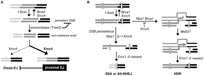
Figure 7 Limiting the persistence of a DSB causes a reduction in repair pathways that result in genetic loss. (A) Proximal EJ is shown to limit Distal-EJ of two tandem DSBs. Xrcc4 and Brca1 are shown to promote EJ of cohesive DSB ends, where repeated cutting by I-SceI results in a persistent DSB. The Trex2 exonuclease is shown as generating non-cohesive DSB ends, which limits the persistence of I-SceI-induced DSBs. Xrcc4 is also shown promoting EJ of non-cohesive ends during both proximal-EJ and Distal-EJ. The bias towards proximal-EJ in WT cells is indicated by the bold arrow (see Figure 5). Similarly, the moderate preference for proximal-EJ via Xrcc4 is indicated by the bold-type. (B) Shown is a model whereby the persistent nature of a DSB is important for SSA and Alt-NHEJ, but not HDR. Nbs1 and Brca1 are shown having an increased role during HDR of a relatively less persistent DSB. End resection that leads to HDR, SSA, and Alt-NHEJ is shown as being suppressed by Xrcc4 and promoted by Nbs1. Msh2 is shown as possibly promoting 3′ end processing prior to nascent DNA synthesis during HDR. Ercc1 is shown to promote the completion of repair pathways if needed, based on the requirement for removal of an extended nonhomologous segment.
Figure 7 Limiting the persistence of a DSB causes a reduction in repair pathways that result in genetic loss. (A) Proximal EJ is shown to limit Distal-EJ of two tandem DSBs. Xrcc4 and Brca1 are shown to promote EJ of cohesive DSB ends, where repeated cutting by I-SceI results in a persistent DSB. The Trex2 exonuclease is shown as generating non-cohesive DSB ends, which limits the persistence of I-SceI-induced DSBs. Xrcc4 is also shown promoting EJ of non-cohesive ends during both proximal-EJ and Distal-EJ. The bias towards proximal-EJ in WT cells is indicated by the bold arrow (see Figure 5). Similarly, the moderate preference for proximal-EJ via Xrcc4 is indicated by the bold-type. (B) Shown is a model whereby the persistent nature of a DSB is important for SSA and Alt-NHEJ, but not HDR. Nbs1 and Brca1 are shown having an increased role during HDR of a relatively less persistent DSB. End resection that leads to HDR, SSA, and Alt-NHEJ is shown as being suppressed by Xrcc4 and promoted by Nbs1. Msh2 is shown as possibly promoting 3′ end processing prior to nascent DNA synthesis during HDR. Ercc1 is shown to promote the completion of repair pathways if needed, based on the requirement for removal of an extended nonhomologous segment.CC BY · PubMed Central

Verification

Claims the paper makes

The claims and statistics of this paper haven’t been checked yet.

Run the check — free, no account needed. It reads the open-access text and checks the sources it cites.

A few free checks a day · papers already verified open instantly · results are public

Claims other papers make about this one

Each is checked against this paper’s own text: does it say what the citing paper says it does?

Self-check

Percentages, sums and reported differences re-computed from the paper’s own numbers.

The self-check needs the paper’s full text.

Citations

Cites

Show all 3 references

Research works printed but not linked: 67

  1. C Wyman, 2006, DNA double-strand break repair: all's well that ends well., Annu Rev Genet, 40, 363, 10.1146/annurev.genet.40.110405.090451 · DOI
  2. MF Greaves, 2003, Origins of chromosome translocations in childhood leukaemia., Nat Rev Cancer, 3, 639, 10.1038/nrc1164 · DOI
  3. Y Ma, 2005, Repair of double-strand DNA breaks by the human nonhomologous DNA end joining pathway: the iterative processing model., Cell Cycle, 4, 1193, 10.4161/cc.4.9.1977 · DOI
  4. JE Haber, 2008, Alternative endings., Proceedings of the National Academy of Sciences, 105, 405, 10.1073/pnas.0711334105 · DOI
  5. M McVey, 2008, MMEJ repair of double-strand breaks (director's cut): deleted sequences and alternative endings., Trends in Genetics, 24, 529, 10.1016/j.tig.2008.08.007 · DOI
  6. LS Symington, 2002, Role of RAD52 epistasis group genes in homologous recombination and double-strand break repair., Microbiol Mol Biol Rev, 66, 630, 10.1128/MMBR.66.4.630-670.2002 · DOI
  7. N Bennardo, 2008, Alternative-NHEJ Is a Mechanistically Distinct Pathway of Mammalian Chromosome Break Repair., PLoS Genetics, 4, e1000110, 10.1371/journal.pgen.1000110 · DOI
  8. L Deriano, 2009, Roles for NBS1 in Alternative Nonhomologous End-Joining of V(D)J Recombination Intermediates., Molecular Cell, 34, 13, 10.1016/j.molcel.2009.03.009 · DOI
  9. L Chen, 2008, Cell Cycle-dependent Complex Formation of BRCA1{middle dot}CtIP{middle dot}MRN Is Important for DNA Double-strand Break Repair., Journal of Biological Chemistry, 283, 7713, 10.1074/jbc.M710245200 · DOI
  10. JM Stark, 2002, ATP hydrolysis by mammalian RAD51 has a key role during homology-directed DNA repair., J Biol Chem, 277, 20185, 10.1074/jbc.M112132200 · DOI
  11. A Tutt, 2001, Mutation in Brca2 stimulates error-prone homology-directed repair of DNA double-strand breaks occurring between repeated sequences., Embo J, 20, 4704, 10.1093/emboj/20.17.4704 · DOI
  12. M Shrivastav, 2008, Regulation of DNA double-strand break repair pathway choice., Cell Res, 18, 134, 10.1038/cr.2007.111 · DOI · Paper page
  13. AJ Pierce, 2001, Ku DNA end-binding protein modulates homologous repair of double-strand breaks in mammalian cells., Genes Dev, 15, 3237, 10.1101/gad.946401 · DOI
  14. J Guirouilh-Barbat, 2004, Impact of the KU80 pathway on NHEJ-induced genome rearrangements in mammalian cells., Mol Cell, 14, 611, 10.1016/j.molcel.2004.05.008 · DOI
  15. J Guirouilh-Barbat, 2007, Defects in XRCC4 and KU80 differentially affect the joining of distal nonhomologous ends., Proceedings of the National Academy of Sciences, 104, 20902, 10.1073/pnas.0708541104 · DOI
  16. M Kalocsay, 2009, Chromosome-wide Rad51 Spreading and SUMO-H2A.Z-Dependent Chromosome Fixation in Response to a Persistent DNA Double-Strand Break., Molecular Cell, 33, 335, 10.1016/j.molcel.2009.01.016 · DOI · Paper page
  17. SE Lee, 1998, Saccharomyces Ku70, mre11/rad50 and RPA proteins regulate adaptation to G2/M arrest after DNA damage., Cell, 94, 399, 10.1016/S0092-8674(00)81482-8 · DOI
  18. CB Bennett, 1993, Lethality induced by a single site-specific double-strand break in a dispensable yeast plasmid., Proc Natl Acad Sci U S A, 90, 5613, 10.1073/pnas.90.12.5613 · DOI
  19. K Lee, 2008, Saccharomyces cerevisiae ATM orthologue suppresses break-induced chromosome translocations., Nature, 454, 543, 10.1038/nature07054 · DOI
  20. M-J Chen, 2007, Cisplatin Depletes TREX2 and Causes Robertsonian Translocations as Seen in TREX2 Knockout Cells., Cancer Res, 67, 9077, 10.1158/0008-5472.CAN-07-1146 · DOI
  21. FW Perrino, 2005, The Human TREX2 3′→5′-Exonuclease Structure Suggests a Mechanism for Efficient Nonprocessive DNA Catalysis., J Biol Chem, 280, 15212, 10.1074/jbc.M500108200 · DOI
  22. JM Stark, 2003, Extensive Loss of Heterozygosity is suppressed during homologous repair of chromosomal breaks., Mol Cell Biol, 23, 733, 10.1128/MCB.23.2.733-743.2003 · DOI
  23. C Richardson, 2000, Coupled Homologous and Nonhomologous Repair of a Double-Strand Break Preserves Genomic Integrity in Mammalian Cells., Molecular and Cellular Biology, 20, 9068, 10.1128/MCB.20.23.9068-9075.2000 · DOI
  24. AJ Pierce, 1999, XRCC3 promotes homology-directed repair of DNA damage in mammalian cells., Genes Dev, 13, 2633, 10.1101/gad.13.20.2633 · DOI
  25. MLG Dronkert, 2001, Repair of DNA interstrand cross-links., Mutation Research/DNA Repair, 486, 217, 10.1016/S0921-8777(01)00092-1 · DOI
  26. T Reya, 2001, Stem cells, cancer, and cancer stem cells., Nature, 414, 105, 10.1038/35102167 · DOI · Paper page
  27. OV Tsodikov, 2005, Crystal structure and DNA binding functions of ERCC1, a subunit of the DNA structure-specific endonuclease XPF-ERCC1., Proceedings of the National Academy of Sciences, 102, 11236, 10.1073/pnas.0504341102 · DOI
  28. A Ahmad, 2008, ERCC1-XPF Endonuclease Facilitates DNA Double-Strand Break Repair., Molecular and Cellular Biology, 28, 5082, 10.1128/MCB.00293-08 · DOI
  29. LJ Niedernhofer, 2001, The structure-specific endonuclease Ercc1-Xpf is required for targeted gene replacement in embryonic stem cells., Embo J, 20, 6540, 10.1093/emboj/20.22.6540 · DOI
  30. MI Aladjem, 1998, ES cells do not activate p53-dependent stress responses and undergo p53-independent apoptosis in response to DNA damage., Curr Biol, 8, 145, 10.1016/S0960-9822(98)70061-2 · DOI
  31. TA Kunkel, 2005, DNA MISMATCH REPAIR*., Annual Review of Biochemistry, 74, 681, 10.1146/annurev.biochem.74.082803.133243 · DOI
  32. N Claij, 2004, Msh2 deficiency does not contribute to cisplatin resistance in mouse embryonic stem cells., Oncogene, 23, 260, 10.1038/sj.onc.1207015 · DOI
  33. JA Smith, 2005, A Role for DNA Mismatch Repair Protein Msh2 in Error-Prone Double-Strand-Break Repair in Mammalian Chromosomes., Genetics, 170, 355, 10.1534/genetics.104.039362 · DOI
  34. P Bertrand, 1998, Physical interaction between components of DNA mismatch repair and nucleotide excision repair., Proceedings of the National Academy of Sciences of the United States of America, 95, 14278, 10.1073/pnas.95.24.14278 · DOI
  35. F Li, 2008, Microarray-Based Genetic Screen Defines SAW1, a Gene Required for Rad1/Rad10-Dependent Processing of Recombination Intermediates., Molecular Cell, 30, 325, 10.1016/j.molcel.2008.02.028 · DOI
  36. N Sugawara, 1997, Role of Saccharomyces cerevisiae Msh2 and Msh3 repair proteins in double-strand break-induced recombination., Proceedings of the National Academy of Sciences of the United States of America, 94, 9214, 10.1073/pnas.94.17.9214 · DOI
  37. A Xie, 2009, Role of mammalian Mre11 in classical and alternative nonhomologous end joining., Nat Struct Mol Biol, 16, 814, 10.1038/nsmb.1640 · DOI
  38. M Dinkelmann, 2009, Multiple functions of MRN in end-joining pathways during isotype class switching., Nat Struct Mol Biol, 16, 808, 10.1038/nsmb.1639 · DOI
  39. E Rass, 2009, Role of Mre11 in chromosomal nonhomologous end joining in mammalian cells., Nat Struct Mol Biol, 16, 819, 10.1038/nsmb.1641 · DOI
  40. YG Yang, 2006, Conditional deletion of Nbs1 in murine cells reveals its role in branching repair pathways of DNA double-strand breaks., Embo J, 25, 5527, 10.1038/sj.emboj.7601411 · DOI
  41. Y Xiao, 1997, Conditional gene targeted deletion by Cre recombinase demonstrates the requirement for the double-strand break repair Mre11 protein in murine embryonic stem cells., Nucleic Acids Research, 25, 2985, 10.1093/nar/25.15.2985 · DOI
  42. G Luo, 2000, Cancer predisposition caused by elevated mitotic recombination in Bloom mice., Nat Genet, 26, 424, 10.1038/82548 · DOI
  43. A Perrin, 1993, Asymmetrical recognition and activity of the I-SceI endonuclease on its site and on intron-exon junctions., Embo J, 12, 2939, 10.1002/j.1460-2075.1993.tb05956.x · DOI
  44. T Ellenberger, 2008, Eukaryotic DNA Ligases: Structural and Functional Insights., Annual Review of Biochemistry, 77, 313, 10.1146/annurev.biochem.77.061306.123941 · DOI
  45. Y Gao, 1998, A Critical Role for DNA End-Joining Proteins in Both Lymphogenesis and Neurogenesis., Cell, 95, 891, 10.1016/S0092-8674(00)81714-6 · DOI · Paper page
  46. JA Downs, 2004, A means to a DNA end: the many roles of Ku., Nat Rev Mol Cell Biol, 5, 367, 10.1038/nrm1367 · DOI
  47. JN Snouwaert, 1999, BRCA1 deficient embryonic stem cells display a decreased homologous recombination frequency and an increased frequency of non-homologous recombination that is corrected by expression of a brca1 transgene., Oncogene, 18, 7900, 10.1038/sj.onc.1203334 · DOI
  48. UK Westermark, 2003, BARD1 participates with BRCA1 in homology-directed repair of chromosome breaks., Mol Cell Biol, 23, 7926, 10.1128/MCB.23.21.7926-7936.2003 · DOI
  49. D-T Bau, 2004, Breast Cancer Risk and the DNA Double-Strand Break End-Joining Capacity of Nonhomologous End-Joining Genes Are Affected by BRCA1., Cancer Research, 64, 5013, 10.1158/0008-5472.CAN-04-0403 · DOI
  50. S Burma, 2006, Role of non-homologous end joining (NHEJ) in maintaining genomic integrity., DNA Repair (Amst), 5, 1042, 10.1016/j.dnarep.2006.05.026 · DOI
  51. LC Dumitrache, 2009, TREX2 exonuclease defective cells exhibit double-strand breaks and chromosomal fragments but not Robertsonian translocations., Mutation Research/Fundamental and Molecular Mechanisms of Mutagenesis, 662, 84, 10.1016/j.mrfmmm.2008.11.012 · DOI
  52. M-J Chen, 2007, Biochemical and cellular characteristics of the 3′→5′ exonuclease TREX2., Nucleic Acids Research, 35, 2682, 10.1093/nar/gkm151 · DOI
  53. O Fernandez-Capetillo, 2004, H2AX: the histone guardian of the genome., DNA Repair, 3, 959, 10.1016/j.dnarep.2004.03.024 · DOI
  54. J Gu, 2007, XRCC4:DNA ligase IV can ligate incompatible DNA ends and can ligate across gaps., Embo J, 26, 1010, 10.1038/sj.emboj.7601559 · DOI
  55. WL de Laat, 1999, Molecular mechanism of nucleotide excision repair., Genes Dev, 13, 768, 10.1101/gad.13.7.768 · DOI · Paper page
  56. GM Adair, 2000, Role of ERCC1 in removal of long non-homologous tails during targeted homologous recombination., Embo J, 19, 5552, 10.1093/emboj/19.20.5552 · DOI
  57. RS Williams, 2008, Mre11 Dimers Coordinate DNA End Bridging and Nuclease Processing in Double-Strand-Break Repair., Cell, 135, 97, 10.1016/j.cell.2008.08.017 · DOI
  58. J Buis, 2008, Mre11 Nuclease Activity Has Essential Roles in DNA Repair and Genomic Stability Distinct from ATM Activation., Cell, 135, 85, 10.1016/j.cell.2008.08.015 · DOI
  59. X Yu, 2000, Nuclear Localization and Cell Cycle-specific Expression of CtIP, a Protein That Associates with the BRCA1 Tumor Suppressor., Journal of Biological Chemistry, 275, 18541, 10.1074/jbc.M909494199 · DOI
  60. TD Stamato, 1988, Cell-cycle-dependent repair of potentially lethal damage in the XR-1 gamma-ray-sensitive Chinese hamster ovary cell., Radiat Res, 115, 325, 10.2307/3577168 · DOI
  61. BP Schlegel, 2006, BRCA1 promotes induction of ssDNA by ionizing radiation., Cancer Res, 66, 5181, 10.1158/0008-5472.CAN-05-3209 · DOI
  62. LJ Reid, 2008, E3 ligase activity of BRCA1 is not essential for mammalian cell viability or homology-directed repair of double-strand DNA breaks., Proceedings of the National Academy of Sciences, 105, 20876, 10.1073/pnas.0811203106 · DOI
  63. RD Johnson, 2000, Sister chromatid gene conversion is a prominent double-strand break repair pathway in mammalian cells., Embo J, 19, 3398, 10.1093/emboj/19.13.3398 · DOI
  64. S Lambert, 2000, Characterization of mammalian RAD51 double strand break repair using non-lethal dominant-negative forms., Embo J, 19, 3090, 10.1093/emboj/19.12.3090 · DOI
  65. JW Szostak, 1983, The double-strand-break repair model for recombination., Cell, 33, 25, 10.1016/0092-8674(83)90331-8 · DOI · Paper page
  66. F Paques, 2007, Meganucleases and DNA double-strand break-induced recombination: perspectives for gene therapy., Curr Gene Ther, 7, 49, 10.2174/156652307779940216 · DOI
  67. ME Moynahan, 2001, BRCA2 is required for homology-directed repair of chromosomal breaks., Mol Cell, 7, 263, 10.1016/S1097-2765(01)00174-5 · DOI

Cited by

Sorted by citations

See all 82

Metadata

Authors
Bennardo, N., Gunn, A., Cheng, A., Hasty, P., & Stark, J. M.
Venue
PLoS Genetics
Published
2009-10-16
Volume
5
Issue
10
Pages
e1000683
ISSN
1553-7404
Cited by
82
References
3
Language
en

About these checks

Registry, retraction, Cites and statistics results are computed, not judged, with one exception: a printed reference that no index lists is read once by a model before it counts as a citation error. Claims are extracted by a model and kept only when their quote appears verbatim in the text; each cited source is then checked, with a model’s help, for whether it says what the claim relies on. Those verdicts can be wrong — read the claim’s quote and the cited source before relying on one.

Statistics and arithmetic are recomputed, not judged.

Confidence bands are shown only for fields calibrated on our annotated test set. None is calibrated yet, so no band is shown.

What counts as a claim ›How we measure confidence ›How we check this ›

Are you an author, or do you see a problem with this page? Request a review

How to cite

Cite CoChat for the checks on this page, and the paper for its own findings.

This page
CoChat. (2026). Citation check: Limiting the Persistence of a Chromosome Break Diminishes Its Mutagenic Potential. https://app.cochat.ai/papers/10.1371/journal.pgen.1000683
The paper
Bennardo, N., Gunn, A., Cheng, A., Hasty, P., & Stark, J. M. (2009). Limiting the Persistence of a Chromosome Break Diminishes Its Mutagenic Potential. PLoS Genetics. https://doi.org/10.1371/journal.pgen.1000683