

The DOI matches its Crossref record; 51 of the 51 references it cites were found in a registry or an index.
Molecular Mechanisms Generating and Stabilizing Terminal 22q13 Deletions in 44 Subjects with Phelan/McDermid Syndrome
Open accessAbstract
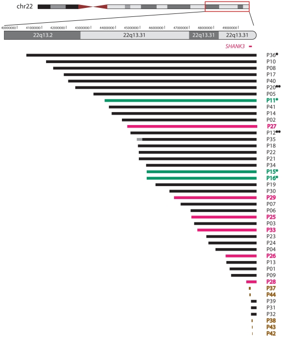
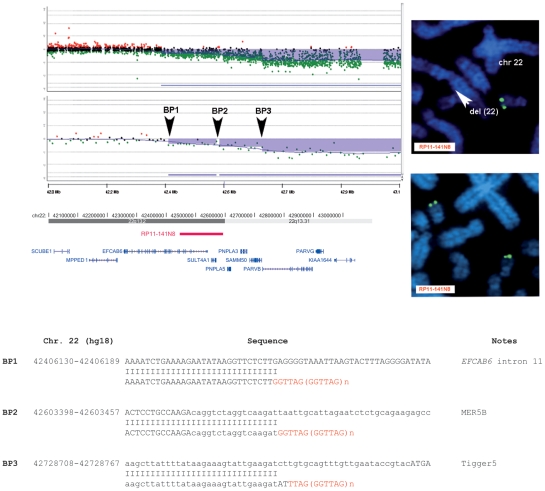
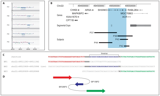
In this study, we used deletions at 22q13, which represent a substantial source of human pathology (Phelan/McDermid syndrome), as a model for investigating the molecular mechanisms of terminal deletions that are currently poorly understood. We characterized at the molecular level the genomic rearrangement in 44 unrelated patients with 22q13 monosomy resulting from simple terminal deletions (72%), ring chromosomes (14%), and unbalanced translocations (7%). We also discovered interstitial deletions between 17-74 kb in 9% of the patients. Haploinsufficiency of the SHANK3 gene, confirmed in all rearrangements, is very likely the cause of the major neurological features associated with PMS. SHANK3 mutations can also result in language and/or social interaction disabilities. We determined the breakpoint junctions in 29 cases, providing a realistic snapshot of the variety of mechanisms driving non-recurrent deletion and repair at chromosome ends. De novo telomere synthesis and telomere capture are used to repair terminal deletions; non-homologous end-joining or microhomology-mediated break-induced replication is probably involved in ring 22 formation and translocations; non-homologous end-joining and fork stalling and template switching prevail in cases with interstitial 22q13.3. For the first time, we also demonstrated that distinct stabilizing events of the same terminal deletion can occur in different early embryonic cells, proving that terminal deletions can be repaired by multistep healing events and supporting the recent hypothesis that rare pathogenic germline rearrangements may have mitotic origin. Finally, the progressive clinical deterioration observed throughout the longitudinal medical history of three subjects over forty years supports the hypothesis of a role for SHANK3 haploinsufficiency in neurological deterioration, in addition to its involvement in the neurobehavioral phenotype of PMS.
Overview
The claims and statistics of this paper haven’t been checked yet.
Run the check — free, no account needed. It reads the open-access text and checks the sources it cites.
Integrity signals
Registry
Cites
Statistics
Figures
Verification
Claims the paper makes
The claims and statistics of this paper haven’t been checked yet.
Run the check — free, no account needed. It reads the open-access text and checks the sources it cites.
Claims other papers make about this one
Each is checked against this paper’s own text: does it say what the citing paper says it does?
In the citing paper
“Similarly, in other cases with almost identical small microdeletions (<100 kb) including SHANK3, neurobehavioral phenotypes were quite variable (Boccuto et al., 2013; Bonaglia et al., 2011; Dhar et al., 2010).”
From Modeling Autism by SHANK Gene Mutations in Mice (2013) · cites this paper as Bonaglia et al., 2011
In the citing paper
“By comparison, for point mutations such as missense mutation and small intragenic deletions identified in SHANK2 and SHANK3, the pathogenic mechanism is less clear (Bonaglia et al., 2011; Durand et al., 2011; Moessner et al., 2007).”
From Modeling Autism by SHANK Gene Mutations in Mice (2013) · cites this paper as Bonaglia et al., 2011
In the citing paper
“The Δex4-9 (Bozdagi et al., 2010) and Δex4-7 mutations in mouse (Peca et al., 2011) are closest to patients with intragenic exon 1-9 deletion and splice mutation of intron 5 in SHANK3 (Bonaglia et al., 2011; Hamdan et al., 2011)”
From Modeling Autism by SHANK Gene Mutations in Mice (2013) · cites this paper as Bonaglia et al., 2011
Self-check
Percentages, sums and reported differences re-computed from the paper’s own numbers.
The self-check needs the paper’s full text.
Citations
Cites
McDonald-McGinn, D. M., Sullivan, K. E., Marino, B., Philip, N., Swillen, A., Vorstman, J. A. S., Zackai, E. H., Emanuel, B. S., Vermeesch, J. R., Morrow, B. E., et al. (2015) · Nature Reviews Disease Primers · doi:10.1038/nrdp.2015.71 · Cited by 2,236
Durand, C. M., Betancur, C., Boeckers, T. M., Bockmann, J., Chaste, P., Fauchereau, F., Nygren, G., Rastam, M., Gillberg, I. C., Anckarsäter, H., et al. (2007) · Nature Genetics · doi:10.1038/ng1933 · Cited by 1,602
Chromosome instability is common in human cleavage-stage embryos
Vanneste, E., Voet, T., Le Caignec, C., Ampe, M., Konings, P., Melotte, C., Debrock, S., Amyere, M., Vikkula, M., Schuit, F., et al. (2009) · Nature Medicine · doi:10.1038/nm.1924 · Cited by 880
Hastings, P. J., Ira, G., & Lupski, J. R. (2009) · PLoS Genetics · doi:10.1371/journal.pgen.1000327 · Cited by 818
Contribution of SHANK3 Mutations to Autism Spectrum Disorder
Moessner, R., Marshall, C. R., Sutcliffe, J. S., Skaug, J., Pinto, D., Vincent, J., Zwaigenbaum, L., Fernandez, B., Roberts, W., Szatmari, P., et al. (2007) · The American Journal of Human Genetics · doi:10.1086/522590 · Cited by 681
Conlin, L. K., Thiel, B. D., Bönnemann, C. G., Medne, L., Ernst, L. M., Zackai, E. H., Deardorff, M. A., Krantz, I. D., Hakonarson, H. H., & Spinner, N. B. (2010) · Human Molecular Genetics · doi:10.1093/hmg/ddq003 · Cited by 475
The 22q13.3 Deletion Syndrome (Phelan-McDermid Syndrome)
Phelan, K., & McDermid, H. E. (2011) · Molecular Syndromology · doi:10.1159/000334260 · Cited by 460
Non-B DNA structure-induced genetic instability and evolution
Zhao, J., Bacolla, A., Wang, G., & Vásquez, K. M. (2009) · Cellular and Molecular Life Sciences · doi:10.1007/s00018-009-0131-2 · Cited by 421
Wilson, H. L., Wong, A. C. C., Shaw, S. R., Tse, W.-Y., Stapleton, G. A., Phelan, M. C., Hu, S., Marshall, J., & McDermid, H. E. (2003) · Journal of Medical Genetics · doi:10.1136/jmg.40.8.575 · Cited by 377
Gauthier, J., Champagne, N., Lafrenière, R. G., Xiong, L., Spiegelman, D., Brustein, E., Lapointe, M., Peng, H., Côté, M., Noreau, A., et al. (2010) · Proceedings of the National Academy of Sciences · doi:10.1073/pnas.0906232107 · Cited by 369
Hamdan, F. F., Gauthier, J., Araki, Y., Lin, D.-T., Yoshizawa, Y., Higashi, K., Park, A.-R., Spiegelman, D., Dobrzeniecka, S., Piton, A., et al. (2011) · The American Journal of Human Genetics · doi:10.1016/j.ajhg.2011.02.001 · Cited by 349
Novel de novo SHANK3 mutation in autistic patients
Gauthier, J., Spiegelman, D., Piton, A., Lafrenière, R. G., Laurent, S., St‐Onge, J., Lapointe, L., Hamdan, F. F., Cossette, P., Mottron, L., et al. (2009) · American Journal of Medical Genetics Part B: Neuropsychiatric Genetics · doi:10.1002/ajmg.b.30822 · Cited by 321
Perfect endings: a review of subtelomeric probes and their use in clinical diagnosis
Knight, S. J. L., & Flint, J. (2000) · Journal of Medical Genetics · doi:10.1136/jmg.37.6.401 · Cited by 297
Heilstedt, H. A., Ballif, B. C., Howard, L. A., Lewis, R. A., Stal, S., Kashork, C. D., Bacino, C. A., Shapira, S. K., & Shaffer, L. G. (2003) · The American Journal of Human Genetics · doi:10.1086/375179 · Cited by 296
Wilkie, A. O. M., Lamb, J., Harris, P. C., Finney, R., & Higgs, D. R. (1990) · Nature · doi:10.1038/346868a0 · Cited by 284
Detection of clinically relevant exonic copy-number changes by array CGH
Boone, P. M., Bacino, C. A., Shaw, C. A., Eng, P. A., Hixson, P. M., Pursley, A. N., Kang, S.-H. L., Yang, Y., Wiszniewska, J., Nowakowska, B., et al. (2010) · Human Mutation · doi:10.1002/humu.21360 · Cited by 245
Identification of a recurrent breakpoint within the SHANK3 gene in the 22q13.3 deletion syndrome
Bonaglia, M. C., Giorda, R., Mani, E., Aceti, G., Anderlid, B.-M., Baroncini, A., Pramparo, T., & Zuffardi, O. (2006) · Journal of Medical Genetics · doi:10.1136/jmg.2005.038604 · Cited by 211
Self-citationPham, E., Crews, L., Ubhi, K., Hansen, L., Adame, A., Cartier, A., Salmon, D., Galasko, D., Michael, S., Savas, J. N., et al. (2010) · The FEBS Journal · doi:10.1111/j.1742-4658.2010.07719.x · Cited by 206
Healing of broken human chromosomes by the addition of telomeric repeats.
Jonathan Flint, C., Craddock, C., Villegas, A., Bentley, D. P., Williams, H. J., Galanello, R., Cao, A., Wood, W. G., Ayyub, H., & Higgs, D. R. (1994) · PubMed Central · Cited by 201
On the Mechanism of Gene Amplification Induced under Stress in Escherichia coli
Slack, A., Thornton, P. C., Magner, D. B., Rosenberg, S. M., & Hastings, P. J. (2006) · PLoS Genetics · doi:10.1371/journal.pgen.0020048 · Cited by 178
Luciani, J. J., de Mas, P., Depetris, D., Mignon-Ravix, C., Bottani, A., Prieur, M., Jonveaux, P., Philippe, A., Bourrouillou, G., de Martinville, B., et al. (2003) · Journal of Medical Genetics · doi:10.1136/jmg.40.9.690 · Cited by 170
Self-citationTelomere maintenance and cancer ? look, no telomerase
Neumann, A. A., & Reddel, R. R. (2002) · Nature reviews. Cancer · doi:10.1038/nrc929 · Cited by 159
Telomere capture stabilizes chromosome breakage
Meltzer, P. S., Guan, X., & Trent, J. M. (1993) · Nature Genetics · doi:10.1038/ng0793-252 · Cited by 158
Rossi, E. G., Riegel, M., Messa, J., Gimelli, S., Maraschio, P., Ciccone, R., Stroppi, M., Riva, P., Perrotta, C. S., Mattina, T., et al. (2007) · Journal of Medical Genetics · doi:10.1136/jmg.2007.054007 · Cited by 158
Self-citationMolecular characterization of inter-telomere and intra-telomere mutations in human ALT cells
Varley, H., Pickett, H. A., Foxon, J. L., Reddel, R. R., & Royle, N. J. (2002) · Nature Genetics · doi:10.1038/ng834 · Cited by 138
22q13.3 deletion syndrome: clinical and molecular analysis using array CGH
Dhar, S. U., del Gaudio, D., German, J. R., Peters, S. U., Ou, Z., Bader, P. I., Berg, J. S., Blazo, M., Brown, C. W., Graham, B. H., et al. (2010) · American Journal of Medical Genetics Part A · doi:10.1002/ajmg.a.33253 · Cited by 138
Chromosome Breakage Hotspots and Delineation of the Critical Region for the 9p-Deletion Syndrome
Christ, L. A., Crowe, C. A., Micale, M. A., Conroy, J. M., & Schwartz, S. (1999) · The American Journal of Human Genetics · doi:10.1086/302606 · Cited by 133
Wong, A. C., Ning, Y., Jonathan Flint, C., Clark, K., Dumanski, J. P., Ledbetter, D. H., & McDermid, H. E. (1997) · Europe PMC (PubMed Central) · Cited by 132
Müller, F., Wicky, C., Spicher, A., & Tobler, H. (1991) · Cell · doi:10.1016/0092-8674(91)90076-b · Cited by 129
Neurobehavioral Profile and Brain Imaging Study of the 22q13.3 Deletion Syndrome in Childhood
Philippe, A., Boddaert, N., Vaivre-Douret, L., Robel, L., Danon-Boileau, L., Malan, V., de Blois, M.-C., Heron, D., Colleaux, L., Golse, B., et al. (2008) · Pediatrics · doi:10.1542/peds.2007-2584 · Cited by 108
Molecular and phenotypic characterization of ring chromosome 22
Jeffries, A. R., Curran, S. R., Elmslie, F., Sharma, A., Wenger, S. L., Hummel, M., & Powell, J. F. (2005) · American Journal of Medical Genetics Part A · doi:10.1002/ajmg.a.30780 · Cited by 106
Disruption of glutamate receptors at Shank-postsynaptic platform in Alzheimer's disease
Gong, Y., Lippa, C. F., Zhu, J., Lin, Q., & Rosso, A. L. (2009) · Brain Research · doi:10.1016/j.brainres.2009.07.056 · Cited by 102
FISH-mapping of a 100-kb terminal 22q13 deletion
Anderlid, B., Schoumans, J., Annerén, G., Tapia‐Páez, I., Dumanski, J. P., Blennow, E., & Nordenskjöld, M. (2002) · Human Genetics · doi:10.1007/s00439-002-0713-7 · Cited by 93
Self-citationLamb, J. A., Harris, P. C., Wilkie, A. O. M., Wood, W. G., Dauwerse, J. G., & Higgs, D. R. (1993) · PubMed · Cited by 88
Bottius, E., Bakhsis, N., & Scherf, A. (1998) · Molecular and Cellular Biology · doi:10.1128/mcb.18.2.919 · Cited by 88
Wilson, H. L., Crolla, J. A., Walker, D., Artifoni, L., Dallapiccola, B., Takano, T., Vasudevan, P., Huang, S., Maloney, V., Yobb, T., et al. (2008) · European Journal of Human Genetics · doi:10.1038/ejhg.2008.107 · Cited by 85
Knijnenburg, J., van Haeringen, A., Hansson, K. B. M., Lankester, A. C., Smit, M. J. M., Belfroid, R. D. M., Bakker, E., Rosenberg, C., Tanke, H. J., & Szuhai, K. (2007) · European Journal of Human Genetics · doi:10.1038/sj.ejhg.5201807 · Cited by 80
Yatsenko, S. A., Brundage, E. K., Roney, E. K., Cheung, S. W., Chinault, A. C., & Lupski, J. R. (2009) · Human Molecular Genetics · doi:10.1093/hmg/ddp114 · Cited by 73
Behavioral and Cerebellar Transmission Deficits in Mice Lacking the Autism-Linked Gene Islet Brain-2
Giza, J., Urbanski, M. J., Prestori, F., Bandyopadhyay, B., Yam, A., Friedrich, V., Kelley, K., D'Angelo, E., & Goldfarb, M. (2010) · The Journal of Neuroscience · doi:10.1523/jneurosci.1161-10.2010 · Cited by 68
Ballif, B. C., Wakui, K., Gajęcka, M., & Shaffer, L. G. (2004) · Human Genetics · doi:10.1007/s00439-003-1029-y · Cited by 63
Delahaye‐Duriez, A., Toutain, A., Aboura, A., Dupont, C., Tabet, A., Benzacken, B., Élion, J., Verloès, A., Pipiras, E., & Drunat, S. (2009) · European Journal of Medical Genetics · doi:10.1016/j.ejmg.2009.05.004 · Cited by 59
Crawford, M. D. (1982) · Journal of Clinical Pathology · doi:10.1136/jcp.35.3.367-d · Cited by 58
Borgaonkar, D. S. (1977) · Cited by 54
New mutations and intellectual function
Lupski, J. R. (2010) · Nature Genetics · doi:10.1038/ng1210-1036 · Cited by 37
Characterization of 5q Deletions by Subtelomeric Probes and Spectral Karyotyping
Ning, Y., Liang, J. C., Nagarajan, L., Schröck, E., & Ried, T. (1998) · Cancer Genetics and Cytogenetics · doi:10.1016/s0165-4608(97)00417-2 · Cited by 31
Computational methods for predicting intramolecular G-quadruplexes in nucleotide sequences
D’Antonio, L., & Bagga, P. S. (2004) · doi:10.1109/csb.2004.63 · Cited by 22
Hannes, F., Houdt, J. V., Quarrell, O., Poot, M., Hochstenbach, R., Fryns, J., & Vermeesch, J. R. (2010) · Human Mutation · doi:10.1002/humu.21368 · Cited by 21
Mosaic 22q13 deletions: evidence for concurrent mosaic segmental isodisomy and gene conversion
Bonaglia, M. C., Giorda, R., Beri, S., Bigoni, S., Sensi, A., Baroncini, A., Capucci, A., Agostini, C. D., Gwilliam, R., Deloukas, P., et al. (2008) · European Journal of Human Genetics · doi:10.1038/ejhg.2008.195 · Cited by 18
Self-citationSubtle trisomy 12q24.3 and subtle monosomy 22q13.3: Three new cases and review
Rodríguez, L., Guardia, N. M., Herens, C., Jamar, M., Verloès, A., López, F., Mun̄oz, J. S., & Martínez‐Frías, M. L. (2003) · American Journal of Medical Genetics Part A · doi:10.1002/ajmg.a.20243 · Cited by 10
Computational methods for predicting intramolecular g-quadruplexes in nucleotide sequences
D’Antonio, L., & Bagga, P. S. (2004) · Proceedings. 2004 IEEE Computational Systems Bioinformatics Conference, 2004. CSB 2004. · doi:10.1109/csb.2004.1332508 · Cited by 10
Plaja, A., Mir�, R., Fuster, C., Perez, C., Sarret, E., Esteve, P., & Egozcue, J. (2001) · American Journal of Medical Genetics · doi:10.1002/1096-8628(20010615)101:2 · Cited by 3
Cited by
Sorted by citations
SHANK proteins: roles at the synapse and in autism spectrum disorder
Monteiro, P., & Feng, G. (2017) · Nature Reviews Neuroscience · doi:10.1038/nrn.2016.183 · Cited by 681
Leblond, C. S., Nava, C., Polge, A., Gauthier, J., Huguet, G., Lumbroso, S., Giuliano, F., Stordeur, C., Depienne, C., Mouzat, K., et al. (2014) · PLoS Genetics · doi:10.1371/journal.pgen.1004580 · Cited by 630
“(2012) [21] found an increase in NMDA currents, while Won et al.”
BackgroundResting state EEG abnormalities in autism spectrum disorders
Wang, J., Barstein, J., Ethridge, L. E., Mosconi, M. W., Takarae, Y., & Sweeney, J. A. (2013) · Journal of Neurodevelopmental Disorders · doi:10.1186/1866-1955-5-24 · Cited by 550
Modeling Autism by SHANK Gene Mutations in Mice
Jiang, Y.-H., & Ehlers, M. D. (2013) · Neuron · doi:10.1016/j.neuron.2013.03.016 · Cited by 488
Constitutive activation of PKA catalytic subunit in adrenal Cushing’s syndrome
Beuschlein, F., Fassnacht, M., Assié, G., Calebiro, D., Stratakis, C. A., Osswald, A., Ronchi, C. L., Wieland, T., Sbiera, S., Faucz, F. R., et al. (2014) · New England Journal of Medicine · doi:10.1056/nejmoa1310359 · Cited by 443
Self-citation
Metadata
- Authors
- Bonaglia, M. C., Giorda, R., Beri, S., De Agostini, C., Novara, F., Fichera, M., Grillo, L., Galesi, O., Vetro, A., Ciccone, R., Bonati, M. T., Giglio, S., Guerrini, R., Osimani, S., Marelli, S., Zucca, C., Grasso, R., Borgatti, R., Mani, E., Motta, C., Molteni, M., Romano, C., Greco, D., Reitano, S., Baroncini, A., Lapi, E., Cecconi, A., Arrigo, G., Patricelli, M. G., Pantaleoni, C., D'Arrigo, S., Riva, D., Sciacca, F., Dalla Bernardina, B., Zoccante, L., Darra, F., Termine, C., Maserati, E., Bigoni, S., Priolo, E., Bottani, A., Gimelli, S., Bena, F., Brusco, A., di Gregorio, E., Bagnasco, I., Giussani, U., Nitsch, L., Politi, P., Martinez-Frias, M. L., Martínez-Fernández, M. L., Martínez Guardia, N., Bremer, A., Anderlid, B.-M., & Zuffardi, O.
- Affiliations
- IRCCS Eugenio Medea (IT)
- University of Pavia (IT)
- Oasi Maria SS (IT)
- IRCCS Istituto Auxologico Italiano (IT)
- Meyer Children's Hospital (IT)
- IRCCS Ospedale San Raffaele (IT)
- Ospedale Santa Maria Annunziata (IT)
- Fondazione IRCCS Istituto Neurologico Carlo Besta (IT)
- University of Verona (IT)
- University of Insubria (IT)
- University of Ferrara (IT)
- Azienda ospedaliera "Bianchi-Melacrino-Morelli" (IT)
- Canton of Geneva (CH)
- University of Turin (IT)
- Ospedale Martini (IT)
- University of Naples Federico II (IT)
- Karolinska Institutet (SE)
- ORCID
- Giorda, Roberto
- Novara, Francesca
- Fichera, Marco
- Grillo, Lucia
- Galesi, Ornella
- Vetro, Annalisa
- Ciccone, Roberto
- Bonati, Maria Teresa
- Giglio, Sabrina
- Guerrini, Renzo
- Osimani, Sara
- Marelli, Susan
- Zucca, Claudio
- Borgatti, Renato
- Mani, Elisa
- Molteni, Massimo
- Romano, Corrado
- Greco, Donatella
- Baroncini, Anna
- Patricelli, Maria Grazia
- Pantaleoni, Chiara
- Riva, Daria
- Sciacca, Francesca
- Zoccante, Leonardo
- Darra, Francesca
- Termine, Cristiano
- Maserati, Emanuela
- Bigoni, Stefania
- Bottani, Armand
- Brusco, Alfredo
- Nitsch, Lucio
- Politi, Pierluigi
- Anderlid, Britt-Marie
- Zuffardi, Orsetta
- Venue
- PLoS Genetics
- Publisher
- Public Library of Science (PLoS)
- Published
- 2011-07-14
- Volume
- 7
- Issue
- 7
- Pages
- e1002173
- ISSN
- 1553-7390 (ISSN-L)
- 1553-7404
- PMC
- PMC3136441
- OpenAlex
- W2144839123
- Cited by
- 224
- References
- 51
- Citations by year
- 2026: 3 · 2025: 2 · 2024: 6 · 2023: 11 · 2022: 15 · 2021: 28 · 2020: 16 · 2019: 10 · 2018: 51 · 2017: 12 · 2016: 11 · 2015: 15 · 2014: 13 · 2013: 10 · 2012: 21
- FWCI
- 8.76
- Type
- journal article
- Language
- en
- Open access
- gold
- doi.org
- Locations
- PLoS Genetics (publishedVersion, cc-by)
- PubMed Central (publishedVersion, cc-by)
- PubMed (publishedVersion)
- Florence Research (University of Florence) (submittedVersion, cc-by)
- Institutional Research Information System University of Turin (University of Turin) (submittedVersion, cc-by)
- DOAJ (DOAJ: Directory of Open Access Journals) (submittedVersion, cc-by-sa)
- Figshare (submittedVersion, cc-by)
- IrInSubria (University of Insubria) (submittedVersion, other-oa)
- UNICA IRIS Institutional Research Information System (University of Cagliari) (submittedVersion)
- Archivio Istituzionale della Ricerca - Università Vita-Salute San Raffaele (submittedVersion)
- Funders
- Regione Lombardia
- Topics
- Life Sciences › Biochemistry, Genetics and Molecular Biology › Genetics › Genomic variations and chromosomal abnormalities
- Life Sciences › Biochemistry, Genetics and Molecular Biology › Molecular Biology › DNA Repair Mechanisms
- Life Sciences › Biochemistry, Genetics and Molecular Biology › Genetics › Genetics and Neurodevelopmental Disorders
- Keywords
- Phelan-McDermid syndrome, ring chromosomes, unbalanced translocation, interstitial deletion, breakpoint junctions, non-homologous end joining, microhomology-mediated break-induced replication, neurological deterioration
- MeSH terms
- Adolescent, Adult, Aged, Aged, 80 and over, Base Sequence, Child, Child, Preschool, Chromosome Deletion, Chromosomes, Human, Pair 22, Female, Humans, Infant, Infant, Newborn, Male, Middle Aged, Molecular Sequence Data, Parents, Ring Chromosomes, Translocation, Genetic, Sequence Deletion, Chromosome Disorders, Comparative Genomic Hybridization, Young Adult
About these checks
Registry, retraction, Cites and statistics results are computed, not judged, with one exception: a printed reference that no index lists is read once by a model before it counts as a citation error. Claims are extracted by a model and kept only when their quote appears verbatim in the text; each cited source is then checked, with a model’s help, for whether it says what the claim relies on. Those verdicts can be wrong — read the claim’s quote and the cited source before relying on one.
Statistics and arithmetic are recomputed, not judged.
Confidence bands are shown only for fields calibrated on our annotated test set. None is calibrated yet, so no band is shown.
What counts as a claim ›How we measure confidence ›How we check this ›
Are you an author, or do you see a problem with this page? Request a review
