

The DOI matches its Crossref record; 60 of the 62 references it cites were found in a registry or an index.
Sex-Related Differences in Gene Expression in Human Skeletal Muscle
Open accessAbstract
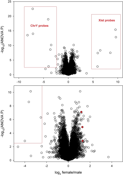
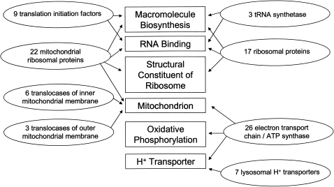
There is sexual dimorphism of skeletal muscle, the most obvious feature being the larger muscle mass of men. The molecular basis for this difference has not been clearly defined. To identify genes that might contribute to the relatively greater muscularity of men, we compared skeletal muscle gene expression profiles of 15 normal men and 15 normal women by using comprehensive oligonucleotide microarrays. Although there were sex-related differences in expression of several hundred genes, very few of the differentially expressed genes have functions that are obvious candidates for explaining the larger muscle mass of men. The men tended to have higher expression of genes encoding mitochondrial proteins, ribosomal proteins, and a few translation initiation factors. The women had >2-fold greater expression than the men (P<0.0001) of two genes that encode proteins in growth factor pathways known to be important in regulating muscle mass: growth factor receptor-bound 10 (GRB10) and activin A receptor IIB (ACVR2B). GRB10 encodes a protein that inhibits insulin-like growth factor-1 (IGF-1) signaling. ACVR2B encodes a myostatin receptor. Quantitative RT-PCR confirmed higher expression of GRB10 and ACVR2B genes in these women. In an independent microarray study of 10 men and 9 women with facioscapulohumeral dystrophy, women had higher expression of GRB10 (2.7-fold, P<0.001) and ACVR2B (1.7-fold, P<0.03). If these sex-related differences in mRNA expression lead to reduced IGF-1 activity and increased myostatin activity, they could contribute to the sex difference in muscle size.
Overview
The claims and statistics of this paper haven’t been checked yet.
Run the check — free, no account needed. It reads the open-access text and checks the sources it cites.
Integrity signals
Registry
Cites
Statistics
Figures
Verification
Claims the paper makes
The claims and statistics of this paper haven’t been checked yet.
Run the check — free, no account needed. It reads the open-access text and checks the sources it cites.
Claims other papers make about this one
Each is checked against this paper’s own text: does it say what the citing paper says it does?
The source backs part of the claim, not all of it — or backs it for a different population, condition or comparison.
In the citing paper
“Previous studies have identified over 3,000 genes that are differentially expressed in male and female skeletal muscle.”
From Sex-Based Differences in Skeletal Muscle Kinetics and Fiber-Type Composition (2015) · cites this paper as [96]
Experimental — accuracy not yet measuredIn the citing paper
“Analysis of mRNA from both male and female vastus lateralis biopsies by microarray shows (when normalized to α-actinin) that the female muscle has 35% more MyHC-I/β, 30% less MyHC-IIa, and 15% less MyHC-IIx mRNA than the corresponding male muscle (96).”
From Sex-Based Differences in Skeletal Muscle Kinetics and Fiber-Type Composition (2015) · cites this paper as [96]
In the citing paper
“In this study, the authors focus on two particular genes that are upregulated in women compared with men and that are known to code for proteins that are in signaling pathways of growth factors known to regulate muscle mass: growth factor receptor-bound protein 10 (GRB10) and activin receptor type-2A (ACVR2A).”
From Sex-Based Differences in Skeletal Muscle Kinetics and Fiber-Type Composition (2015) · cites this paper as [96]
In the citing paper
“The identification of over 3,000 genes differentially regulated in male and female muscle highlights the complex differences that occur in skeletal muscle from both sexes (96).”
From Sex-Based Differences in Skeletal Muscle Kinetics and Fiber-Type Composition (2015) · cites this paper as [96]
Self-check
Percentages, sums and reported differences re-computed from the paper’s own numbers.
The self-check needs the paper’s full text.
Citations
Cites
Subramanian, A., Tamayo, P., Mootha, V. K., Mukherjee, S., Ebert, B. L., Gillette, M. A., Paulovich, A., Pomeroy, S. L., Golub, T. R., Lander, E. S., et al. (2005) · Proceedings of the National Academy of Sciences · doi:10.1073/pnas.0506580102 · Cited by 53,625
In silico prediction of protein-protein interactions in human macrophages
Souiai, O., Guerfali, F. Z., Miled, S. B., Brun, C., & Benkahla, A. (2014) · BMC Research Notes · doi:10.1186/1756-0500-7-157 · Cited by 10,785
Exploration, normalization, and summaries of high density oligonucleotide array probe level data
Irizarry, R. A. (2003) · Biostatistics · doi:10.1093/biostatistics/4.2.249 · Cited by 10,603 · cited 5×
“This method was chosen after comparing it the standard Affymetrix method, a modified version of the Affymetrix comparison analysis algorithm that was used for our analyses of age-related differences [53], and RMA [54], because it had the best performance in accounting for non-specific hybridization (expression scores…”
InfluentialSignificance analysis of microarrays applied to the ionizing radiation response
Tusher, V. G., Tibshirani, R., & Chu, G. (2001) · Proceedings of the National Academy of Sciences · doi:10.1073/pnas.091062498 · Cited by 10,568
Regulation of skeletal muscle mass in mice by a new TGF-p superfamily member
McPherron, A. C., Lawler, A. M., & Lee, S.-J. (1997) · Nature · doi:10.1038/387083a0 · Cited by 4,013 · cited 1×
“Myostatin has a major role in determining muscle size. Constitutive myostatin knockout in mice leads to a hypermuscular phenotype caused by an increased number of fibers and fiber enlargement”
BackgroundSandri, M., Sandri, C., Gilbert, A., Skurk, C., Calabria, E., Picard, A., Walsh, K., Schiaffino, S., Lecker, S. H., & Goldberg, A. L. (2004) · Cell · doi:10.1016/s0092-8674(04)00400-3 · Cited by 3,012 · cited 1×
“In its active non-phosphorylated state, this transcription factor stimulates expression of the ubiquitin ligase atrogin-1 (also known as MAFbx or F-box protein 32) [47].”
Identifying biological themes within lists of genes with EASE
Hosack, D. A., Dennis, G., Sherman, B. T., Lane, H. C., & Lempicki, R. A. (2003) · Genome biology · doi:10.1186/gb-2003-4-10-r70 · Cited by 1,961 · cited 4×
“The EASE program (Version 2.0) was provided by the National Institutes of Health [27].”
InfluentialMultiple types of skeletal muscle atrophy involve a common program of changes in gene expression
Lecker, S. H., Jagoe, R. T., Gilbert, A., Gomes, M., Baracos, V., Bailey, J., Price, S. R., Mitch, W. E., & Goldberg, A. L. (2004) · The FASEB Journal · doi:10.1096/fj.03-0610com · Cited by 1,539 · cited 1×
“Atrogin-1 is upregulated under conditions associated with muscle atrophy [48].”
Myostatin Mutation Associated with Gross Muscle Hypertrophy in a Child
Schuelke, M., Wagner, K. R., Stolz, L. E., Hübner, C., Riebel, T., Kömen, W., Braun, T., Tobin, J. F., & Lee, S.-J. (2004) · New England Journal of Medicine · doi:10.1056/nejmoa040933 · Cited by 1,399
Skeletal muscle hypertrophy and atrophy signaling pathways
Glass, D. J. (2005) · The International Journal of Biochemistry & Cell Biology · doi:10.1016/j.biocel.2005.04.018 · Cited by 1,106 · cited 1×
“It is well accepted that IGF-1 has an anabolic effect on muscle [28].”
Cited by
Sorted by citations
Fiber Types in Mammalian Skeletal Muscles
Schiaffino, S., & Reggiani, C. (2011) · Physiological Reviews · doi:10.1152/physrev.00031.2010 · Cited by 3,022
“Thus, in men, the levels of MyHC-slow transcripts are lower than in women and the levels of MyHC-2A and -2X transcripts are higher (855).”
BackgroundSex differences in human fatigability: mechanisms and insight to physiological responses
Hunter, S. K. (2014) · Acta Physiologica · doi:10.1111/apha.12234 · Cited by 595
Sex-Based Differences in Skeletal Muscle Kinetics and Fiber-Type Composition
Haizlip, K. M., Harrison, B. C., & Leinwand, L. A. (2015) · Physiology · doi:10.1152/physiol.00024.2014 · Cited by 521
“Analysis of mRNA from both male and female vastus lateralis biopsies by microarray shows (when normalized to -actinin) that the female muscle has 35% more MyHC-I/ , 30% less MyHC-IIa, and 15% less MyHC-IIx mRNA than the corresponding male muscle (96).”
BackgroundInfluentialAnsdell, P., Thomas, K., Hicks, K. M., Hunter, S. K., Howatson, G., & Goodall, S. (2020) · Experimental Physiology · doi:10.1113/ep088548 · Cited by 403
“Sex differences in genetic expression and phenotype within skeletal muscle at rest are well established (Lindholm et al., 2014; Welle et al., 2008); however, whether sex mediates the phenotypic adaptation to endurance training is not clear.”
BackgroundThe role of sex in the genomics of human complex traits
Khramtsova, E. A., Davis, L. K., & Stranger, B. E. (2019) · Nature Reviews Genetics · doi:10.1038/s41576-018-0083-1 · Cited by 303
Lundsgaard, A.-M., & Kiens, B. (2014) · Frontiers in Endocrinology · doi:10.3389/fendo.2014.00195 · Cited by 289
“The immunohistochemical findings are reflected at the transcriptional level of the myosin heavy chains (MHC), as MHCI mRNA are reported lower in the vastus lateralis muscle of men than women (77), while MHCIIA and -IIX mRNA are higher in men (78).”
BackgroundCentral and Peripheral Fatigue in Physical Exercise Explained: A Narrative Review
Tornero-Aguilera, J. F., Jiménez-Morcillo, J., Rubio-Zarapuz, A., & Clemente‐Suárez, V. J. (2022) · International Journal of Environmental Research and Public Health · doi:10.3390/ijerph19073909 · Cited by 304
HUNTER, S. K., S. ANGADI, S., BHARGAVA, A., HARPER, J., HIRSCHBERG, A. L., D. LEVINE, B., L. MOREAU, K., J. NOKOFF, N., STACHENFELD, N. S., & BERMON, S. (2023) · Medicine & Science in Sports & Exercise · doi:10.1249/mss.0000000000003300 · Cited by 241
Sex- and Gender-Based Pharmacological Response to Drugs
Mauvais‐Jarvis, F., Berthold, H. K., Campesi, I., Carrero, J. J., Dhakal, S., Franconi, F., Gouni‐Berthold, I., Heiman, M. L., Kautzky‐Willer, A., Klein, S. L., et al. (2021) · Pharmacological Reviews · doi:10.1124/pharmrev.120.000206 · Cited by 203
“This is associated with over 3000 genes differentially expressed in male and female muscle (Welle et al., 2008; Haizlip et al., 2015).”
BackgroundVascularized and Innervated Skeletal Muscle Tissue Engineering
Gilbert‐Honick, J., & Grayson, W. L. (2019) · Advanced Healthcare Materials · doi:10.1002/adhm.201900626 · Cited by 197
Zhang, Y., Klein, K., Sugathan, A., Nassery, N., Dombkowski, A. A., Zanger, U. M., & Waxman, D. J. (2011) · PLoS ONE · doi:10.1371/journal.pone.0023506 · Cited by 192
“Indeed, all but two of the X-chromosome genes (PGRMC1, PHF6; Table 1) that showed female-biased expression in human liver also show female-bias in human muscle [38], supporting the conclusion that the female-biased expression of these genes reflects (partial) escape from X-inactivation, rather than hormone-based sex-b…”
BackgroundSex differences in the human peripheral blood transcriptome
Jansen, R., Batista, S., Brooks, A. I., Tischfield, J. A., Willemsen, G. A. H. M., van Grootheest, G., Hottenga, J., Milaneschi, Y., Mbarek, H., Madar, V., et al. (2014) · BMC Genomics · doi:10.1186/1471-2164-15-33 · Cited by 172
“Surprisingly few studies aimed at identifying and investigating sex-biased genes in humans, and only in small sample sizes (N < 250 [19-22]).”
BackgroundMethodResultDogan, C., Antonio, M. D., Hamroun, D., Varet, H., Fabbro, M., Rougier, F., Amarof, K., Arné‐Bes, M., Bédat‐Millet, A., Béhin, A., et al. (2016) · PLoS ONE · doi:10.1371/journal.pone.0148264 · Cited by 160
“Male and female skeletal muscles display considerable differences in their metabolic properties and gene expression patterns [46,47].”
BackgroundSex Differences in Endurance Running.
Besson, T., Macchi, R., Rossi, J., Morio, C. Y. M., Kunimasa, Y., Nicol, C., Vercruyssen, F., & Millet, G. Y. (2022) · Sports Medicine · doi:10.1007/s40279-022-01651-w · Cited by 158
“…difference that mainly explains the difference in maximal strength is muscle mass, due to larger diameter muscle fibers in males rather than the number of fibers [138], this larger size being due to sex-related differences in human skeletal muscle gene (e.g. GRB10 and ACVR2B) expression [139].”
Postmenopausal osteoporosis coexisting with sarcopenia: the role and mechanisms of estrogen
Lu, L., & Tian, L. (2023) · Journal of Endocrinology · doi:10.1530/joe-23-0116 · Cited by 175
Smith, G. I., Atherton, P., Villareal, D. T., Frimel, T. N., Rankin, D., Rennie, M. J., & Mittendorfer, B. (2008) · PLoS ONE · doi:10.1371/journal.pone.0001875 · Cited by 168
“The differences we observed between older men and women, namely a ,30% greater basal rate of mixed MPS in women than in men and resistance of MPS to feeding a liquid mixed meal (providing a total of ,10 g of protein) in women, are consistent with a recent study in which comprehensive oligonucleotide microarrays were u…”
ResultSex differences in human performance
Hunter, S. K., & Senefeld, J. W. (2024) · The Journal of Physiology · doi:10.1113/jp284198 · Cited by 172
“…males and females are ultimately due to sex-related differences in human skeletal muscle gene expression and the interaction with sex-specific hormones and training status (Chapman et al., 2020; Landen et al., 2021; Liu et al., 2010; Maher et al., 2009; Roth et al., 2002; Welle et al., 2008).”
Landen, S., Hiam, D., Voisin, S., Jacques, M., Lamon, S., & Eynon, N. (2023) · The Journal of Physiology · doi:10.1113/jp279499 · Cited by 137
“Multiple studies (Welle et al. 2008; Maher et al. 2009; Liu et al. 2010; Lindholm et al. 2014; Gershoni & Pietrokovski, 2017; Chapman et al. 2020; Lopes-Ramos et al. 2020; Oliva et al. 2020) have analysed sex differences in the skeletal muscle transcriptome.”
BackgroundSkeletal muscle gene expression in response to resistance exercise: sex specific regulation
Liu, D., Sartor, M. A., Nader, G. A., Gutmann, L., Treutelaar, M. K., Pistilli, E. E., IglayReger, H. B., Burant, C. F., Hoffman, E. P., & Gordon, P. M. (2010) · BMC Genomics · doi:10.1186/1471-2164-11-659 · Cited by 122
“It is plausible to speculate that sex-related gene transcriptional regulation contributes to the sexual dimorphism observed in the skeletal muscle phenotypes [16].”
BackgroundNookaew, I., Svensson, P., Jacobson, P., Jernås, M., Taube, M., Larsson, I., Andersson‐Assarsson, J. C., Sjöström, L., Froguel, P., Walley, A. J., et al. (2012) · The Journal of Clinical Endocrinology & Metabolism · doi:10.1210/jc.2012-2764 · Cited by 124
“Expression profiles from skeletal muscle (GSE9676) and liver (GSE9588) were from Welle et al (17) and Schadt et al (18), respectively.”
Sex Differences in Cancer Cachexia
Zhong, X.-L., & Zimmers, T. (2020) · Current Osteoporosis Reports · doi:10.1007/s11914-020-00628-w · Cited by 98
Do Sex Differences in Physiology Confer a Female Advantage in Ultra-Endurance Sport?
Tiller, N. B., Elliott-Sale, K. J., Knechtle, B., Wilson, P. B., Roberts, J., & Millet, G. Y. (2021) · Sports Medicine · doi:10.1007/s40279-020-01417-2 · Cited by 126
Sex Differences in Inflammation and Muscle Wasting in Aging and Disease
Peruta, C. D., Lozanoska‐Ochser, B., Renzini, A., Moresi, V., Riera, C. S., Bouché, M., & Coletti, D. (2023) · International Journal of Molecular Sciences · doi:10.3390/ijms24054651 · Cited by 118
“Physiological Mass [30] Energy metabolism [32–34] Mitochondrial content [42] Protein turnover [47,50] Insulin sensitivity [66,88] Muscle regeneration [61,62,65]”
BackgroundSex Differences in Global mRNA Content of Human Skeletal Muscle
Maher, A. C., Fu, M., Isfort, R. J., Varbanov, A. R., Qu, X. A., & Tarnopolsky, M. A. (2009) · PLoS ONE · doi:10.1371/journal.pone.0006335 · Cited by 108
“(2008) have previously shown that there are significant differences in skeletal muscle mRNA content between men and women, as well as showing sex differences are significantly greater than age and/or strength training effects on mRNA content [38,39].”
BackgroundResultInfluentialLindholm, M. E., Huss, M., Solnestam, B. W., Kjellqvist, S., Lundeberg, J., & Sundberg, C. J. (2014) · The FASEB Journal · doi:10.1096/fj.14-255000 · Cited by 99
“(27) found 3000 genes to be differentially expressed between males and females, a number similar to that in our study.”
BackgroundResultSex influences DNA methylation and gene expression in human skeletal muscle myoblasts and myotubes
Davegårdh, C., Hall, E., Broholm, C., Henriksen, T. I., Pedersen, M., Pedersen, B. K., Schéele, C., & Ling, C. (2019) · Stem Cell Research & Therapy · doi:10.1186/s13287-018-1118-4 · Cited by 92
Sexually dimorphic characteristics of the small intestine and colon of prepubescent C57BL/6 mice
Steegenga, W. T., Mischke, M., Lute, C., Boekschoten, M. V., Pruis, M. G. M., Lendvai, Á., Verkade, H. J., Boekhorst, J., Timmerman, H. M., Plösch, T., et al. (2014) · Biology of Sex Differences · doi:10.1186/s13293-014-0011-9 · Cited by 90
“Genome-wide gene expression analysis has revealed that sex differences occur in a wide variety of organs and tissues in humans as well as in rodents, including the liver, heart, kidney, brain, muscle, placenta, gonads and adipose tissue [17-25].”
Background(13376925), S. L., Hiam, D., Voisin, S. A. C., (13283841), M. J., Lamon, S., & (13308696), N. E. (2024) · Figshare · Cited by 85
Understanding sex differences in the regulation of cancer-induced muscle wasting
Montalvo, R. N., Counts, B. R., & Carson, J. A. (2018) · Current Opinion in Supportive and Palliative Care · doi:10.1097/spc.0000000000000380 · Cited by 78
Myonuclear content regulates cell size with similar scaling properties in mice and humans
Hansson, K., Eftestøl, E., Bruusgaard, J. C., Juvkam, I. S., Cramer, A., Malthe‐Sørenssen, A., Millay, D. P., & Gundersen, K. (2020) · Nature Communications · doi:10.1038/s41467-020-20057-8 · Cited by 85
Genetic and epigenetic sex-specific adaptations to endurance exercise
Landen, S., Voisin, S., Craig, J. M., McGee, S. L., Lamon, S., & Eynon, N. (2019) · Epigenetics · doi:10.1080/15592294.2019.1603961 · Cited by 76
β - catenin is central to DUX4 -driven network rewiring in facioscapulohumeral muscular dystrophy
Banerji, C. R. S., Knopp, P., Moyle, L. A., Simone Severini, B., Orrell, R. W., Teschendorff, A. E., & Zammit, P. S. (2014) · Journal of The Royal Society Interface · doi:10.1098/rsif.2014.0797 · Cited by 75
“We also used human skeletal muscle studies into gene expression in muscle diseases other than FSHD (GSE3307 [19]), ageing (GSE5086 [24] and GSE9676 [25]) and atrophy (GSE5110 [26] and GSE8872 [27]).”
MethodWomen Have Higher Protein Content of β-Oxidation Enzymes in Skeletal Muscle than Men
Maher, A. C., Akhtar, M., Vockley, J., & Tarnopolsky, M. A. (2010) · PLoS ONE · doi:10.1371/journal.pone.0012025 · Cited by 72
“Several studies have shown that sex differences alone are greater predictors of substrate selection than are age, menstrual cycle phase, 17b-estradiol supplementation, endurance and strength training effects on mRNA content [20–23].”
BackgroundSkeletal Muscle Transcriptomic Comparison between Long-Term Trained and Untrained Men and Women
Chapman, M. A., Arif, M., Emanuelsson, E. B., Reitzner, S. M., Lindholm, M. E., Mardinoğlu, A., & Sundberg, C. J. (2020) · Cell Reports · doi:10.1016/j.celrep.2020.107808 · Cited by 72
Beef tenderness and intramuscular fat proteomic biomarkers: Effect of gender and rearing practices
Picard, B. B., Gagaoua, M., Al-Jammas, M., & Bonnet, M. (2019) · Journal of Proteomics · doi:10.1016/j.jprot.2019.03.010 · Cited by 66
Lloyd, E. M., Pinniger, G. J., Murphy, R. M., & Grounds, M. D. (2023) · Acta Physiologica · doi:10.1111/apha.14012 · Cited by 67
Stretch, C., Khan, S., Asgarian, N., Eisner, R., Vaisipour, S., Damaraju, S., Graham, K., Bathe, O. F., Steed, H. L., Greiner, R., et al. (2013) · PLoS ONE · doi:10.1371/journal.pone.0065380 · Cited by 62
“These observations explain the lack of concordance between the findings of two prior studies of sexual dimorphism [2,4] with each other, and with our results.”
ResultChopard, A., Cunff, M. L., Danger, R., Lamirault, G., Bihouée, A., Teusan, R., Jasmin, B. J., Marini, J. F., & Leger, J. J. (2009) · Physiological Genomics · doi:10.1152/physiolgenomics.00036.2009 · Cited by 69
Micro-RNAs in ALS muscle: Differences in gender, age at onset and disease duration
Pegoraro, V., Merico, A., & Angelini, C. I. (2017) · Journal of the Neurological Sciences · doi:10.1016/j.jns.2017.07.008 · Cited by 63
“It is well known that normal male and female skeletal muscles display considerable differences in their metabolic properties and gene expression patterns [45].”
Extraordinary fast-twitch fiber abundance in elite weightlifters
Serrano, N., Colenso‐Semple, L. M., Lazauskus, K. K., Siu, J. W., Bagley, J. R., Lockie, R. G., Costa, P. B., & Galpin, A. J. (2019) · PLoS ONE · doi:10.1371/journal.pone.0207975 · Cited by 60
Lavin, K. M., Coen, P. M., Baptista, L. C. P., Bell, M. B., Drummer, D. J., Harper, S. A., Lixandrão, M. E., McAdam, J. S., O’Bryan, S. M., Ramos, S. V., et al. (2022) · Comprehensive physiology · doi:10.1002/cphy.c200033 · Cited by 78
Microarray Analysis Reveals Novel Features of the Muscle Aging Process in Men and Women
Liu, D., Sartor, M. A., Nader, G. A., Pistilli, E. E., Tanton, L. C., Lilly, C., Gutmann, L., IglayReger, H. B., Visich, P. S., Hoffman, E. P., et al. (2013) · The Journals of Gerontology Series A · doi:10.1093/gerona/glt015 · Cited by 58
de Jager, N., Hudson, N. J., Reverter, A., Wang, Y., Nagaraj, S. H., Cafe, L. M., Greenwood, P. L., Barnard, R. T., Kongsuwan, K., & Dalrymple, B. P. (2011) · Physiological Genomics · doi:10.1152/physiolgenomics.00226.2010 · Cited by 57
Landen, S., Jacques, M., Hiam, D., Alvarez‐Romero, J., Harvey, N. R., Haupt, L. M., Griffiths, L. R., Ashton, K. J., Lamon, S., Voisin, S. A. C., et al. (2021) · Clinical Epigenetics · doi:10.1186/s13148-021-01188-1 · Cited by 60
Age-Related DNA Methylation Changes: Potential Impact on Skeletal Muscle Aging in Humans
Gensous, N., Bacalini, M. G., Franceschi, C., Meskers, C. G. M., Maier, A. B., & Garagnani, P. (2019) · Frontiers in Physiology · doi:10.3389/fphys.2019.00996 · Cited by 55
“It was previously reported substantial differences in gene expression or proteinprotein interactions in human SM according to gender during aging (Roth et al., 2002; Welle et al., 2008; Liu et al., 2010, 2013; Shafiee et al., 2018), and a recently published study also observed for the first time differences in DNA met…”
BackgroundHakimov, H. A., Walters, S. A., Wright, T. C., Meidinger, R. G., Verschoor, C. P., Gadish, M. A., Chiu, D., Strömvik, M. V., Forsberg, C. W., & Golovan, S. P. (2009) · PROTEOMICS · doi:10.1002/pmic.200900049 · Cited by 53
“There are few omics comparisons of sexually dimorphic gene expression mainly in mouse and human muscle transcriptome [114–116].”
BackgroundDamas, F., Ugrinowitsch, C., Libardi, C. A., Jannig, P. R., Hector, A. J., McGlory, C., Lixandrão, M. E., VECHIN, F. C., Montenegro, H., Tricoli, V., et al. (2018) · European Journal of Applied Physiology · doi:10.1007/s00421-018-3984-y · Cited by 51
Sexual dimorphism of monocyte transcriptome in individuals with chronic low-grade inflammation
So, J., Tai, A., Lichtenstein, A. H., Wu, D., & Lamon‐Fava, S. (2021) · Biology of Sex Differences · doi:10.1186/s13293-021-00387-y · Cited by 51
Refalo, M. C., Helms, E. R., Hamilton, D. L., & Fyfe, J. J. (2023) · Sports Medicine - Open · doi:10.1186/s40798-023-00554-y · Cited by 56
MacNeil, L., Melov, S., Hubbard, A., Baker, S. K., & Tarnopolsky, M. A. (2010) · PLoS ONE · doi:10.1371/journal.pone.0010695 · Cited by 49
“Specifically, women express a higher abundance of mRNA for several genes involved in fat metabolism that include trifunctional protein b and lipoprotein lipase [36], and greater expression of the negative regulators of the anabolism growth factor receptor-bound 10 and activin A receptor IIB [37].”
BackgroundKaspric, N., Picard, B. B., Reichstadt, M., Tournayre, J., & Bonnet, M. (2015) · PLoS ONE · doi:10.1371/journal.pone.0128086 · Cited by 50
“The presence of these proteins or PLOS ONE | DOI:10.1371/journal.pone.0128086 May 22, 2015 18 / 24 their mRNA in adipose and muscular cells was reported for VCAM1 [63,64], GABARAPL1 [65,66], GABARAPL2 [64,67], GABARAP [64,68], MAP1LC3A [64,69], and MAP1LC3B [64,70].”
BackgroundInfluentialSvensson, J., Movérare‐Skrtic, S., Windahl, S. H., Swanson, C., & Sjögren, K. (2010) · Journal of Molecular Endocrinology · doi:10.1677/jme-09-0165 · Cited by 46
“Interestingly, the expression of Grb10 in muscle has been shown to be gender dependent with higher expression in women than in men, suggesting a role for Grb10 in the sexual dimorphism of skeletal muscle mass (Welle et al. 2008).”
BackgroundHunter, S. K., Angadi, S. S., Bhargava, A., Harper, J., Hirschberg, A. L., Levine, B. D., Moreau, K. L., Nokoff, N. J., Stachenfeld, N. S., & Bermon, S. (2023) · Translational Journal of the American College of Sports Medicine · doi:10.1249/tjx.0000000000000236 · Cited by 43
O’Reilly, J., Ono‐Moore, K. D., Chintapalli, S. V., Rutkowsky, J. M., Tolentino, T., Lloyd, K. C. K., Olfert, I. M., & Adams, S. H. (2021) · Physiological Reports · doi:10.14814/phy2.15031 · Cited by 44
Lee, J. K., Park, Y. S., Lee, K., Youn, S. I., Won, Y., Min, S.-H., Ahn, S., Park, D. J., & Kim, H. (2021) · Journal of Cachexia Sarcopenia and Muscle · doi:10.1002/jcsm.12793 · Cited by 42
Davies, R. W., Carson, B. P., & Jakeman, P. M. (2018) · Frontiers in Physiology · doi:10.3389/fphys.2018.01480 · Cited by 41
“, 1993) and a higher proportion of type I fibers relative to type II (Staron et al., 2000; Roepstorff et al., 2006; Welle et al., 2008), greater muscle capillarisation (Roepstorff et al.”
BackgroundSystems biology asks new questions about sex differences
Arnold, A. P., van Nas, A., & Lusis, A. J. (2009) · Trends in Endocrinology and Metabolism · doi:10.1016/j.tem.2009.06.007 · Cited by 42
“The earliest microarray studies on non-gonadal tissues reported that only tens to hundreds of genes are expressed consistently at different levels in males and females [10–17].”
BackgroundOnoki, T., Izumi, Y., Takahashi, M., Murakami, S., Matsumaru, D., Ohta, N., Wati, S. M., Hatanaka, N., Katsuoka, F., Okutsu, M., et al. (2021) · Redox Biology · doi:10.1016/j.redox.2021.101966 · Cited by 39
“× 150 mm, 3 μm particle size, Merck) coupled with a Q Exactive instrument (PFPP-LC/HRMS/MS) [57].”
MethodShero, J. A., Lindholm, M. E., Sandri, M. F., & Stanford, K. I. (2025) · Circulation Research · doi:10.1161/circresaha.124.325614 · Cited by 45
Female-specific regulation of skeletal muscle mass by USP19 in young mice
Ogawa, M., Kitakaze, T., Harada, N., & Yamaji, R. (2015) · Journal of Endocrinology · doi:10.1530/joe-15-0128 · Cited by 38
“Sex hormones are involved in the differential regulation of gene expression in human skeletal muscle cells (Deasy et al. 2008, Welle et al. 2008).”
BackgroundGenetic Architecture of Sexual Dimorphism in Humans
Rigby, N., & Kulathinal, R. J. (2015) · Journal of Cellular Physiology · doi:10.1002/jcp.24979 · Cited by 43
Sexual dimorphism in laryngeal muscle fibers and ultrasonic vocalizations in the adult rat
Lenell, C., & Johnson, A. M. (2017) · The Laryngoscope · doi:10.1002/lary.26561 · Cited by 38
“In the limb muscles, however, patterns of sexual dimorphism have been identified, such as men having larger muscle fiber cross-sectional areas and a higher ratio of type II (fast twitch) muscle fibers than women.(16,17) These sexually dimorphic muscle properties result in differences in muscle performance between sexe…”
Lenell, C., Sandage, M. J., & Johnson, A. M. (2019) · Journal of Speech Language and Hearing Research · doi:10.1044/2018_jslhr-s-18-0179 · Cited by 38
Dennis, R. A., Zhu, H., KORTEBEIN, P. M., Bush, H., Harvey, J., Sullivan, D. H., & Peterson, C. A. (2009) · Physiological Genomics · doi:10.1152/physiolgenomics.00056.2009 · Cited by 38
“The application of these results is limited in several ways: 1) The sample size was small and lacked the power to identify possible confounding variables such as sex, which can affect muscle phenotype and gene expression (37); 2) The study involved only older adults, and thus, whether expression of these genes has pre…”
BackgroundEarly inflammatory and myogenic responses to resistance exercise in the elderly
Mathers, J. L., Farnfield, M. M., GARNHAM, A. P., Caldow, M. K., Cameron‐Smith, D., & Peake, J. M. (2012) · Muscle & Nerve · doi:10.1002/mus.23317 · Cited by 39
“tein expression in skeletal muscle will help to clarify the potential functional importance of differences in myostatin expression between men and women.(2,4,46) The difference in myogenin mRNA expression”
BackgroundResultExercise Does Not Influence Myostatin and Follistatin Messenger RNA Expression in Young Women
Jensky, N. E., Sims, J. K., Dieli-Conwright, C. M., Sattler, F. R., Rice, J. C., & Schroeder, E. T. (2010) · The Journal of Strength and Conditioning Research · doi:10.1519/jsc.0b013e3181c8664f · Cited by 37
“These findings may indicate that increased myostatin activity contributes to gender differences in muscle size (31).”
BackgroundInfluentialRune, A., Salehzadeh, F., Szekeres, F. L. M., Kühn, I., Osler, M. E., & Al‐Khalili, L. (2009) · Acta Physiologica · doi:10.1111/j.1748-1716.2009.02010.x · Cited by 37
“Although earlier studies have addressed sex differences in mRNA expression from skeletal muscle biopsies (Welle et al. 2008), this question has not previously been addressed in human skeletal muscle cell cultures.”
BackgroundGender differences in contractile and passive properties of mdx extensor digitorum longus muscle
Hakim, C. H., & Duan, D. (2011) · Muscle & Nerve · doi:10.1002/mus.22275 · Cited by 36
“It is well established that male and female skeletal muscles display considerable differences in their metabolic properties and gene expression patterns.(25,26) However, Glenmark et al.”
BackgroundSalamone, I. M., Quattrocelli, M., Barefield, D. Y., Page, P., Tahtah, I., Hadhazy, M., Tomar, G., & McNally, E. M. (2022) · Journal of Clinical Investigation · doi:10.1172/jci149828 · Cited by 35
Sex differences in muscle protein expression and DNA methylation in response to exercise training
Landen, S., Jacques, M., Hiam, D., Alvarez‐Romero, J., Schittenhelm, R. B., Shah, A. D., Huang, C., Steele, J. R., Harvey, N. R., Haupt, L. M., et al. (2023) · Biology of Sex Differences · doi:10.1186/s13293-023-00539-2 · Cited by 35
Pöllänen, E., Fey, V., Törmäkangas, T., Ronkainen, P. H. A., Taaffe, D. R., Takala, T., Koskinen, S., Cheng, S., Puolakka, J. O., Kujala, U. M., et al. (2010) · AGE · doi:10.1007/s11357-010-9140-1 · Cited by 37
“One important and interesting aspect of aging-related phenomena discovered in previous microarray studies is the enhanced negative transcriptional regulation of oxidative phosphorylation and other mitochondrial functions with advancing age (Welle et al. 2003; Welle et al. 2004; Zahn et al. 2006; Melov et al. 2007; Wel…”
BackgroundMethodInfluentialSex- and age-dependent gene expression in human liver: An implication for drug-metabolizing enzymes
Uno, Y., Takata, R., Kito, G., Yamazaki, H., Nakagawa, K., Nakamura, Y., Kamataki, T., & Katagiri, T. (2016) · Drug Metabolism and Pharmacokinetics · doi:10.1016/j.dmpk.2016.10.409 · Cited by 36
van Vugt, J. L. A., van Putten, Y., van der Kall, I. M., Buettner, S., d’Ancona, F. C. H., Dekker, H. M., Kimenai, H. J. A. N., de Bruin, R. W. F., Warlé, M. C., & IJzermans, J. N. M. (2018) · European Journal of Clinical Nutrition · doi:10.1038/s41430-018-0287-7 · Cited by 33
Peterson, M. P., Rosvall, K. A., Taylor, C. A., Lopez, J. A., Choi, J.-H., Ziegenfus, C., Tang, H., Colbourne, J. K., & Ketterson, E. D. (2013) · Journal of Experimental Biology · doi:10.1242/jeb.089813 · Cited by 32
“For example, Lipid phosphate phosphohydrolase 1, a gene involved in glycerolipid synthesis and lipid uptake (Kai et al., 1997), was expressed at a higher level in control males than females, consistent with sex differences in metabolic activity (Fernando et al., 2010; Wikelski et al., 1999).”
BackgroundInfluentialSex differences in diaphragmatic fatigue: Effects of hypoxia during inspiratory loading
Archiza, B., Reinhard, P. A., Welch, J. F., & Sheel, A. W. (2020) · The Journal of Physiology · doi:10.1113/jp280704 · Cited by 33
“Although not known in the diaphragm muscle, women have a larger proportional area of type I fibres (‘slow’ oxidative fibres and more fatigue resistant) than men in several skeletal muscles (Carter et al. 2001; Welle et al. 2008), whereas men present a greater cross-sectional area of type II fibres (‘fast’ glycolytic f…”
BackgroundSex differences in forkhead box O3a signaling response to hindlimb unloading in rat soleus muscle
Yoshihara, T., Natsume, T., Tsuzuki, T., Chang, S.-W., Kakigi, R., Sugiura, T., & Naıto, H. (2018) · The Journal of Physiological Sciences · doi:10.1007/s12576-018-0640-6 · Cited by 31
Brockbals, L., Garrett-Rickman, S., Fu, S., Ueland, M., McNevin, D. B., & Padula, M. P. (2023) · Analytical and Bioanalytical Chemistry · doi:10.1007/s00216-023-04822-4 · Cited by 29
FAT/CD36 Participation in Human Skeletal Muscle Lipid Metabolism: A Systematic Review
Ramos‐Jiménez, A., Zavala-Lira, R. A., Moreno-Brito, V., & González-Rodríguez, E. (2022) · Journal of Clinical Medicine · doi:10.3390/jcm12010318 · Cited by 29
Koruyucu, A. N., & Aşantoğrol, F. (2023) · Dentomaxillofacial Radiology · doi:10.1093/dmfr/twad013 · Cited by 28
Rienecker, K. D. A., Chavasse, A. T., Moorwood, K., Ward, A., & Isles, A. R. (2019) · Genes Brain & Behavior · doi:10.1111/gbb.12571 · Cited by 25
Exercise as a therapy for cancer-induced muscle wasting
Halle, J. L., Counts, B. R., & Carson, J. A. (2020) · Sports Medicine and Health Science · doi:10.1016/j.smhs.2020.11.004 · Cited by 28
The impact of biological sex on alternative splicingPreprint
Karlebach, G., Veiga, D. F. T., Mays, A. D., Chatzipantsiou, C., Barja, P. P., Chatzou, M., Kesarwani, A. K., Daniš, D., Kararigas, G., Zhang, X., et al. (2018) · bioRxiv (Cold Spring Harbor Laboratory) · doi:10.1101/490904 · Cited by 28
“A number of the GO terms could reflect known sex differences, such as the enrichment of extracellular matrix (ECM) in breast tissue, which is known to display sex-biased differential expression of multiple ECM proteins,(18) or translation initiation factor activity, which was differential in five tissues in our examin…”
BackgroundAn integrative approach to the pulmonary physiology of exercise: when does biological sex matter?
Archiza, B., Leahy, M. G., Kipp, S., & Sheel, A. W. (2021) · European Journal of Applied Physiology · doi:10.1007/s00421-021-04690-9 · Cited by 26
Shafiee, G., Asgari, Y., Soltani, A., Larijani, B. A., & Heshmat, R. (2018) · PeerJ · doi:10.7717/peerj.5239 · Cited by 25
Sex differences in metabolic pathways are regulated by Pfkfb3 and Pdk4 expression in rodent muscle
Christianto, A., Baba, T., Takahashi, F., Inui, K., Inoue, M., Suyama, M., Ono, Y., Ohkawa, Y., & Morohashi, K.-I. (2021) · Communications Biology · doi:10.1038/s42003-021-02790-y · Cited by 26
Clinical correlations and long‐term follow‐up in 100 patients with sarcoglycanopathies
Guimarães‐Costa, R., Fernández‐Eulate, G., Wahbi, K., Leturcq, F., Malfatti, E., Béhin, A., Léonard-Louis, S., Desguerre, I., Barnérias, C., Nouguès, M., et al. (2020) · European Journal of Neurology · doi:10.1111/ene.14592 · Cited by 23
“The relationship between sex and phenotype has previously been reported in prevalent myopathies, such as myotonic dystrophy type 1 [27], as well as a sexual dimorphism of skeletal muscle metabolic and gene expression profiles [28].”
BackgroundMogilski, J. K. (2020) · SAGE Publications Ltd eBooks · doi:10.4135/9781529739442.n8 · Cited by 22
Sexual dimorphisms in skeletal muscle: current concepts and research horizons
Emmert, M. E., Emmert, A. S., Goh, Q., & Cornwall, R. (2024) · Journal of Applied Physiology · doi:10.1152/japplphysiol.00529.2023 · Cited by 24
Sex differences in skeletal muscle metabolism in exercise and type 2 diabetes mellitus
MacGregor, K. A., Ellefsen, S., Pillon, N. J., Hammarström, D., & Krook, A. (2024) · Nature Reviews Endocrinology · doi:10.1038/s41574-024-01058-9 · Cited by 23
“The higher ratio of type 2 to type 1 fibre mass in men has been attributed to increased testosterone-mediated hypertrophy of type 2 fibres 34,35,46 .”
Wu, Z., Wang, Y., Ye, Z., Guan, Y., Ye, X., Chen, Z., Li, C., Chen, G., Zhu, Y., Du, J., et al. (2021) · Frontiers in Physiology · doi:10.3389/fphys.2021.718068 · Cited by 20
de Zevallos, J. O., Hogwood, A. C., Kruse, K., Guzman, J. D., Buckley, M., WELTMAN, A. L., & Allen, J. D. (2023) · Journal of Applied Physiology · doi:10.1152/japplphysiol.00220.2023 · Cited by 23
Ji, J. S., Liu, L., Shu, C., Yan, L. L., & Zeng, Y. (2022) · The Journals of Gerontology: Series A · doi:10.1093/gerona/glab378 · Cited by 19
Subcellular effects of myocyte-specific androgen receptor overexpression in mice
Musa, M., Fernando, S. M., Chatterjee, D., & Monks, D. A. (2011) · Journal of Endocrinology · doi:10.1530/joe-11-0071 · Cited by 19
Hyperammonaemia and associated factors in unprovoked convulsive seizures: A cross-sectional study
Sato, K., Arai, N., Omori, A., Hida, A., Kimura, A., & Takeuchi, S. (2016) · Seizure · doi:10.1016/j.seizure.2016.09.015 · Cited by 19
Omosule, C. L., Gremminger, V. L., AGUILLARD, A., Jeong, Y., Harrelson, E. N., Miloscio, L., Mastaitis, J. W., Rafique, A., Kleiner, S., Pfeiffer, F. M., et al. (2020) · Journal of Bone and Mineral Research · doi:10.1002/jbmr.4223 · Cited by 21
“Previous reports suggest skeletal muscles of male C57 mice have less processed myostatin (−40% to 60%)((68)) and ACVR2B expression((69)) compared with female mice, which may result in male and female mice requiring different anti-myostatin treatment dosages to elicit equivalent responses.”
BackgroundYang, W., Park, J.-H., Park, S.-Y., & Park, Y. (2022) · Metabolites · doi:10.3390/metabo12100965 · Cited by 21
Yang, S., Yang, F., Gao, Y., Su, Y., Sun, W., Jia, S., Wang, Y., & Lam, W. (2022) · International Journal of Environmental Research and Public Health · doi:10.3390/ijerph19137802 · Cited by 17
Comparative Meta-Analysis of Transcriptomics Data during Cellular Senescence andIn VivoTissue Ageing
Voutetakis, K., Chatziioannou, A. A., Gonos, E. S., & Trougakos, I. P. (2015) · Oxidative Medicine and Cellular Longevity · doi:10.1155/2015/732914 · Cited by 17
“On the basis of our findings, gene expression profile in males is characterized by overexpression of genes involved in transcription, mRNA processing, and translation process (these genes were suppressed in females) [80], something which might represent a reparative mechanism in the muscle to counteract the age-relate…”
BackgroundDreher, S. I., Goj, T., von Toerne, C., Hoene, M., Irmler, M., Ouni, M., Jähnert, M., Beckers, J., de Angelis, M. H., Peter, A., et al. (2025) · Molecular Metabolism · doi:10.1016/j.molmet.2025.102185 · Cited by 17
“Investigations in the fi eld reported transcriptional and epigenetic differences [21 e 23], or compared the skeletal muscle proteome of both sexes in an untrained and a trained group [24].”
Applegate, M. E., France, C. R., Russ, D. W., Leitkam, S. T., & Thomas, J. S. (2018) · JMIR Serious Games · doi:10.2196/10522 · Cited by 17
“There is some evidence that females have a greater ratio of type I oxidative muscle fibers in the trunk extensors [53], which would have a greater concentration of beta-2 adrenergic receptors, enhancing vasodilation [54]; there is also evidence that females have a greater degree of capillarization…”
Background
Metadata
- Authors
- Welle, S., Tawil, R., & Thornton, C. A.
- Affiliations
- University of Rochester Medicine (US)
- Corresponding author
- Welle, Stephen
- Venue
- PLoS ONE
- Publisher
- Public Library of Science (PLoS)
- Published
- 2008-01-02
- Volume
- 3
- Issue
- 1
- Pages
- e1385
- ISSN
- 1932-6203 (ISSN-L)
- PMC
- PMC2148100
- OpenAlex
- W2094746410
- Cited by
- 224
- References
- 62
- Citations by year
- 2026: 8 · 2025: 22 · 2024: 19 · 2023: 27 · 2022: 24 · 2021: 23 · 2020: 9 · 2019: 12 · 2018: 16 · 2017: 5 · 2016: 8 · 2015: 6 · 2014: 9 · 2013: 5 · 2012: 4
- FWCI
- 3.42
- Type
- journal article
- Language
- en
- Open access
- gold
- doi.org
- Locations
- PLoS ONE (publishedVersion, cc-by)
- Europe PMC (PubMed Central) (publishedVersion, cc-by)
- PubMed Central (publishedVersion, cc-by)
- PubMed (publishedVersion)
- DOAJ (DOAJ: Directory of Open Access Journals) (submittedVersion, cc-by-sa)
- Funders
- University of Rochester
- National Institutes of Health
- National Institute on Aging (AG18254, AG17891, P30AG018254, R01AG017891)
- National Institute of Arthritis and Musculoskeletal and Skin Diseases (AR/NS48143, K24AR048143)
- National Center for Research Resources (RR-00044, M01RR000044)
- Topics
- Life Sciences › Biochemistry, Genetics and Molecular Biology › Molecular Biology › Muscle Physiology and Disorders
- Life Sciences › Biochemistry, Genetics and Molecular Biology › Genetics › Genetics and Physical Performance
- Health Sciences › Nursing › Nutrition and Dietetics › Muscle metabolism and nutrition
- Keywords
- sex differences, skeletal muscle, gene expression, sexual dimorphism, muscle mass, Grb10, ACVR2B, IGF-1 signaling, facioscapulohumeral muscular dystrophy, mitochondrial proteins, ribosomal proteins, growth factor pathways
- MeSH terms
- Adult, Aged, Female, Gene Expression Regulation, Humans, Male, Middle Aged, Sex Factors, Muscle, Skeletal, Oligonucleotide Array Sequence Analysis
About these checks
Registry, retraction, Cites and statistics results are computed, not judged, with one exception: a printed reference that no index lists is read once by a model before it counts as a citation error. Claims are extracted by a model and kept only when their quote appears verbatim in the text; each cited source is then checked, with a model’s help, for whether it says what the claim relies on. Those verdicts can be wrong — read the claim’s quote and the cited source before relying on one.
Statistics and arithmetic are recomputed, not judged.
Confidence bands are shown only for fields calibrated on our annotated test set. None is calibrated yet, so no band is shown.
What counts as a claim ›How we measure confidence ›How we check this ›
Are you an author, or do you see a problem with this page? Request a review
