

The DOI matches its Crossref record; 65 of the 65 references it cites were found in a registry or an index.
Viewing Ageing Eyes: Diverse Sites of Amyloid Beta Accumulation in the Ageing Mouse Retina and the Up-Regulation of Macrophages
Open accessAbstract
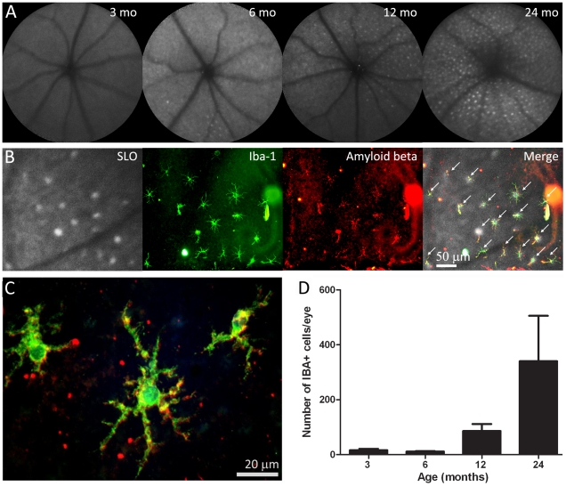
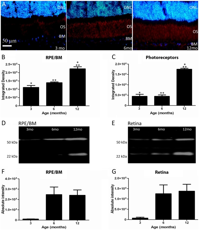
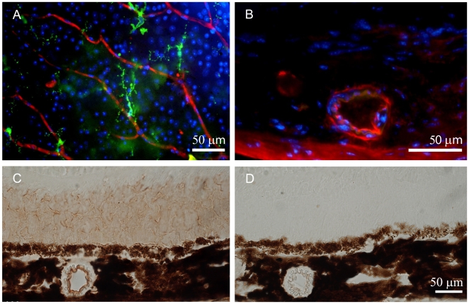
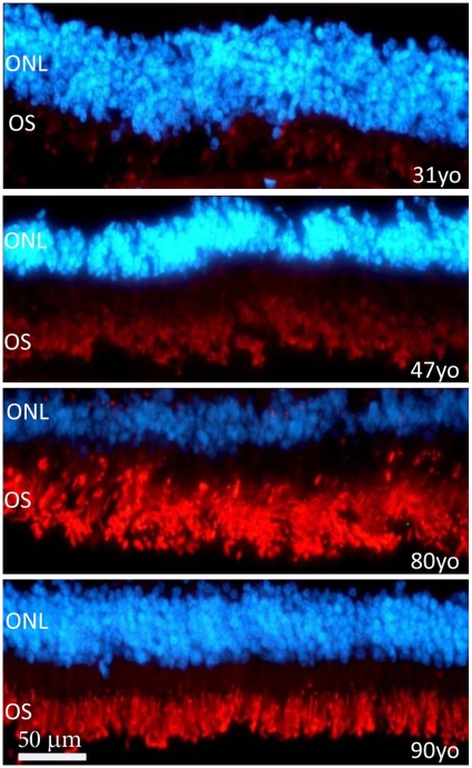
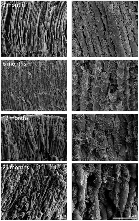
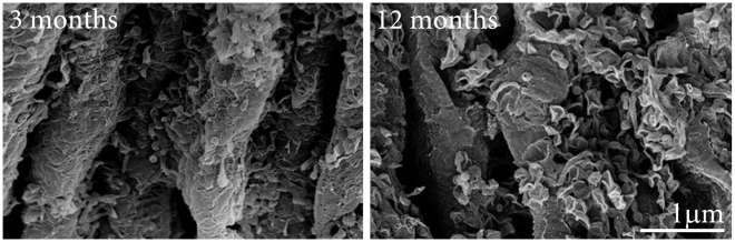
BACKGROUND: Amyloid beta (Aβ) accumulates in the ageing central nervous system and is associated with a number of age-related diseases, including age-related macular degeneration (AMD) in the eye. AMD is characterised by accumulation of extracellular deposits called drusen in which Aβ is a key constituent. Aβ activates the complement cascade and its deposition is associated with activated macrophages. So far, little is known about the quantitative measurements of Aβ accumulation and definitions of its relative sites of ocular deposition in the normal ageing mouse. METHODOLOGY/PRINCIPAL FINDINGS: We have traced Aβ accumulation quantitatively in the ageing mouse retina using immunohistochemistry and Western blot analysis. We reveal that it is not only deposited at Bruch's membrane and along blood vessels, but unexpectedly, it also coats photoreceptor outer segments. While Aβ is present at all sites of deposition from 3 months of age, it increases markedly from 6 months onward. Progressive accumulation of deposits on outer segments was confirmed with scanning electron microscopy, revealing age-related changes in their morphology. Such progress of accumulation of Aβ on photoreceptor outer segments with age was also confirmed in human retinae using immunohistochemistry. We also chart the macrophage response to increases in Aβ showing up-regulation in their numbers using both confocal laser imaging of the eye in vivo followed by in vitro immunostaining. With age macrophages become bloated with cellular debris including Aβ, however, their increasing numbers fail to stop Aβ accumulation. CONCLUSIONS: Increasing Aβ deposition in blood vessels and Bruch's membrane will impact upon retinal perfusion and clearance of cellular waste products from the outer retina, a region of very high metabolic activity. This accumulation of Aβ may contribute to the 30% reduction of photoreceptors found throughout life and the shortening of those that remain. The coating of Aβ on outer segments may also have an impact upon visual function with age.
Overview
The claims and statistics of this paper haven’t been checked yet.
Run the check — free, no account needed. It reads the open-access text and checks the sources it cites.
Integrity signals
Registry
Cites
Statistics
Figures
Verification
Claims the paper makes
The claims and statistics of this paper haven’t been checked yet.
Run the check — free, no account needed. It reads the open-access text and checks the sources it cites.
Claims other papers make about this one
Each is checked against this paper’s own text: does it say what the citing paper says it does?
In the citing paper
“With advancing age, the outer retina of the eye displays elevated inflammation and widespread deposition of extracellular debris, including amyloid species [55, 88].”
From Fecal microbiota transfer between young and aged mice reverses hallmarks of the aging gut, eye, and brain (2022) · cites this paper as [55]
Self-check
Percentages, sums and reported differences re-computed from the paper’s own numbers.
The self-check needs the paper’s full text.
Citations
Cites
1 retracted
RETRACTED ARTICLE: A specific amyloid-β protein assembly in the brain impairs memory
Lesné, S., Koh, M. T., Kotilinek, L., Kayed, R., Glabe, C. G., Yang, A., Gallagher, M., & Ashe, K. H. (2006) · Nature · doi:10.1038/nature04533 · Cited by 2,769 · cited 1×
“Much has been undertaken to study Aβ depositions in the brain of mice models of AD,”
BackgroundRetractedMicroglial Dysfunction and Defective β-Amyloid Clearance Pathways in Aging Alzheimer's Disease Mice
Hickman, S. E., Allison, E. K., & El Khoury, J. (2008) · The Journal of Neuroscience · doi:10.1523/jneurosci.0616-08.2008 · Cited by 1,329
Activation of microglial cells by β-amyloid protein and interferon-γ
Meda, L., Cassatella, M. A., Szendrei, G. I., Otvos, L., Baron, P., Villalba, M., Ferrari, D., & Rossi, F. (1995) · Nature · doi:10.1038/374647a0 · Cited by 1,314
Hageman, G. (2001) · Progress in Retinal and Eye Research · doi:10.1016/s1350-9462(01)00010-6 · Cited by 1,265
Leibowitz, H. M., Krueger, D. E., Maunder, L. R., Milton, R. C., KINI, M. M., Kahn, H. A., Nickerson, R. J., Pool, J., Colton, T., Ganley, J. P., et al. (1980) · PubMed · Cited by 1,250
The Five-year Incidence and Progression of Age-related Maculopathy
Klein, R., Klein, R., Jensen, S. C., & Meuer, S. M. (1997) · Ophthalmology · doi:10.1016/s0161-6420(97)30368-6 · Cited by 1,154
A role for local inflammation in the formation of drusen in the aging eye
Anderson, D. H., Mullins, R. F., Hageman, G. S., & Johnson, L. V. (2002) · American Journal of Ophthalmology · doi:10.1016/s0002-9394(02)01624-0 · Cited by 1,087
The epidemiology of age-related macular degeneration
Klein, R., Peto, T., Bird, A., & Vannewkirk, M. R. (2004) · American Journal of Ophthalmology · doi:10.1016/j.ajo.2003.11.069 · Cited by 932
Adult mouse astrocytes degrade amyloid-β in vitro and in situ
Wyss-Coray, T., Loike, J. D., Brionne, T. C., Lu, E., Anankov, R., Yan, F., Silverstein, S. C., & Husemann, J. (2003) · Nature Medicine · doi:10.1038/nm838 · Cited by 909
Ageing and degeneration in the macular region: A clinico-pathological study
Sarks, S. H. (1976) · British Journal of Ophthalmology · doi:10.1136/bjo.60.5.324 · Cited by 891
Research works printed but not linked: 1
- [58]Graymore CN 1969 General aspects of the metabolism of the retina. Davson H In the Eye. 2nd ed New York Academic Press 601 645
Cited by
Sorted by citations
Parker, A., Romano, S., Ansorge, R., Aboelnour, A., Le Gall, G., Savva, G. M., Pontifex, M. G., Telatin, A., Baker, D., Jones, E., et al. (2022) · Microbiome · doi:10.1186/s40168-022-01243-w · Cited by 368
Self-citationSpectral-Domain OCT Measurements in Alzheimer’s Disease
Chan, V. T. T., Sun, Z., Tang, S., Chen, L. J., Wong, A., Tham, C. C. Y., Wong, T. Y., Chen, C., Ikram, M. K., Whitson, H. E., et al. (2018) · Ophthalmology · doi:10.1016/j.ophtha.2018.08.009 · Cited by 368
Parallel findings in age-related macular degeneration and Alzheimer’s disease
Ohno-Matsui, K. (2011) · Progress in Retinal and Eye Research · doi:10.1016/j.preteyeres.2011.02.004 · Cited by 280
Miller, J. W. (2012) · American Journal of Ophthalmology · doi:10.1016/j.ajo.2012.10.018 · Cited by 254
Age related macular degeneration and drusen: Neuroinflammation in the retina
Buschini, E., Piras, A., Nuzzi, R., & Vercelli, A. (2011) · Progress in Neurobiology · doi:10.1016/j.pneurobio.2011.05.011 · Cited by 230
The Aging Eye: Common Degenerative Mechanisms Between the Alzheimer's Brain and Retinal Disease
Sivak, J. M. (2013) · Investigative Ophthalmology & Visual Science · doi:10.1167/iovs.12-10827 · Cited by 226
The Eye As a Biomarker for Alzheimer's Disease
Lim, J. K. H., Li, Q.-X., He, Z., Vingrys, A. J., Wong, V. H. Y., Currier, N. V., Mullen, J., Bui, B. V., & Nguyen, C. T. O. (2016) · Frontiers in Neuroscience · doi:10.3389/fnins.2016.00536 · Cited by 221
Begum, R., Powner, M. B., Hudson, N., Hogg, C. R., & Jeffery, G. (2013) · PLoS ONE · doi:10.1371/journal.pone.0057828 · Cited by 179
Self-citationGharbiya, M., Trebbastoni, A., Parisi, F., Manganiello, S., Cruciani, F., D’Antonio, F., Vico, U. D., Imbriano, L., Campanelli, A., & de Lena, C. (2014) · Journal of Alzheimer s Disease · doi:10.3233/jad-132039 · Cited by 174
Zanzottera, E. C., Messinger, J. D., Ach, T., Smith, R. T., Freund, K. B., & Curcio, C. A. (2015) · Investigative Opthalmology & Visual Science · doi:10.1167/iovs.15-16431 · Cited by 164
Past, present and future role of retinal imaging in neurodegenerative disease
Kashani, A. H., Asanad, S., Chan, J. W., Singer, M., Zhang, J., Sharifi, M., Khansari, M. M., Abdolahi, F., Shi, Y., Biffi, A., et al. (2021) · Progress in Retinal and Eye Research · doi:10.1016/j.preteyeres.2020.100938 · Cited by 169
Dementia of the eye: the role of amyloid beta in retinal degeneration
Ratnayaka, J. A., Serpell, L. C., & Lotery, A. J. (2015) · Eye · doi:10.1038/eye.2015.100 · Cited by 160
Grimaldi, A., Pediconi, N., Oieni, F., Pizzarelli, R., Rosito, M., Giubettini, M., Santini, T., Limatola, C., Ruocco, G., Ragozzino, D., et al. (2019) · Frontiers in Neuroscience · doi:10.3389/fnins.2019.00925 · Cited by 153
Liu, R. T., Gao, J., Cao, S., Sandhu, N., Cui, J. Z., Chou, C. L., Fang, E., & Matsubara, J. A. (2013) · Investigative Ophthalmology & Visual Science · doi:10.1167/iovs.12-10849 · Cited by 152
Pugazhendhi, A., Hubbell, M., Jairam, M. P., & Ambati, B. K. (2021) · International Journal of Molecular Sciences · doi:10.3390/ijms22031170 · Cited by 148
Gupta, V. B., Chitranshi, N., den Haan, J., Mirzaei, M., You, Y., Lim, J. K. H., Basavarajappa, D. K., Godinez, A., Angelantonio, S. D., Sachdev, P. S., et al. (2020) · Progress in Retinal and Eye Research · doi:10.1016/j.preteyeres.2020.100899 · Cited by 149
Amyloid-beta and tau protein beyond Alzheimer’s disease
Abyadeh, M., Gupta, V., Paulo, J. A., Mahmoudabad, A. G., Shadfar, S., Mirshahvaladi, S., Gupta, V. B., Nguyen, C. T. O., Finkelstein, D. I., You, Y., et al. (2023) · Neural Regeneration Research · doi:10.4103/1673-5374.386406 · Cited by 126
Choroidal Neovascularization: Mechanisms of Endothelial Dysfunction
Yeo, N. J. Y., Chan, E. J. J., & Cheung, C. (2019) · Frontiers in Pharmacology · doi:10.3389/fphar.2019.01363 · Cited by 120
Recharging mitochondrial batteries in old eyes. Near infra-red increases ATP
Gkotsi, D., Begum, R., Salt, T. E., Lascaratos, G., Hogg, C. R., Chau, K., Schapira, A. H. V., & Jeffery, G. (2014) · Experimental Eye Research · doi:10.1016/j.exer.2014.02.023 · Cited by 108
Self-citationLee, V., Rekhi, E., Kam, J. H., & Jeffery, G. (2012) · Neurobiology of Aging · doi:10.1016/j.neurobiolaging.2011.12.002 · Cited by 105
Self-citationNLRP3 Inflammasome: Activation and Regulation in Age‐Related Macular Degeneration
Gao, J., Liu, R. T., Cao, S., Cui, J. Z., Wang, A., To, E. C., & Matsubara, J. A. (2015) · Mediators of Inflammation · doi:10.1155/2015/690243 · Cited by 105
Stefanova, N. A., Kozhevnikova, O. S., Vitovtov, A. O., Maksimova, K. Y., Logvinov, S. V., Rudnitskaya, E. A., Korbolina, E. E., Muraleva, N. A., & Kolosova, N. G. (2014) · Cell Cycle · doi:10.4161/cc.28255 · Cited by 97
Kokkinopoulos, I., Colman, A., Hogg, C. R., Heckenlively, J., & Jeffery, G. (2012) · Neurobiology of Aging · doi:10.1016/j.neurobiolaging.2012.04.014 · Cited by 90
Self-citationGao, J., Cui, J. Z., To, E., Cao, S., & Matsubara, J. A. (2018) · Journal of Neuroinflammation · doi:10.1186/s12974-018-1062-3 · Cited by 83
Interplay between aging and other factors of the pathogenesis of age-related macular degeneration
Błasiak, J., Sobczuk, P., Pawłowska, E., & Kaarniranta, K. (2022) · Ageing Research Reviews · doi:10.1016/j.arr.2022.101735 · Cited by 86
Kam, J. H., Lenassi, E., Malik, T. H., Pickering, M. C., & Jeffery, G. (2013) · American Journal Of Pathology · doi:10.1016/j.ajpath.2013.04.008 · Cited by 82
Self-citationAmyloid Beta Immunoreactivity in the Retinal Ganglion Cell Layer of the Alzheimer’s Eye
Lee, S., Jiang, K., McIlmoyle, B. J., To, E. C., Xu, Q., Hirsch‐Reinshagen, V., Mackenzie, I. R., Hsiung, G. R., Eadie, B. D., Šarunic, M. V., et al. (2020) · Frontiers in Neuroscience · doi:10.3389/fnins.2020.00758 · Cited by 77
Interconnection Between Brain and Retinal Neurodegenerations
Jindal, V. (2014) · Molecular Neurobiology · doi:10.1007/s12035-014-8733-6 · Cited by 75
Elovanoids counteract oligomeric β-amyloid-induced gene expression and protect photoreceptors
Do, K. V., Kautzmann, M.-A. I., Jun, B., Gordon, W. C., Nshimiyimana, R., Yang, R., Petasis, N. A., & Bazan, N. G. (2019) · Proceedings of the National Academy of Sciences · doi:10.1073/pnas.1912959116 · Cited by 69
Gupta, V., Gupta, V. B., Chitranshi, N., Gangoda, S. V. S., Wall, R. V., Abbasi, M., Golzan, M., Dheer, Y., Shah, T. M., Avolio, A. P., et al. (2016) · Cellular and Molecular Life Sciences · doi:10.1007/s00018-016-2295-x · Cited by 70
Nizari, S., Guo, L., Davis, B. M., Normando, E. M., Galvão, J., Turner, L., Bizrah, M., Dehabadi, M. H., Tian, K., & Cordeiro, M. F. (2016) · Cell Death and Disease · doi:10.1038/cddis.2016.397 · Cited by 69
Cao, L., Wang, H., Wang, F., Xu, D., Liu, F., & Liu, C. (2013) · Investigative Ophthalmology & Visual Science · doi:10.1167/iovs.13-11612 · Cited by 68
Choroidal Thickness in Patients with Mild Cognitive Impairment and Alzheimer’s Type Dementia
Bulut, M., Yaman, A., Erol, M. K., Kurtuluş, F., Toslak, D., Doğan, B., Çoban, D. T., & Başar, E. K. (2016) · Journal of Ophthalmology · doi:10.1155/2016/7291257 · Cited by 66
Role of the choroid in age-related macular degeneration: A current review
Ebrahimi, K., & Farazdaghi, M. (2019) · Journal of Ophthalmic and Vision Research · doi:10.4103/jovr.jovr_125_18 · Cited by 70
Shiose, S., Chen, Y., Okano, K., Roy, S., Kohno, H., Tang, J. C. O., Pearlman, E. M., Maeda, T., Palczewski, K., & Maeda, A. (2011) · Journal of Biological Chemistry · doi:10.1074/jbc.m111.228551 · Cited by 66
Shah, T. M., Gupta, S. M., Chatterjee, P., Campbell, M., & Martins, R. N. (2017) · Molecular Psychiatry · doi:10.1038/mp.2016.251 · Cited by 62
Uchida, A., Pillai, J. A., Bermel, R. A., Bonner‐Jackson, A., Rae-Grant, A. D., Fernandez, H. H., Bena, J., Jones, S. E., Leverenz, J. B., Srivastava, S. K., et al. (2018) · Investigative Ophthalmology & Visual Science · doi:10.1167/iovs.17-23240 · Cited by 62
Salobrar‐García, E., Mendez-Hernandez, C. D., de Hoz, R., Ramirez, A. I., López‐Cuenca, I., Fernández‐Albarral, J. A., Rojas, P., Wang, S., García-Feijoo, J., Gil, P., et al. (2020) · Journal of Personalized Medicine · doi:10.3390/jpm10040231 · Cited by 57
Damage-Associated Molecular Patterns (DAMPs) in Retinal Disorders
Mahaling, B., Low, S. W. Y., Beck, M., Kumar, D., Ahmed, S., Connor, T. B., Ahmad, B. U., & Chaurasia, S. S. (2022) · International Journal of Molecular Sciences · doi:10.3390/ijms23052591 · Cited by 56
Attenuation of Choroidal Thickness in Patients With Alzheimer Disease
Trebbastoni, A., Marcelli, M., Mallone, F., D’Antonio, F., Imbriano, L., Campanelli, A., de Lena, C., & Gharbiya, M. (2016) · Alzheimer Disease & Associated Disorders · doi:10.1097/wad.0000000000000176 · Cited by 52
Hu, M. L., Quinn, J., & Xue, K. (2021) · Life · doi:10.3390/life11070635 · Cited by 51
In vivo Retinal Fluorescence Imaging With Curcumin in an Alzheimer Mouse Model
Sidiqi, A. M., Wahl, D. J., Lee, S., Ma, D., To, E., Cui, J. Z., To, E. C., Beg, M. F., Šarunic, M. V., & Matsubara, J. A. (2020) · Frontiers in Neuroscience · doi:10.3389/fnins.2020.00713 · Cited by 49
Pretorius, E., Mbotwe, S., & Kell, D. B. (2017) · Scientific Reports · doi:10.1038/s41598-017-09860-4 · Cited by 46
Kumar, A., Zhao, L., Fariss, R. N., McMenamin, P. G., & Wong, W. T. (2014) · Investigative Ophthalmology & Visual Science · doi:10.1167/iovs.13-13522 · Cited by 45
Calaza, K. D. C., Kam, J. H., Hogg, C. R., & Jeffery, G. (2015) · Neurobiology of Aging · doi:10.1016/j.neurobiolaging.2015.06.010 · Cited by 45
Self-citationTGF-β1 prevents rat retinal insult induced by amyloid-β (1–42) oligomers
Fisichella, V., Giurdanella, G., Platania, C. B. M., Romano, G. L., Leggio, G. M., Salomone, S., Drago, F., Caraci, F., & Bucolo, C. (2016) · European Journal of Pharmacology · doi:10.1016/j.ejphar.2016.02.002 · Cited by 45
Retinal biomarkers provide “insight” into cortical pharmacology and disease
Nguyen, C. T. O., Hui, F., Charng, J., Velaedan, S., van Koeverden, A. K., Lim, J. K. H., He, Z., Wong, V. H. Y., Vingrys, A. J., Bui, B. V., et al. (2017) · Pharmacology & Therapeutics · doi:10.1016/j.pharmthera.2017.02.009 · Cited by 47
Wang, J., Ohno‐Matsui, K., & Morita, I. (2012) · Biochemical and Biophysical Research Communications · doi:10.1016/j.bbrc.2012.05.085 · Cited by 46
Amyloidosis in Retinal Neurodegenerative Diseases
Masuzzo, A., Dinet, V., Cavanagh, C., Mascarelli, F., & Krantic, S. M. (2016) · Frontiers in Neurology · doi:10.3389/fneur.2016.00127 · Cited by 45
Reduction of amyloid-beta levels in mouse eye tissues by intra-vitreally delivered neprilysin
Parthasarathy, R., Chow, K. M., Derafshi, Z., Fautsch, M. P., Hetling, J. R., Rodgers, D. W., Hersh, L. B., & Pepperberg, D. R. (2015) · Experimental Eye Research · doi:10.1016/j.exer.2015.06.027 · Cited by 44
Zhao, T., Gao, J., Van, J., To, E. C., Wang, A., Cao, S., Cui, J. Z., Guo, J., Lee, M., McGeer, P. L., et al. (2015) · Journal of Neuroinflammation · doi:10.1186/s12974-015-0337-1 · Cited by 45
Markers of the ageing macrophage: a systematic review and meta-analysis
Moss, C. E., Phipps, H., Wilson, H. L., & Kiss‐Toth, E. (2023) · Frontiers in Immunology · doi:10.3389/fimmu.2023.1222308 · Cited by 42
Ratnayaka, J. A., Lynn, S. A., Keeling, E., Munday, R., Gabha, G., Griffiths, H., & Lotery, A. J. (2017) · Neural Regeneration Research · doi:10.4103/1673-5374.205083 · Cited by 41
Dry age-related macular degeneration: A currently unmet clinical need
Girmens, J. (2012) · Intractable & Rare Diseases Research · doi:10.5582/irdr.2012.v1.3.103 · Cited by 41
Ashraf, G., McGuinness, M. B., Khan, M. A., Obtinalla, C., Hadoux, X., & van Wijngaarden, P. (2023) · Alzheimer s & Dementia Diagnosis Assessment & Disease Monitoring · doi:10.1002/dad2.12421 · Cited by 42
Microglial changes in the early aging stage in a healthy retina and an experimental glaucoma model
Ramirez, A. I., Fernández‐Albarral, J. A., de Hoz, R., López‐Cuenca, I., Salobrar‐García, E., Rojas, P., Valiente‐Soriano, F. J., Avilés‐Trigueros, M., Villegas‐Pérez, M. P., Vidal‐Sanz, M., et al. (2020) · Progress in brain research · doi:10.1016/bs.pbr.2020.05.024 · Cited by 37
Biological basis for amyloidogenesis in Alzheimer’S disease
Андреева, Т. В., Lukiw, W. J., & Rogaev, E. I. (2017) · Biochemistry (Moscow) · doi:10.1134/s0006297917020043 · Cited by 38
Kozhevnikova, O. S., Korbolina, E. E., Stefanova, N. A., Muraleva, N. A., Orlov, Y. L., & Kolosova, N. G. (2013) · Biogerontology · doi:10.1007/s10522-013-9439-2 · Cited by 35
Melville, H., Carpiniello, M. F., Hollis, K., Staffaroni, A., & Golestaneh, N. (2013) · Journal of Translational Medicine · doi:10.1186/1479-5876-11-53 · Cited by 35
Prasad, T., Zhu, P., Verma, A., Chakrabarty, P., Rosario, A. M., Golde, T. E., & Li, Q. (2017) · Scientific Reports · doi:10.1038/s41598-017-03397-2 · Cited by 35
Age- and glaucoma-induced changes to the ocular glymphatic system
Wang, X., Delle, C., Peng, W., Plá, V., Giannetto, M. J., Kusk, P., Sigurdsson, B., Sakurai, S., Sweeney, A. M., Sun, Q., et al. (2023) · Neurobiology of Disease · doi:10.1016/j.nbd.2023.106322 · Cited by 34
Obata, R., & Yanagi, Y. (2014) · PLoS ONE · doi:10.1371/journal.pone.0091873 · Cited by 32
Paraoan, L., Sharif, U., Carlsson, E., Supharattanasitthi, W., Mahmud, N. M., Kamalden, T. A., Hiscott, P. S., Jackson, M. J., & Grierson, I. (2020) · Progress in Retinal and Eye Research · doi:10.1016/j.preteyeres.2020.100859 · Cited by 31
Parsons, C. G., Ruitenberg, M., Freitag, C. E., Sroka, K., Russ, H., & Rammes, G. (2015) · Neuropharmacology · doi:10.1016/j.neuropharm.2014.12.038 · Cited by 31
Translational opportunities for amyloid-targeting fluorophores
Cao, K., & Yang, J. (2018) · Chemical Communications · doi:10.1039/c8cc03619e · Cited by 32
Catchpole, I. R., Germaschewski, V., Kam, J. H., von Leithner, P. L., Ford, S., Gough, G., Adamson, P., Overend, P., Hilpert, J., López, F. J., et al. (2013) · PLoS ONE · doi:10.1371/journal.pone.0065518 · Cited by 29
Self-citationYeo, I. J., Lee, M. J., Baek, A., Miller, Z. C., Bhattarai, D., Baek, Y. M., Jeong, H. J., Kim, Y. K., Kim, D., Hong, J. T., et al. (2019) · Scientific Reports · doi:10.1038/s41598-019-54846-z · Cited by 29
Bastian, F. O., Elzer, P. H., & Wu, X. (2012) · Experimental and Molecular Pathology · doi:10.1016/j.yexmp.2012.04.017 · Cited by 28
Zhang, J., Gao, F., Ma, Y., Xue, T., & Shen, Y. (2021) · iScience · doi:10.1016/j.isci.2021.103327 · Cited by 28
Lei, L., Tzekov, R. T., Tang, S., & Kaushal, S. (2012) · PubMed Central · Cited by 28
Dong, Z., Li, J., Gan, Y., Sun, X., Leng, Y., & Ge, J. (2018) · International Journal of Ophthalmology · doi:10.18240/ijo.2018.05.06 · Cited by 27
Retinal Manifestations of Alzheimer’s Disease
Dehabadi, M. H., Davis, B. M., Wong, T. K. Y., & Cordeiro, M. F. (2014) · Neurodegenerative Disease Management · doi:10.2217/nmt.14.19 · Cited by 28
Fundamental differences in patterns of retinal ageing between primates and mice
Kam, J. H., Weinrich, T. W., Shinhmar, H., Powner, M. B., Roberts, N. W., Aboelnour, A., & Jeffery, G. (2019) · Scientific Reports · doi:10.1038/s41598-019-49121-0 · Cited by 27
Self-citationDinet, V., Bruban, J., Chalour, N., Maoui, A., An, N., Jonet, L., Buret, A., Béhar‐Cohen, F., Klein, C., Tréton, J., et al. (2012) · Aging Cell · doi:10.1111/j.1474-9726.2012.00834.x · Cited by 26
Optical coherence tomography reveals light-dependent retinal responses in Alzheimer’s disease
Bissig, D., Zhou, C. G., Le, V., & Bernard, J. T. (2020) · NeuroImage · doi:10.1016/j.neuroimage.2020.117022 · Cited by 27
Evidence of Synaptic and Neurochemical Remodeling in the Retina of Aging Degus
Chang, L., Ardiles, Á. O., Tapia‐Rojas, C., Araya, J., Inestrosa, N. C., Palacios, A. G., & Acosta, M. L. (2020) · Frontiers in Neuroscience · doi:10.3389/fnins.2020.00161 · Cited by 27
Taylor-Walker, G., Lynn, S. A., Keeling, E., Munday, R., Johnston, D. A., Page, A. M., Scott, J. A., Goverdhan, S., Lotery, A. J., & Ratnayaka, J. A. (2016) · Experimental Eye Research · doi:10.1016/j.exer.2016.10.013 · Cited by 24
Sen, S., Saxena, R., Vibha, D., Tripathi, M., Sharma, P. K., Phuljhele, S., Tandon, R., & Kumar, P. (2020) · Seminars in Ophthalmology · doi:10.1080/08820538.2020.1748203 · Cited by 24
High-resolution contrast-enhanced optical coherence tomography in mice retinae
Sen, D., SoRelle, E. D., Liba, O., Dalal, R., Paulus, Y. M., Kim, T.-W., Moshfeghi, D. M., & de la Zerda, A. (2016) · Journal of Biomedical Optics · doi:10.1117/1.jbo.21.6.066002 · Cited by 23
Akula, M., McNamee, S. M., Love, Z., Nasraty, N., Chan, N. P. M., Whalen, M., Avola, M. O., Olivares, A. M., Leehy, B. D., Jelcick, A. S., et al. (2024) · Gene Therapy · doi:10.1038/s41434-024-00455-z · Cited by 22
Topical cyclodextrin reduces amyloid beta and inflammation improving retinal function in ageing mice
Kam, J. H., Lynch, A., Begum, R., Cunea, A., & Jeffery, G. (2015) · Experimental Eye Research · doi:10.1016/j.exer.2015.03.023 · Cited by 21
Self-citationLoss of thioredoxin function in retinas of mice overexpressing amyloid β
Lamoke, F., Ripandelli, G., Webster, S. J., Montemari, A. L., Maraschi, A., Martin, P. M., Marcus, D. M., Liou, G. I., & Bartoli, M. (2012) · Free Radical Biology and Medicine · doi:10.1016/j.freeradbiomed.2012.04.010 · Cited by 20
Retinal hyperspectral imaging in the 5xFAD mouse model of Alzheimer’s disease
Lim, J. K. H., Li, Q., Ryan, T. M., Bedggood, P. A., Metha, A. B., Vingrys, A. J., Bui, B. V., & Nguyen, C. T. O. (2021) · Scientific Reports · doi:10.1038/s41598-021-85554-2 · Cited by 20
Oligomeric Aβ1-42 Induces an AMD-Like Phenotype and Accumulates in Lysosomes to Impair RPE Function
Lynn, S. A., Johnston, D. A., Scott, J. A., Munday, R., Desai, R. S., Keeling, E., Weaterton, R., Simpson, A. P., Davis, D., Freeman, T., et al. (2021) · Cells · doi:10.3390/cells10020413 · Cited by 19
Evaluation of choroidal thickness in prodromal Alzheimer’s disease defined by amyloid PET
López‐de‐Eguileta, A., Lage, C., López‐García, S., Pozueta, A., García‐Martínez, M., Kazimierczak, M., Bravo, M., de Arcocha-Torres, M., Banzo, I., Jiménez-Bonilla, J. F., et al. (2020) · PLoS ONE · doi:10.1371/journal.pone.0239484 · Cited by 21
Harvey, H., & Durant, S. (2014) · Experimental Brain Research · doi:10.1007/s00221-014-4078-7 · Cited by 16
Autophagy activated by SIRT6 regulates Aβ induced inflammatory response in RPEs
Feng, Y., Liang, J., Zhai, Y., Sun, J., Wang, J., She, X., Gu, Q., Liu, Y., Zhu, H., Luo, X., et al. (2018) · Biochemical and Biophysical Research Communications · doi:10.1016/j.bbrc.2018.01.159 · Cited by 16
Nettleton, J. A., & Salem, N. (2019) · Annals of Nutrition and Metabolism · doi:10.1159/000495906 · Cited by 17
El‐Darzi, N., Mast, N., Buchner, D. A., Saadane, A., Dailey, B., Trichonas, G., & Pikuleva, I. A. (2022) · Frontiers in Pharmacology · doi:10.3389/fphar.2022.902254 · Cited by 16
Jo, D. H., Cho, C. S., Kim, J. H., & Kim, J. H. (2020) · Neurobiology of Aging · doi:10.1016/j.neurobiolaging.2020.07.013 · Cited by 14
Naaman, E., Ya’ari, S., Itzkovich, C., Safuri, S., Macsi, F., Kellerman, L., Mimouni, M., Mann, I., Gazit, E., Adler‐Abramovich, L., et al. (2020) · Scientific Reports · doi:10.1038/s41598-020-77712-9 · Cited by 14
Kam, J. H., Morgan, J. E., & Jeffery, G. (2016) · Experimental Eye Research · doi:10.1016/j.exer.2016.07.002 · Cited by 14
Self-citationSerum Leptin and Age-Related Macular Degeneration
Seshasai, S., Liao, J., Toh, Q. C., Cheng, C., Cheung, C. M. G., Sethi, S. K., Wong, T. Y., & Sabanayagam, C. (2015) · Investigative Ophthalmology & Visual Science · doi:10.1167/iovs.14-15933 · Cited by 13
Ageing-associated phenotypes in mice
Xie, K., & Ehninger, D. (2023) · Mechanisms of Ageing and Development · doi:10.1016/j.mad.2023.111852 · Cited by 14
670 nm LED ameliorates inflammation in the CFH−/− mouse neural retina
Kokkinopoulos, I. (2013) · Journal of Photochemistry and Photobiology B Biology · doi:10.1016/j.jphotobiol.2013.03.003 · Cited by 12
Aboelnour, A., Kam, J. H., Elnasharty, M., Sayed-Ahmed, A., & Jeffery, G. (2016) · Experimental Eye Research · doi:10.1016/j.exer.2016.05.015 · Cited by 11
Self-citationPaolo, M. D., Corsi, F., Cerri, C., Bisti, S., Piano, I., & Gargini, C. (2023) · Pharmaceuticals · doi:10.3390/ph16091307 · Cited by 9
Rinaldi, M., Pezone, A., Quadrini, G. I., Abbadessa, G., Laezza, M. P., Passaro, M. L., Porcellini, A., & Costagliola, C. (2024) · Frontiers in Aging Neuroscience · doi:10.3389/fnagi.2024.1371745 · Cited by 9
Melatonin and abeta, macular degeneration and alzheimers disease: same disease, different outcomes?
Bajić, V., & Panfoli, I. (2012) · Medical Hypothesis Discovery & Innovation in Ophthalmology · Cited by 9
Retinal manifestations and their diagnostic significance in Alzheimer's disease
Abhyankar, S. D., Little, K., Stitt, A. W., & Bhatwadekar, A. D. (2025) · Journal of Alzheimer s Disease Reports · doi:10.1177/25424823251361937 · Cited by 8
Metadata
- Authors
- Hoh Kam, J., Lenassi, E., & Jeffery, G.
- Affiliations
- Ljubljana University Medical Centre (SI)
- University College London (GB)
- Corresponding author
- Jeffery, Glen
- ORCID
- Jeffery, Glen
- Venue
- PLoS ONE
- Publisher
- Public Library of Science (PLoS)
- Published
- 2010-10-01
- Volume
- 5
- Issue
- 10
- Pages
- e13127
- ISSN
- 1932-6203 (ISSN-L)
- PMC
- PMC2948519
- OpenAlex
- W1982572468
- Cited by
- 131
- References
- 65
- Citations by year
- 2026: 6 · 2025: 6 · 2024: 5 · 2023: 7 · 2022: 6 · 2021: 8 · 2020: 12 · 2019: 9 · 2018: 7 · 2017: 7 · 2016: 14 · 2015: 10 · 2014: 8 · 2013: 11 · 2012: 12
- FWCI
- 9.07
- Type
- journal article
- Language
- en
- Open access
- gold
- doi.org
- Locations
- PLoS ONE (publishedVersion, cc-by)
- PubMed Central (publishedVersion, cc-by)
- PubMed (publishedVersion)
- DOAJ (DOAJ: Directory of Open Access Journals) (submittedVersion, cc-by-sa)
- UCL Discovery (University College London) (submittedVersion)
- Funders
- Rosetrees Trust
- Topics
- Health Sciences › Medicine › Ophthalmology › Retinal Diseases and Treatments
- Health Sciences › Medicine › Radiology, Nuclear Medicine and Imaging › Retinal Imaging and Analysis
- Life Sciences › Neuroscience › Cellular and Molecular Neuroscience › Alzheimer's disease research and treatments
- Keywords
- amyloid-beta, retinal aging, age-related macular degeneration, drusen, photoreceptor outer segments, Bruch's membrane, retinal macrophages, complement activation, mouse retina, photoreceptor degeneration, retinal blood flow
- MeSH terms
- Aging, Animals, Humans, Immunohistochemistry, Macrophage Activation, Macrophages, Macular Degeneration, Retina, Blotting, Western, Up-Regulation, Amyloid beta-Peptides, Mice
About these checks
Registry, retraction, Cites and statistics results are computed, not judged, with one exception: a printed reference that no index lists is read once by a model before it counts as a citation error. Claims are extracted by a model and kept only when their quote appears verbatim in the text; each cited source is then checked, with a model’s help, for whether it says what the claim relies on. Those verdicts can be wrong — read the claim’s quote and the cited source before relying on one.
Statistics and arithmetic are recomputed, not judged.
Confidence bands are shown only for fields calibrated on our annotated test set. None is calibrated yet, so no band is shown.
What counts as a claim ›How we measure confidence ›How we check this ›
Are you an author, or do you see a problem with this page? Request a review
