

The DOI matches its Crossref record; 65 of the 65 references it cites were found in a registry or an index.
Viewing Ageing Eyes: Diverse Sites of Amyloid Beta Accumulation in the Ageing Mouse Retina and the Up-Regulation of Macrophages
Open accessAbstract
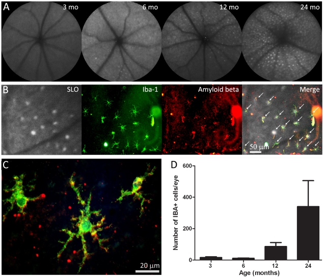
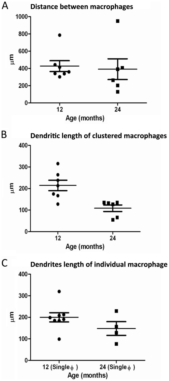
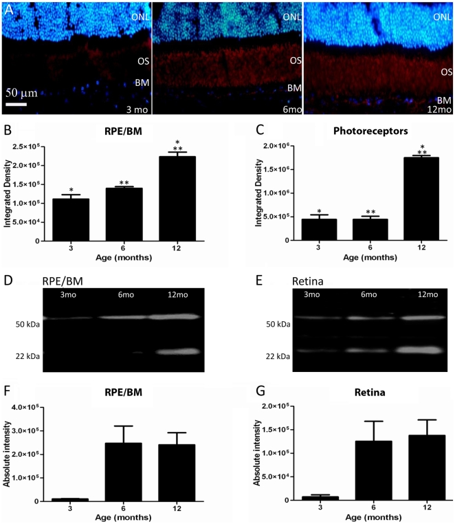
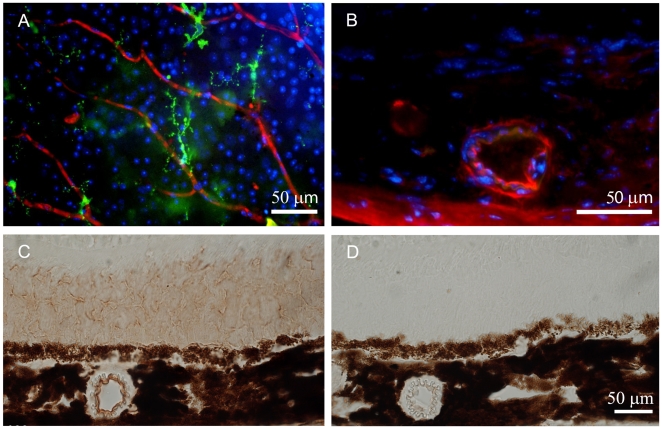
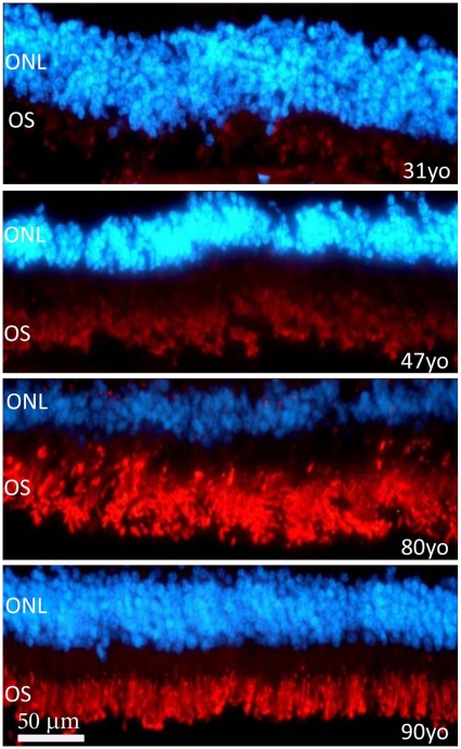
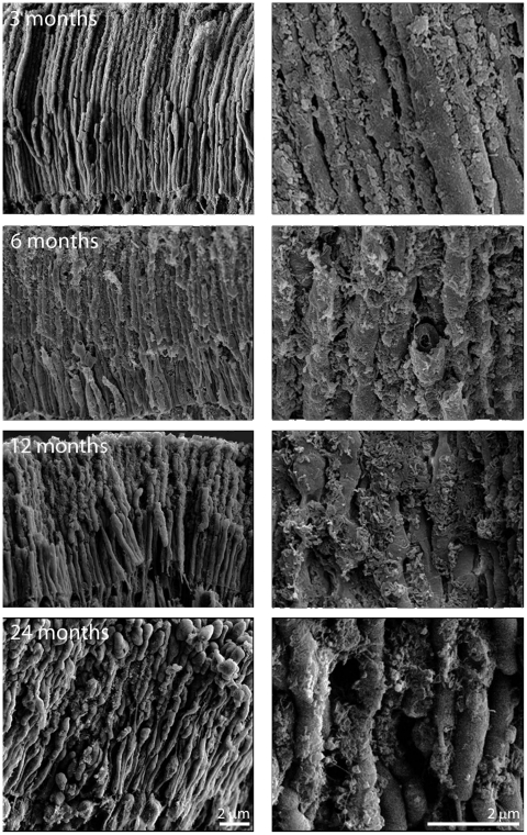
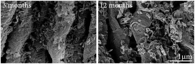
BACKGROUND: Amyloid beta (Aβ) accumulates in the ageing central nervous system and is associated with a number of age-related diseases, including age-related macular degeneration (AMD) in the eye. AMD is characterised by accumulation of extracellular deposits called drusen in which Aβ is a key constituent. Aβ activates the complement cascade and its deposition is associated with activated macrophages. So far, little is known about the quantitative measurements of Aβ accumulation and definitions of its relative sites of ocular deposition in the normal ageing mouse. METHODOLOGY/PRINCIPAL FINDINGS: We have traced Aβ accumulation quantitatively in the ageing mouse retina using immunohistochemistry and Western blot analysis. We reveal that it is not only deposited at Bruch's membrane and along blood vessels, but unexpectedly, it also coats photoreceptor outer segments. While Aβ is present at all sites of deposition from 3 months of age, it increases markedly from 6 months onward. Progressive accumulation of deposits on outer segments was confirmed with scanning electron microscopy, revealing age-related changes in their morphology. Such progress of accumulation of Aβ on photoreceptor outer segments with age was also confirmed in human retinae using immunohistochemistry. We also chart the macrophage response to increases in Aβ showing up-regulation in their numbers using both confocal laser imaging of the eye in vivo followed by in vitro immunostaining. With age macrophages become bloated with cellular debris including Aβ, however, their increasing numbers fail to stop Aβ accumulation. CONCLUSIONS: Increasing Aβ deposition in blood vessels and Bruch's membrane will impact upon retinal perfusion and clearance of cellular waste products from the outer retina, a region of very high metabolic activity. This accumulation of Aβ may contribute to the 30% reduction of photoreceptors found throughout life and the shortening of those that remain. The coating of Aβ on outer segments may also have an impact upon visual function with age.
Overview
The claims and statistics of this paper haven’t been checked yet.
Run the check — free, no account needed. It reads the open-access text and checks the sources it cites.
Integrity signals
Registry
Cites
Statistics
Figures
Verification
Claims the paper makes
The claims and statistics of this paper haven’t been checked yet.
Run the check — free, no account needed. It reads the open-access text and checks the sources it cites.
Claims other papers make about this one
Each is checked against this paper’s own text: does it say what the citing paper says it does?
In the citing paper
“With advancing age, the outer retina of the eye displays elevated inflammation and widespread deposition of extracellular debris, including amyloid species [55, 88].”
From Fecal microbiota transfer between young and aged mice reverses hallmarks of the aging gut, eye, and brain (2022) · cites this paper as [55]
Self-check
Percentages, sums and reported differences re-computed from the paper’s own numbers.
The self-check needs the paper’s full text.
Citations
Cites
1 retracted
RETRACTED ARTICLE: A specific amyloid-β protein assembly in the brain impairs memory
Lesné, S., Koh, M. T., Kotilinek, L., Kayed, R., Glabe, C. G., Yang, A., Gallagher, M., & Ashe, K. H. (2006) · Nature · doi:10.1038/nature04533 · Cited by 2,769 · cited 1×
“Much has been undertaken to study Aβ depositions in the brain of mice models of AD,”
BackgroundRetractedMicroglial Dysfunction and Defective β-Amyloid Clearance Pathways in Aging Alzheimer's Disease Mice
Hickman, S. E., Allison, E. K., & El Khoury, J. (2008) · The Journal of Neuroscience · doi:10.1523/jneurosci.0616-08.2008 · Cited by 1,329
Activation of microglial cells by β-amyloid protein and interferon-γ
Meda, L., Cassatella, M. A., Szendrei, G. I., Otvos, L., Baron, P., Villalba, M., Ferrari, D., & Rossi, F. (1995) · Nature · doi:10.1038/374647a0 · Cited by 1,314
Hageman, G. (2001) · Progress in Retinal and Eye Research · doi:10.1016/s1350-9462(01)00010-6 · Cited by 1,265
Leibowitz, H. M., Krueger, D. E., Maunder, L. R., Milton, R. C., KINI, M. M., Kahn, H. A., Nickerson, R. J., Pool, J., Colton, T., Ganley, J. P., et al. (1980) · PubMed · Cited by 1,250
The Five-year Incidence and Progression of Age-related Maculopathy
Klein, R., Klein, R., Jensen, S. C., & Meuer, S. M. (1997) · Ophthalmology · doi:10.1016/s0161-6420(97)30368-6 · Cited by 1,154
A role for local inflammation in the formation of drusen in the aging eye
Anderson, D. H., Mullins, R. F., Hageman, G. S., & Johnson, L. V. (2002) · American Journal of Ophthalmology · doi:10.1016/s0002-9394(02)01624-0 · Cited by 1,087
The epidemiology of age-related macular degeneration
Klein, R., Peto, T., Bird, A., & Vannewkirk, M. R. (2004) · American Journal of Ophthalmology · doi:10.1016/j.ajo.2003.11.069 · Cited by 932
Adult mouse astrocytes degrade amyloid-β in vitro and in situ
Wyss-Coray, T., Loike, J. D., Brionne, T. C., Lu, E., Anankov, R., Yan, F., Silverstein, S. C., & Husemann, J. (2003) · Nature Medicine · doi:10.1038/nm838 · Cited by 909
Ageing and degeneration in the macular region: A clinico-pathological study
Sarks, S. H. (1976) · British Journal of Ophthalmology · doi:10.1136/bjo.60.5.324 · Cited by 891
Complement activation by beta-amyloid in Alzheimer disease.
Rogers, J., Cooper, N. R., Webster, S., Schultz, J., McGeer, P. L., Styren, S. D., Civin, W. H., Brachova, L., Bradt, B., & Ward, P. (1992) · Proceedings of the National Academy of Sciences · doi:10.1073/pnas.89.21.10016 · Cited by 817
Curcio, C. A., Millican, C. L., Allen, K. A., & Kalina, R. E. (1993) · PubMed · Cited by 678
Combadière, C., Feumi, C., Raoul, W., Keller, N., Rodéro, M., Pézard, A., Lavalette, S., Houssier, M., Jonet, L., Picard, E., et al. (2007) · Journal of Clinical Investigation · doi:10.1172/jci31692 · Cited by 618
TGF-β1 promotes microglial amyloid-β clearance and reduces plaque burden in transgenic mice
Wyss-Coray, T., Lin, C., Yan, F., Yu, G.-Q., Rohde, M., McConlogue, L., Masliah, E., & Mucke, L. (2001) · Nature Medicine · doi:10.1038/87945 · Cited by 587
Aging human RPE: morphometric analysis of macular, equatorial, and peripheral cells.
Feeney-Burns, L., Hilderbrand, E. S., & Eldridge, S. (1984) · PubMed · Cited by 561
Retinal Abnormalities in Early Alzheimer’s Disease
Berisha, F., Feke, G. T., Trempe, C. L., McMeel, J. W., & Schepens, C. L. (2007) · Investigative Ophthalmology & Visual Science · doi:10.1167/iovs.06-1029 · Cited by 505
A Cell Surface Receptor Complex for Fibrillar β-Amyloid Mediates Microglial Activation
Bamberger, M. E., Harris, M. E., McDonald, D. R., Husemann, J., & Landreth, G. E. (2003) · The Journal of Neuroscience · doi:10.1523/jneurosci.23-07-02665.2003 · Cited by 501
Johnson, L. V., Leitner, W. P., Rivest, A. J., Staples, M. K., Radeke, M. J., & Anderson, D. H. (2002) · Proceedings of the National Academy of Sciences · doi:10.1073/pnas.192203399 · Cited by 447
Phagocytosis of Retinal Rod and Cone Photoreceptors
Kevany, B. M., & Palczewski, K. (2010) · Physiology · doi:10.1152/physiol.00038.2009 · Cited by 426 · cited 2×
“This may be indicative of less efficient patterns of phagocytosis by RPE cells, which remove the end of the photoreceptors daily to compensate for the addition of new photoreceptor disks at the outer segments base [34,35].”
BackgroundMicroglia Mediate the Clearance of Soluble Aβ through Fluid Phase Macropinocytosis
Mandrekar, S., Jiang, Q., Lee, C. Y. D., Koenigsknecht-Talboo, J., Holtzman, D. M., & Landreth, G. E. (2009) · The Journal of Neuroscience · doi:10.1523/jneurosci.5572-08.2009 · Cited by 415
İşeri, P., Altıntaş, Ö., Tokay, T., & Yüksel, N. (2006) · Journal of Neuro-Ophthalmology · doi:10.1097/01.wno.0000204645.56873.26 · Cited by 402
Lipofuscin: Mechanisms of formation and increase with age
TERMAN, A., & BRUNK, U. T. (1998) · APMIS · doi:10.1111/j.1699-0463.1998.tb01346.x · Cited by 387
THE RENEWAL OF ROD AND CONE OUTER SEGMENTS IN THE RHESUS MONKEY
Young, R. W. (1971) · The Journal of Cell Biology · doi:10.1083/jcb.49.2.303 · Cited by 375
Bressler, S. B. (1990) · Archives of Ophthalmology · doi:10.1001/archopht.1990.01070120090035 · Cited by 325
Anderson, D. H., Talaga, K. C., Rivest, A. J., Barron, E., Hageman, G. S., & Johnson, L. V. (2004) · Experimental Eye Research · doi:10.1016/j.exer.2003.10.011 · Cited by 319
Histopathology of Age-Related Macular Degeneration
Lim, J. I. (2007) · CRC Press eBooks · doi:10.3109/9781420019865-6 · Cited by 310
Amyloid-β Deposits Lead to Retinal Degeneration in a Mouse Model of Alzheimer Disease
Ning, A., Cui, J., To, E., Ashe, K. H., & Matsubara, J. (2008) · Investigative Opthalmology & Visual Science · doi:10.1167/iovs.08-1849 · Cited by 303
Epidemiology of Age-Related Macular Degeneration
Klein, R. (2005) · Springer eBooks · doi:10.1007/3-540-26977-0_5 · Cited by 287
Lipofuscin and Aging: A Matter of Toxic Waste
Gray, D. A., & Woulfe, J. (2005) · Science of Aging Knowledge Environment · doi:10.1126/sageke.2005.5.re1 · Cited by 279
Xu, H., Chen, M., Manivannan, A., Lois, N., & Forrester, J. V. (2008) · Aging Cell · doi:10.1111/j.1474-9726.2007.00351.x · Cited by 277
Dentchev, T., Milam, A. H., Lee, V. M. Y., Trojanowski, J. Q., & Dunaief, J. L. (2003) · American Journal of Ophthalmology · doi:10.1016/s0002-9394(03)00804-3 · Cited by 258
Dentchev, T., Milam, A. H., Lee, V. M. ‐., Trojanowski, J. Q., & Dunaief, J. L. (2003) · PubMed · Cited by 252
Perez, S. E., Lumayag, S., Kovacs, B., Mufson, E. J., & Xu, S. (2009) · Investigative Ophthalmology & Visual Science · doi:10.1167/iovs.08-2384 · Cited by 236
Assessment of Visual Impairment in Patients With Alzheimer's Disease
Sadun, A. A., Borchert, M., DeVita, E., Hinton, D. R., & Bassi, C. J. (1987) · American Journal of Ophthalmology · doi:10.1016/0002-9394(87)90001-8 · Cited by 224
Ma, W., Zhao, L., Fontainhas, A. M., Fariss, R. N., & Wong, W. T. (2009) · PLoS ONE · doi:10.1371/journal.pone.0007945 · Cited by 222
Boissonneault, V., Filali, M., Lessard, M., Relton, J., Wong, G., & Rivest, S. (2008) · Brain · doi:10.1093/brain/awn331 · Cited by 221
DRUSEN AND THEIR RELATIONSHIP TO SENILE MACULAR DEGENERATION
Sarks, S. H. (1980) · Australian and New Zealand Journal of Ophthalmology · doi:10.1111/j.1442-9071.1980.tb01670.x · Cited by 219
The microglial phagocytic role with specific plaque types in the Alzheimer disease brain
D’Andrea, M. R., Cole, G. M., & Ard, M. D. (2004) · Neurobiology of Aging · doi:10.1016/j.neurobiolaging.2003.12.026 · Cited by 215
Luhmann, U. F. O., Robbie, S. J., Munro, P., Barker, S. E., Durán, Y., Luong, V., Fitzke, F. W., Bainbridge, J. W., Ali, R. R., & MacLaren, R. E. (2009) · Investigative Ophthalmology & Visual Science · doi:10.1167/iovs.09-3462 · Cited by 212
Drusen as Risk Factors in Age-Related Macular Disease
Pauleikhoff, D., Barondes, M. J., Minassian, D. C., Chisholm, I. H., & Bird, A. C. (1990) · American Journal of Ophthalmology · doi:10.1016/s0002-9394(14)75576-x · Cited by 192
Ophthalmologic manifestations of Alzheimer's disease
Katz, B., & Rimmer, S. (1989) · Survey of Ophthalmology · doi:10.1016/0039-6257(89)90127-6 · Cited by 183
Soluble and Mature Amyloid Fibrils in Drusen Deposits
Isas, J. M., Luibl, V., Johnson, L. V., Kayed, R., Wetzel, R. B., Glabe, C. G., Langen, R., & Chen, J. (2010) · Investigative Ophthalmology & Visual Science · doi:10.1167/iovs.09-4207 · Cited by 159
Structure–function analysis of rods and cones in juvenile, adult, and aged C57BL/6 and Balb/c mice
GRESH, J., Goletz, P. W., Crouch, R. K., & Rohrer, B. (2003) · Visual Neuroscience · doi:10.1017/s0952523803202108 · Cited by 145
Amyloid precursor protein processing and retinal pathology in mouse models of Alzheimer’s disease
Dutescu, R. M., Li, Q., Crowston, J. G., Masters, C. L., Baird, P. N., & Culvenor, J. G. (2009) · Graefe s Archive for Clinical and Experimental Ophthalmology · doi:10.1007/s00417-009-1060-3 · Cited by 139
Medrano, C. J., & Fox, D. A. (1995) · Experimental Eye Research · doi:10.1016/s0014-4835(05)80122-8 · Cited by 125
Amyloid‐β(1‐42) alters structure and function of retinal pigmented epithelial cells
Bruban, J., Glotin, A., Dinet, V., Chalour, N., Sennlaub, F., Jonet, L., An, N., Faussat, A. M., & Mascarelli, F. (2009) · Aging Cell · doi:10.1111/j.1474-9726.2009.00456.x · Cited by 118
Coimbra, A., & de Andrade, C. A. F. G. (1971) · Brain · doi:10.1093/brain/94.2.207 · Cited by 111
Amyloid enhancing factor-loaded macrophages in amyloid fibril formation.
Shirahama, T., Miura, K., Ju, S., Kisilevsky, R., Gruys, E., & Cohen, A. S. (1990) · PubMed · Cited by 105
Histopathology of age-related macular degeneration
Kang, S. J., & Grossniklaus, H. E. (2012) · CRC Press eBooks · doi:10.1201/b14548-2 · Cited by 99
The influence of age on the spatial and temporal contrast sensitivity function
Wright, C. E., & Drasdo, N. (1985) · Documenta Ophthalmologica · doi:10.1007/bf00159172 · Cited by 95
Liesegang, T. J. (2002) · American Journal of Ophthalmology · doi:10.1016/s0002-9394(02)01493-9 · Cited by 94
The amyloid beta-protein of Alzheimer's disease is chemotactic for mononuclear phagocytes
Davis, J. B., McMurray, H. F., & Schubert, D. (1992) · Biochemical and Biophysical Research Communications · doi:10.1016/0006-291x(92)92317-q · Cited by 93
COIMBRA, A., & ANDRADE, C. (1971) · Brain · doi:10.1093/brain/94.2.199 · Cited by 78
Mureşan, V., Varvel, N. H., Lamb, B. T., & Muresan, Z. L. (2009) · Journal of Neuroscience · doi:10.1523/jneurosci.2558-08.2009 · Cited by 72
Raoul, W., Feumi, C., Keller, N., Lavalette, S., Houssier, M., Béhar‐Cohen, F., Combadière, C., & Sennlaub, F. (2008) · Ophthalmic Research · doi:10.1159/000119860 · Cited by 61
Takechi, R., Galloway, S., Pallebage‐Gamarallage, M. M., Wellington, C. L., Johnsen, R. D., & Mamo, J. C. L. (2009) · Histochemistry and Cell Biology · doi:10.1007/s00418-009-0567-3 · Cited by 51
Cunea, A., & Jeffery, G. (2007) · Visual Neuroscience · doi:10.1017/s0952523807070204 · Cited by 51
Self-citationSatoh, A. K., Tokunaga, F., Kawamura, S., & Ozaki, K. (1998) · Trends in Cell Biology · doi:10.1016/0962-8924(93)80020-p · Cited by 48
Jackson, G. R., McGwin, G., Phillips, J. M., Klein, R., & Owsley, C. J. (2004) · Investigative Ophthalmology & Visual Science · doi:10.1167/iovs.04-0019 · Cited by 47
β-Amyloid(25–35) induces the production of interleukin-8 from human monocytes
Meda, L., Bonaiuto, C., Szendrei, G. I., Ceska, M., Rossi, F., & Cassatella, M. A. (1995) · Journal of Neuroimmunology · doi:10.1016/0165-5728(95)00021-s · Cited by 44
Lewis, H., Straatsma, B. R., & Foos, R. Y. (1986) · Ophthalmology · doi:10.1016/s0161-6420(86)33615-7 · Cited by 39
Jackson, G. R., McGwin, G., Phillips, J. M., Klein, R., & Owsley, C. J. (2005) · Vision Research · doi:10.1016/j.visres.2005.09.003 · Cited by 37
Optometric management of patients with Alzheimer's disease.
Baker, D. R., Mendez, M. F., Townsend, J. C., Ilsen, P. F., & Bright, D. C. (1997) · PubMed · Cited by 26
Sergott, R. C. (2007) · Yearbook of Ophthalmology · doi:10.1016/s0084-392x(08)70160-6 · Cited by 16
Histochemically Recognizable Changes of Amyloid Fibrillogenic Macrophages
Shirahama, T., Miura, K., Ju, S., Cohen, A. S., Gruys, E., & Kisilevsky, R. (1991) · Springer eBooks · doi:10.1007/978-94-011-3284-8_130 · Cited by 2
Research works printed but not linked: 1
- [58]Graymore CN 1969 General aspects of the metabolism of the retina. Davson H In the Eye. 2nd ed New York Academic Press 601 645
Cited by
Sorted by citations
Parker, A., Romano, S., Ansorge, R., Aboelnour, A., Le Gall, G., Savva, G. M., Pontifex, M. G., Telatin, A., Baker, D., Jones, E., et al. (2022) · Microbiome · doi:10.1186/s40168-022-01243-w · Cited by 368
Self-citationSpectral-Domain OCT Measurements in Alzheimer’s Disease
Chan, V. T. T., Sun, Z., Tang, S., Chen, L. J., Wong, A., Tham, C. C. Y., Wong, T. Y., Chen, C., Ikram, M. K., Whitson, H. E., et al. (2018) · Ophthalmology · doi:10.1016/j.ophtha.2018.08.009 · Cited by 368
Parallel findings in age-related macular degeneration and Alzheimer’s disease
Ohno-Matsui, K. (2011) · Progress in Retinal and Eye Research · doi:10.1016/j.preteyeres.2011.02.004 · Cited by 280
Miller, J. W. (2012) · American Journal of Ophthalmology · doi:10.1016/j.ajo.2012.10.018 · Cited by 254
Age related macular degeneration and drusen: Neuroinflammation in the retina
Buschini, E., Piras, A., Nuzzi, R., & Vercelli, A. (2011) · Progress in Neurobiology · doi:10.1016/j.pneurobio.2011.05.011 · Cited by 230
Metadata
- Authors
- Hoh Kam, J., Lenassi, E., & Jeffery, G.
- Affiliations
- Ljubljana University Medical Centre (SI)
- University College London (GB)
- Corresponding author
- Jeffery, Glen
- ORCID
- Jeffery, Glen
- Venue
- PLoS ONE
- Publisher
- Public Library of Science (PLoS)
- Published
- 2010-10-01
- Volume
- 5
- Issue
- 10
- Pages
- e13127
- ISSN
- 1932-6203 (ISSN-L)
- PMC
- PMC2948519
- OpenAlex
- W1982572468
- Cited by
- 131
- References
- 65
- Citations by year
- 2026: 6 · 2025: 6 · 2024: 5 · 2023: 7 · 2022: 6 · 2021: 8 · 2020: 12 · 2019: 9 · 2018: 7 · 2017: 7 · 2016: 14 · 2015: 10 · 2014: 8 · 2013: 11 · 2012: 12
- FWCI
- 9.07
- Type
- journal article
- Language
- en
- Open access
- gold
- doi.org
- Locations
- PLoS ONE (publishedVersion, cc-by)
- PubMed Central (publishedVersion, cc-by)
- PubMed (publishedVersion)
- DOAJ (DOAJ: Directory of Open Access Journals) (submittedVersion, cc-by-sa)
- UCL Discovery (University College London) (submittedVersion)
- Funders
- Rosetrees Trust
- Topics
- Health Sciences › Medicine › Ophthalmology › Retinal Diseases and Treatments
- Health Sciences › Medicine › Radiology, Nuclear Medicine and Imaging › Retinal Imaging and Analysis
- Life Sciences › Neuroscience › Cellular and Molecular Neuroscience › Alzheimer's disease research and treatments
- Keywords
- amyloid-beta, retinal aging, age-related macular degeneration, drusen, photoreceptor outer segments, Bruch's membrane, retinal macrophages, complement activation, mouse retina, photoreceptor degeneration, retinal blood flow
- MeSH terms
- Aging, Animals, Humans, Immunohistochemistry, Macrophage Activation, Macrophages, Macular Degeneration, Retina, Blotting, Western, Up-Regulation, Amyloid beta-Peptides, Mice
About these checks
Registry, retraction, Cites and statistics results are computed, not judged, with one exception: a printed reference that no index lists is read once by a model before it counts as a citation error. Claims are extracted by a model and kept only when their quote appears verbatim in the text; each cited source is then checked, with a model’s help, for whether it says what the claim relies on. Those verdicts can be wrong — read the claim’s quote and the cited source before relying on one.
Statistics and arithmetic are recomputed, not judged.
Confidence bands are shown only for fields calibrated on our annotated test set. None is calibrated yet, so no band is shown.
What counts as a claim ›How we measure confidence ›How we check this ›
Are you an author, or do you see a problem with this page? Request a review
