

The DOI matches its Crossref record; 60 of the 60 references it cites were found in a registry or an index.
Imaging of Functional Connectivity in the Mouse Brain
Open accessAbstract
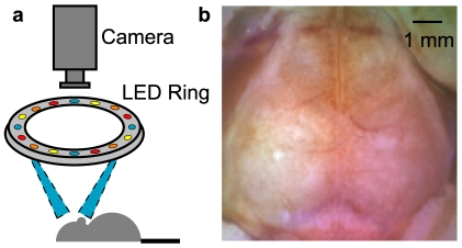
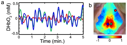
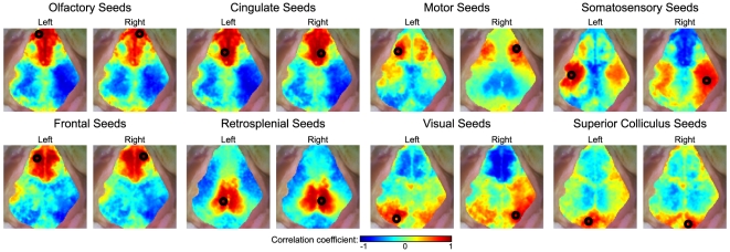
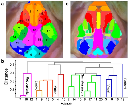
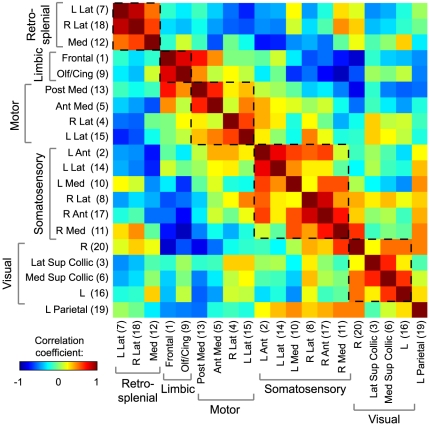
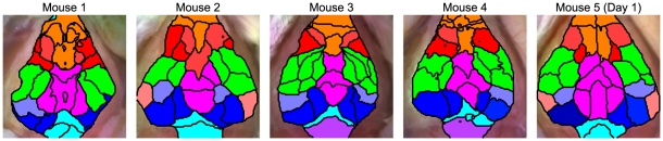
Functional neuroimaging (e.g., with fMRI) has been difficult to perform in mice, making it challenging to translate between human fMRI studies and molecular and genetic mechanisms. A method to easily perform large-scale functional neuroimaging in mice would enable the discovery of functional correlates of genetic manipulations and bridge with mouse models of disease. To satisfy this need, we combined resting-state functional connectivity mapping with optical intrinsic signal imaging (fcOIS). We demonstrate functional connectivity in mice through highly detailed fcOIS mapping of resting-state networks across most of the cerebral cortex. Synthesis of multiple network connectivity patterns through iterative parcellation and clustering provides a comprehensive map of the functional neuroarchitecture and demonstrates identification of the major functional regions of the mouse cerebral cortex. The method relies on simple and relatively inexpensive camera-based equipment, does not require exogenous contrast agents and involves only reflection of the scalp (the skull remains intact) making it minimally invasive. In principle, fcOIS allows new paradigms linking human neuroscience with the power of molecular/genetic manipulations in mouse models.
Overview
The claims and statistics of this paper haven’t been checked yet.
Run the check — free, no account needed. It reads the open-access text and checks the sources it cites.
Integrity signals
Registry
Cites
Statistics
Figures
Verification
Claims the paper makes
The claims and statistics of this paper haven’t been checked yet.
Run the check — free, no account needed. It reads the open-access text and checks the sources it cites.
Claims other papers make about this one
Each is checked against this paper’s own text: does it say what the citing paper says it does?
The cited source says what the claim attributes to it.
In the citing paper
“We have shown previously that fcOIS is a sensitive imaging modality capable of measuring functional connectivity between brain regions in healthy mice (White et al., 2011;Bero et al., 2012)”
From Anti-ApoE Antibody Given after Plaque Onset Decreases Aβ Accumulation and Improves Brain Function in a Mouse Model of Aβ Amyloidosis (2014) · cites this paper as White et al., 2011
The cited source says what the claim attributes to it.
In the citing paper
“Compared with PBS-treated mice, bilateral functional connectivity in the HJ6.3 group appeared to be more globally restored and to resemble that of healthy, wild-type mice (Fig. 7C), as described previously (White et al., 2011;Bero et al., 2012).”
From Anti-ApoE Antibody Given after Plaque Onset Decreases Aβ Accumulation and Improves Brain Function in a Mouse Model of Aβ Amyloidosis (2014) · cites this paper as White et al., 2011
The check wasn’t confident either way, so it gives no verdict — never disputed.
In the citing paper
“and that there is functional disruption in APP/PS1 mice after A deposition (White et al., 2011;Bero et al., 2012).”
From Anti-ApoE Antibody Given after Plaque Onset Decreases Aβ Accumulation and Improves Brain Function in a Mouse Model of Aβ Amyloidosis (2014) · cites this paper as White et al., 2011
Experimental — accuracy not yet measured
Self-check
Percentages, sums and reported differences re-computed from the paper’s own numbers.
The self-check needs the paper’s full text.
Citations
Cites
The Mouse Brain in Stereotaxic Coordinates
Paxinos, G. (2001) · Cited by 12,859
Functional connectivity in the motor cortex of resting human brain using echo‐planar mri
Biswal, B., Zerrin Yetkin, F., Haughton, V. M., & Hyde, J. S. (1995) · Magnetic Resonance in Medicine · doi:10.1002/mrm.1910340409 · Cited by 9,812
The Brain's Default Network: Anatomy, Function, and Relevance to Disease
Buckner, R. L., Andrews‐Hanna, J. R., & Schacter, D. L. (2008) · Annals of the New York Academy of Sciences · doi:10.1196/annals.1440.011 · Cited by 9,761
The human brain is intrinsically organized into dynamic, anticorrelated functional networks
Fox, M. D., Snyder, A. Z., Vincent, J. L., Corbetta, M., Van Essen, D. C., & Raichle, M. E. (2005) · Proceedings of the National Academy of Sciences · doi:10.1073/pnas.0504136102 · Cited by 8,553
Self-citationSpontaneous fluctuations in brain activity observed with functional magnetic resonance imaging
Fox, M. D., & Raichle, M. E. (2007) · Nature Reviews Neuroscience · doi:10.1038/nrn2201 · Cited by 6,700
Functional connectivity in the resting brain: a network analysis of the default mode hypothesis
Greicius, M. D., Krasnow, B., Reiss, A. L., & Menon, V. (2003) · Proceedings of the National Academy of Sciences · doi:10.1073/pnas.0135058100 · Cited by 6,358
Talairach, J., Tournoux, P., & Rayport, M. (1988) · Thieme eBooks · Cited by 5,058
Buckner, R. L., Sepulcre, J., Talukdar, T., Krienen, F. M., Liu, H., Hedden, T., Andrews-Hanna, J. R., Sperling, R. A., & Johnson, K. A. (2009) · The Journal of Neuroscience · doi:10.1523/jneurosci.5062-08.2009 · Cited by 2,862
Neurodegenerative Diseases Target Large-Scale Human Brain Networks
Seeley, W. W., Crawford, R. K., Zhou, J., Miller, B. L., & Greicius, M. D. (2009) · Neuron · doi:10.1016/j.neuron.2009.03.024 · Cited by 2,350
Resting-State Functional Connectivity Reflects Structural Connectivity in the Default Mode Network
Greicius, M. D., Supekar, K., Menon, V., & Dougherty, R. F. (2009) · Cerebral Cortex · doi:10.1093/cercor/bhn059 · Cited by 2,175
Research works printed but not linked: 5
- A Devor, 2003, Coupling of total hemoglobin concentration, oxygenation, and neural activity in rat somatosensory cortex., Neuron, 39, 353, 10.1016/S0896-6273(03)00403-3 · DOI · Paper page
- A Devor, 2005, Coupling of the cortical hemodynamic response to cortical and thalamic neuronal activity., PNAS, 102, 3822, 10.1073/pnas.0407789102 · DOI
- MD Fox, 2007, Spontaneous fluctuations in brain activity observed with functional magnetic resonance imaging., Nature Reviews Neuroscience, 8, 700, 10.1038/nrn2201 · DOI · Paper page
- MD Fox, 2009, The Global Signal and Observed Anticorrelated Resting State Brain Networks., Journal of Neurophysiology, 101, 3270, 10.1152/jn.90777.2008 · DOI · Paper page
- AK Dunn, 2005, Spatial extent of oxygen metabolism and hemodynamic changes during functional activation of the rat somatosensory cortex., NeuroImage, 27, 279, 10.1016/j.neuroimage.2005.04.024 · DOI · Paper page
Cited by
Sorted by citations
Items 101–200 of 252
Optics based biomedical imaging: Principles and applications
Wang, D., & Xia, J. (2019) · Journal of Applied Physics · doi:10.1063/1.5088490 · Cited by 22
Vazquez, A. L., Murphy, M. C., & Kim, S. (2014) · Brain Connectivity · doi:10.1089/brain.2014.0276 · Cited by 21
Altered resting-state functional connectivity after cortical spreading depression in mice
Li, B., Zhou, F., Luo, Q., & Li, P. (2012) · NeuroImage · doi:10.1016/j.neuroimage.2012.08.024 · Cited by 21
Mesoscale brain explorer, a flexible python-based image analysis and visualization tool
Haupt, D., Vanni, M. P., Bolaños, F., Mitelut, C., LeDue, J. M., & Murphy, T. H. (2017) · Neurophotonics · doi:10.1117/1.nph.4.3.031210 · Cited by 21
Bertoglio, D., Jonckers, E., Ali, I. Y., Verhoye, M., der Linden, A. V., & Dedeurwaerdere, S. (2019) · Neurobiology of Disease · doi:10.1016/j.nbd.2019.02.012 · Cited by 21
Mapping of language and motor function during awake neurosurgery with intraoperative optical imaging
Oelschlägel, M., Meyer, T., Morgenstern, U., Wahl, H., Gerber, J. C., Reiß, G., Koch, E., Steiner, G. R., Kirsch, M., Schackert, G., et al. (2020) · Neurosurgical FOCUS · doi:10.3171/2019.11.focus19759 · Cited by 21
Network analysis of neuroimaging in mice
Scharwächter, L., Schmitt, F. J., Pallast, N., Fink, G. R., & Aswendt, M. (2022) · NeuroImage · doi:10.1016/j.neuroimage.2022.119110 · Cited by 22
Open-source statistical and data processing tools for wide-field optical imaging data in mice
Brier, L. M., & Culver, J. P. (2023) · Neurophotonics · doi:10.1117/1.nph.10.1.016601 · Cited by 20
Self-citationRecent progress in translational research on neurovascular and neurodegenerative disorders
Demuth, H., Dijkhuizen, R. M., Farr, T. D., Gelderblom, M., Horsburgh, K. J., Iadecola, C., McLeod, D. D., Michalski, D., Murphy, T. H., Orbe, J., et al. (2017) · Restorative Neurology and Neuroscience · doi:10.3233/rnn-160690 · Cited by 20
Self-citationThe Brain Network in a Model of Thalamocortical Dysrhythmia
Zobeiri, M., van Luijtelaar, G., Budde, T., & Sysoev, I. V. (2018) · Brain Connectivity · doi:10.1089/brain.2018.0621 · Cited by 20
Liou, J.-Y., Baird-Daniel, E., Zhao, M., Daniel, A. G. S., Schevon, C. A., Ma, H., & Schwartz, T. H. (2019) · Brain · doi:10.1093/brain/awz246 · Cited by 20
White, B. R., Padawer-Curry, J. A., Cohen, A. S., Licht, D. J., & Yodh, A. G. (2019) · Biomedical Optics Express · doi:10.1364/boe.10.005952 · Cited by 20
Self-citationPalanisamy, A., Giri, T. K., Jiang, J., Bice, A. R., Quirk, J. D., Conyers, S. B., Maloney, S. E., Raghuraman, N., Bauer, A. Q., Garbow, J. R., et al. (2020) · JCI Insight · doi:10.1172/jci.insight.133172 · Cited by 20
Self-citationResting-state functional connectivity imaging of the mouse brain using photoacoustic tomography
Avanaki, K., Xia, J., Wan, H., Bauer, A. Q., Culver, J. P., & Wang, L. V. (2014) · Proceedings of SPIE, the International Society for Optical Engineering/Proceedings of SPIE · doi:10.1117/12.2036606 · Cited by 19
Self-citationWang, X., Padawer-Curry, J. A., Bice, A. R., Kim, B., Rosenthal, Z. P., Lee, J., Goyal, M. S., Macauley, S. L., & Bauer, A. Q. (2024) · Cell Reports · doi:10.1016/j.celrep.2024.114723 · Cited by 23
Self-citationLee, H., Jung, S., Lee, P., & Jeong, Y. (2017) · Experimental Neurology · doi:10.1016/j.expneurol.2017.07.007 · Cited by 19
Mapping Structure-Function Relationships in the Brain
Snyder, A. Z., & Bauer, A. Q. (2018) · Biological Psychiatry Cognitive Neuroscience and Neuroimaging · doi:10.1016/j.bpsc.2018.10.005 · Cited by 21
Self-citationMeyer-Baese, L., Jaeger, D., & Keilholz, S. D. (2025) · Journal of Neurophysiology · doi:10.1152/jn.00418.2024 · Cited by 18
Dipole Source Localization of Mouse Electroencephalogram Using the Fieldtrip Toolbox
Lee, C., Oostenveld, R., Lee, S. H., Kim, L. H., Sung, H., & Choi, J. H. (2013) · PLoS ONE · doi:10.1371/journal.pone.0079442 · Cited by 18
Conti, E., Mascaro, A. L. A., & Pavone, F. S. (2019) · Methods and Protocols · doi:10.3390/mps2010011 · Cited by 18
Avanaki, K., Xia, J., & Wang, L. V. (2013) · Proceedings of SPIE, the International Society for Optical Engineering/Proceedings of SPIE · doi:10.1117/12.2004737 · Cited by 17
Structured illumination diffuse optical tomography for noninvasive functional neuroimaging in mice
Reisman, M. D., Markow, Z. E., Bauer, A. Q., & Culver, J. P. (2017) · Neurophotonics · doi:10.1117/1.nph.4.2.021102 · Cited by 17
Self-citationCrouzet, C., Phan, T. P., Wilson, R. H., Shin, T. J., & Choi, B. (2023) · Neurophotonics · doi:10.1117/1.nph.10.2.020601 · Cited by 18
Network analysis of mesoscale optical recordings to assess regional, functional connectivity
Lim, D. H., LeDue, J. M., & Murphy, T. H. (2015) · Neurophotonics · doi:10.1117/1.nph.2.4.041405 · Cited by 17
Nietz, A. K., Streng, M. L., Popa, L. S., Carter, R. E., Flaherty, E. B., Aronson, J. D., & Ebner, T. J. (2022) · Cerebral Cortex · doi:10.1093/cercor/bhac523 · Cited by 14
Gong, X., Jin, T., Wang, Y., Zhang, R., Qi, W., & Xi, L. (2022) · Journal of Neural Engineering · doi:10.1088/1741-2552/ac5fcc · Cited by 14
Classification of Spatiotemporal Neural Activity Patterns in Brain Imaging Data
Song, M. J., Kang, M., Lee, H., Jeong, Y., & Paik, S. (2018) · Scientific Reports · doi:10.1038/s41598-018-26605-z · Cited by 14
Hoehn, M., & Aswendt, M. (2012) · Experimental Neurology · doi:10.1016/j.expneurol.2012.04.014 · Cited by 14
Functional connectivity in the mouse brain imaged by B-mode photoacoustic microscopy
Avanaki, K., Xing, W., Xia, J., & Wang, L. V. (2014) · Proceedings of SPIE, the International Society for Optical Engineering/Proceedings of SPIE · doi:10.1117/12.2036638 · Cited by 13
Vasquez, B., Campos, B., Cao, A., Theint, A. T., & Zeiger, W. (2023) · eNeuro · doi:10.1523/eneuro.0046-23.2023 · Cited by 13
Padawer-Curry, J. A., Jahnavi, J., Breimann, J. S., Licht, D. J., Yodh, A. G., Cohen, A. S., & White, B. R. (2021) · Journal of the Optical Society of America A · doi:10.1364/josaa.410447 · Cited by 13
Self-citationIntrinsic Brain Activity and Resting State Networks
Snyder, A. Z. (2016) · Springer eBooks · doi:10.1007/978-1-4939-3474-4_133 · Cited by 13
Self-citationKraft, A. W., Mitra, A., Bauer, A. Q., Snyder, A. Z., Raichle, M. E., Culver, J. P., & Lee, J. (2017) · Proceedings of the National Academy of Sciences · doi:10.1073/pnas.1711789114 · Cited by 13
Self-citationRole of inhibitory control in modulating focal seizure spreadPreprint
Liou, J.-Y., Ma, H., Wenzel, M., Zhao, M., Baird-Daniel, E., Smith, E. H., Daniel, A. G. S., Emerson, R. G., Yuste, R., Schwartz, T. H., et al. (2017) · bioRxiv (Cold Spring Harbor Laboratory) · doi:10.1101/146407 · Cited by 14
Visual Deprivation during Mouse Critical Period Reorganizes Network-Level Functional Connectivity
Chen, S., Rahn, R. M., Bice, A. R., Bice, S. H., Padawer-Curry, J. A., Hengen, K. B., Dougherty, J. D., & Culver, J. P. (2024) · Journal of Neuroscience · doi:10.1523/jneurosci.1019-23.2024 · Cited by 14
Self-citationDvořáková, L., Stenroos, P., Salo, R. A., Paasonen, E., Tanila, H., Michaeli, S., Mangia, S., Zehnder, T., Mueggler, T., Künnecke, B., et al. (2024) · Imaging Neuroscience · doi:10.1162/imag_a_00384 · Cited by 12
Clarke, J. V., Brier, L. M., Rahn, R. M., Diwan, D., Yuan, J. Y., Bice, A. R., Imai, S., Vellimana, A. K., Culver, J. P., & Zipfel, G. J. (2022) · Journal of Cerebral Blood Flow & Metabolism · doi:10.1177/0271678x221079902 · Cited by 12
Self-citationLin, C., Chiu, T.-C., Wang, Y., Chuang, C., & Gramann, K. (2017) · Brain Research · doi:10.1016/j.brainres.2017.11.016 · Cited by 11
An open source statistical and data processing toolbox for wide-field optical imaging in micePreprint
Brier, L. M., & Culver, J. P. (2021) · bioRxiv (Cold Spring Harbor Laboratory) · doi:10.1101/2021.04.07.438885 · Cited by 12
Self-citationEndothelial ether lipids link the vasculature to blood pressure, behavior, and neurodegeneration
Spears, L. D., Adak, S., Dong, G., Wei, X., Spyropoulos, G., Zhang, Q., Li, Y., Chu, F., Hu, D., Lodhi, I. J., et al. (2021) · Journal of Lipid Research · doi:10.1016/j.jlr.2021.100079 · Cited by 14
Self-citationKim, G. S., Lee, H., & Jeong, Y. (2021) · NeuroImage · doi:10.1016/j.neuroimage.2021.118740 · Cited by 14
Holschneider, D. P., Wang, Z., & Pang, R. D. (2014) · Frontiers in Neuroinformatics · doi:10.3389/fninf.2014.00061 · Cited by 11
Wavelength censoring for spectroscopy in optical functional neuroimaging
White, B. R., Padawer-Curry, J. A., Ko, T. S., Baker, W. B., Breimann, J. S., Cohen, A. S., Licht, D. J., & Yodh, A. G. (2020) · Physics in Medicine and Biology · doi:10.1088/1361-6560/abd418 · Cited by 11
Self-citationBrier, L. M., Zhang, X., Bice, A. R., Gaines, S. H., Landsness, E. C., Lee, J., Anastasio, M. A., & Culver, J. P. (2021) · Cerebral Cortex · doi:10.1093/cercor/bhab282 · Cited by 11
Self-citationTurley, J. A., Zalewska, K., Nilsson, M., Walker, F. R., & Johnson, S. J. (2017) · Scientific Reports · doi:10.1038/s41598-017-06864-y · Cited by 10
Electrically coupled inhibitory interneurons constrain long-range connectivity of cortical networks
Kraft, A. W., Mitra, A., Rosenthal, Z. P., Dosenbach, N. U. F., Bauer, A. Q., Snyder, A. Z., Raichle, M. E., Culver, J. P., & Lee, J. (2020) · NeuroImage · doi:10.1016/j.neuroimage.2020.116810 · Cited by 10
Self-citationCortical connectivity is embedded in resting state at columnar resolution
Card, N. S., & Gharbawie, O. A. (2022) · Progress in Neurobiology · doi:10.1016/j.pneurobio.2022.102263 · Cited by 11
Mohtasebi, M., Irwin, D., Singh, D., Mazdeyasna, S., Liu, X., Haratbar, S. R., Fathi, F., Huang, C., Saatman, K. E., Chen, L., et al. (2023) · Journal of Biomedical Optics · doi:10.1117/1.jbo.28.12.121204 · Cited by 10
Li, M., Gui, S., Huang, Q., Shi, L., Lu, J., & Li, P. (2019) · Neurophotonics · doi:10.1117/1.nph.6.4.045014 · Cited by 11
Yoshida, Y., Nakao, M., & Katayama, N. (2018) · Physiological Measurement · doi:10.1088/1361-6579/aac033 · Cited by 10
Control of neurovascular coupling by ATP-sensitive potassium channels
Bowen, R. M., York, N., Padawer-Curry, J. A., Bauer, A. Q., Lee, J., & Nichols, C. G. (2025) · Journal of Cerebral Blood Flow & Metabolism · doi:10.1177/0271678x251313906 · Cited by 10
Self-citationWide-Field Optical Imaging in Mouse Models of Ischemic Stroke
Padawer-Curry, J. A., Bowen, R. M., Jarang, A., Wang, X., Lee, J., & Bauer, A. Q. (2023) · Methods in molecular biology · doi:10.1007/978-1-0716-2926-0_11 · Cited by 9
Self-citationLi, B., Liu, R., Huang, Q., Lu, J., Luo, Q., & Li, P. (2014) · NeuroImage · doi:10.1016/j.neuroimage.2014.01.004 · Cited by 9
Irmen, R. E., Turner, S. M., Snipes, J. A., Schloss, K. H., Wang, X., Williams, H. C., Velmurugan, G. V., Hunt, J. B., Li, J., Sullivan, P. G., et al. (2026) · npj Dementia · doi:10.1038/s44400-025-00054-8 · Cited by 11
Self-citationMecp2 deletion results in profound alterations of developmental and adult functional connectivity
Rahn, R. M., Yen, A., Chen, S., Gaines, S. H., Bice, A. R., Brier, L. M., Swift, R. G., Lee, L., Maloney, S. E., Culver, J. P., et al. (2023) · Cerebral Cortex · doi:10.1093/cercor/bhad050 · Cited by 9
Self-citationSeitzman, B. A., Reynoso, F. J., Mitchell, T. J., Bice, A. R., Jarang, A., Wang, X., Mpoy, C., Strong, L., Rogers, B. E., Yuede, C. M., et al. (2023) · GeroScience · doi:10.1007/s11357-023-00944-w · Cited by 9
Self-citationDoran, P. R., Fomin-Thunemann, N., Tang, R. P., Balog, D., Zimmerman, B., Kılıç, K., Martin, E. A., Kura, S., Fisher, H. P., Chabbott, G., et al. (2024) · Neurophotonics · doi:10.1117/1.nph.11.3.034310 · Cited by 8
Intrinsic Brain Activity and Resting State Networks
Snyder, A. Z. (2022) · Springer eBooks · doi:10.1007/978-3-030-88832-9_133 · Cited by 8
Self-citationBélanger, S., de Souza, B. O. F., Casanova, C., & Lesage, F. (2016) · Neuroscience Letters · doi:10.1016/j.neulet.2016.01.067 · Cited by 8
Santos, M. D., Kereselidze, D., Gloaguen, C., Benadjaoud, M. A., Tack, K., Lestaevel, P., & Durand, C. (2018) · Scientific Reports · doi:10.1038/s41598-018-35579-x · Cited by 11
HemoSYS: A Toolkit for Image-based Systems Biology of Tumor Hemodynamics
Senarathna, J., Prasad, A., Bhargava, A., Gil, S., Thakor, N. V., & Pathak, A. P. (2020) · Scientific Reports · doi:10.1038/s41598-020-58918-3 · Cited by 8
Chemogenetic Stimulation of Tonic Locus Coeruleus Activity Strengthens the Default Mode NetworkPreprint
Oyarzabal, E. A., Hsu, L., Das, M., Chao, T.-H. H., Zhou, J., Song, S., Zhang, W., Smith, K. G., Sciolino, N. R., Evsyukova, I. Y., et al. (2021) · bioRxiv (Cold Spring Harbor Laboratory) · doi:10.1101/2021.10.28.463794 · Cited by 8
Poster Viewing Sessions PB01-B01 to PB03-V09
Badji, A., de la Colina, A. N., Sabra, D., Karakuzu, A., Bherer, L., Lamarre-Cliche, M., Stikov, N., Gauthier, C. J., Cohen‐Adad, J., & Girouard, H. (2019) · Journal of Cerebral Blood Flow & Metabolism · doi:10.1177/0271678x19851020 · Cited by 8
Rho-Kinase Inhibition Improves the Outcome of Focal Subcortical White Matter Lesions
Aykan, S. A., Xie, H., Zheng, Y. Z., Chung, D. Y., Kura, S., Lai, J. H., Erdogan, T. D., Morais, A., Tamim, I., Yagmur, D., et al. (2022) · Stroke · doi:10.1161/strokeaha.121.037358 · Cited by 7
Systems Analysis of the Neuroinflammatory and Hemodynamic Response to Traumatic Brain Injury
Brothers, R. O., Bitarafan, S., Pybus, A. F., Wood, L. B., & Buckley, E. M. (2022) · Journal of Visualized Experiments · doi:10.3791/61504 · Cited by 7
Yeon, C., Im, J., Kim, M., Kim, Y. R., & Chung, E. (2022) · Experimental Neurobiology · doi:10.5607/en22015 · Cited by 7
In vivo voltage-sensitive dye imaging of mouse cortical activity with mesoscopic optical tomography
Tang, Q., Tsytsarev, V., Yan, F., Wang, C., Erzurumlu, R. S., & Chen, Y. (2020) · Neurophotonics · doi:10.1117/1.nph.7.4.041402 · Cited by 7
White, B. R., Chan, C., Vandekar, S., & Shinohara, R. T. (2022) · Neurophotonics · doi:10.1117/1.nph.9.4.041405 · Cited by 7
Self-citationBowen, R. M., Lee, J., Wang, B., Lohse, K. R., Miao, H., Padawer-Curry, J. A., Albertson, A. J., Landsness, E. C., Bauer, A. Q., & Lee, J. (2024) · Neurophotonics · doi:10.1117/1.nph.12.s1.s14604 · Cited by 9
Self-citationZimmer, A. R., Bourourou, M., Lesage, F., & Hamel, É. (2025) · Alzheimer s & Dementia · doi:10.1002/alz.70402 · Cited by 7
Gong, L., Huang, X., Hu, Z., Chen, C., Zhang, Z., Liao, H., Xiao, Y., Fan, J., Zeng, L., Chen, S., et al. (2023) · Neurophotonics · doi:10.1117/1.nph.11.1.015001 · Cited by 6
Warm, D., Bassetti, D., Gellért, L., Yang, J., Luhmann, H. J., & Sinning, A. (2025) · NeuroImage · doi:10.1016/j.neuroimage.2025.121088 · Cited by 6
Evaluation of a transparent cranial implant for multi-wavelength intrinsic optical signal imaging
Davoodzadeh, N., Cano‐Velázquez, M. S., Halaney, D. L., Jonak, C. R., Binder, D. K., & Aguilar, G. (2019) · Neural Imaging and Sensing · doi:10.1117/12.2511035 · Cited by 6
Mixtures and Psychological Inference with Resting State fMRI
McCaffrey, J. B., & Danks, D. J. (2017) · The British Journal for the Philosophy of Science · doi:10.1093/bjps/axx053 · Cited by 6
Nietz, A. K., Popa, L. S., Carter, R. E., Gerhart, M. L., Manikonda, K., Ranum, L. P. W., & Ebner, T. J. (2025) · Neurobiology of Disease · doi:10.1016/j.nbd.2025.106795 · Cited by 5
Study of data analysis methods in functional connectivity photoacoustic tomography (fcPAT)
Khodaee, A., & Avanaki, K. (2017) · Proceedings of SPIE, the International Society for Optical Engineering/Proceedings of SPIE · doi:10.1117/12.2254341 · Cited by 5
Mesoscale functional connectivity in macaque visual areas
Cai, X., Xu, H., Han, C., Li, P., Wang, J., Zhang, R., Tang, R., Chen, F., Yan, K., Song, Q., et al. (2023) · NeuroImage · doi:10.1016/j.neuroimage.2023.120019 · Cited by 5
Xiao, D., Yan, Y., & Murphy, T. H. (2023) · eLife · doi:10.7554/elife.87691 · Cited by 6
Naturalistic movies and encoding analysis define areal borders in marmoset third-tier visual cortex
Shimaoka, D., Wong, Y. T., Rosa, M. G. P., & Price, N. S. C. (2024) · Progress in Neurobiology · doi:10.1016/j.pneurobio.2024.102657 · Cited by 5
Khanal, N., Padawer-Curry, J. A., Voss, T., Schulte, K. A., Bice, A. R., & Bauer, A. Q. (2024) · Brain stimulation · doi:10.1016/j.brs.2024.10.010 · Cited by 10
Self-citationOptical Devices for the Diagnosis and Management of Spinal Cord Injuries: A Review
Sharma, S., Kalyani, N., Dutta, T., Velázquez-González, J. S., Llamas‐Garro, I., Ung, B., Bas, J., Dubey, R., & Mishra, S. K. (2024) · Biosensors · doi:10.3390/bios14060296 · Cited by 4
Dogs’ olfactory resting-state functional connectivity is modulated by age and brain shape
Nakaimuki, A., Paska, B., Cuaya, L. V., Hernández-Pérez, R., Czeibert, K., Szabó, D., Enikö Kubinyi, B., & Andics, A. (2025) · Scientific Reports · doi:10.1038/s41598-025-95123-6 · Cited by 4
Functional connectivity alterations in a murine model of optic neuritis
Wright, P. W., Archambault, A. S., Peek, S. L., Bauer, A. Q., Culican, S. M., Ances, B. M., Culver, J. P., & Wu, G. F. (2017) · Experimental Neurology · doi:10.1016/j.expneurol.2017.05.004 · Cited by 4
Self-citationWhite, B. R., Chan, C., Adepoju, T. E., Shinohara, R. T., & Vandekar, S. (2023) · Neurophotonics · doi:10.1117/1.nph.10.1.015004 · Cited by 4
Self-citationRahn, R. M., Maloney, S. E., Brier, L. M., Dougherty, J. D., & Culver, J. P. (2019) · eNeuro · doi:10.1523/eneuro.0238-19.2019 · Cited by 4
Self-citationPaik, S.-H., Erdogan, S., Phillips, Z., V, Kim, Y.-K., Song, K., Park, S. E., Choi, Y., Youn, I., & Kim, B.-M. (2019) · Biomedical Optics Express · doi:10.1364/boe.10.001736 · Cited by 4
Sulfonylureas target the neurovascular response to decrease Alzheimer’s pathologyPreprint
Macauley, S. L., Stanley, M., Caesar, E. E., Moritz, W. R., Bice, A. R., Cruz‐Diaz, N., Carroll, C. M., Day, S. M., Grizzanti, J., Mahan, T. E., et al. (2021) · bioRxiv (Cold Spring Harbor Laboratory) · doi:10.1101/2021.08.11.455969 · Cited by 6
Self-citationBridging the gap: umIT makes complex imaging data accessible to scientists of all backgrounds
de Souza, B. O. F., Samantzis, M., Albert, C., Bélanger, S., Bouchard, J., Balbi, M., & Vanni, M. P. (2025) · Neurophotonics · doi:10.1117/1.nph.12.s1.s14616 · Cited by 3
The protocol for mesoscopic wide-field optical imaging in mice: from zero to hero
Kislukhina, E. N., Lizunova, N. V., Surin, A. M., & Валериевна, Б. З. (2025) · Biology Methods and Protocols · doi:10.1093/biomethods/bpaf090 · Cited by 3
A cost-effective functional connectivity photoacoustic tomography (fcPAT) of the mouse brain
Hariri, A., Fatima, A., & Avanaki, K. (2017) · Proceedings of SPIE, the International Society for Optical Engineering/Proceedings of SPIE · doi:10.1117/12.2254345 · Cited by 3
Brier, L. M., Chen, S., Sherafati, A., Bice, A. R., Lee, J. M., & Culver, J. P. (2022) · Cerebral Cortex · doi:10.1093/cercor/bhac291 · Cited by 4
Self-citationMesoscale calcium imaging in vivo: evolution and contribution to developmental neuroscience
Guillamón-Vivancos, T., Vandael, D., Torres, D., López‐Bendito, G., & Martini, F. J. (2023) · Frontiers in Neuroscience · doi:10.3389/fnins.2023.1210199 · Cited by 3
Topological functional network analysis of cortical blood flow in hyperacute ischemic rats
Niu, B., Wu, H., Li, Y., Klugah‐Brown, B., Hanna, G. P., Yao, Y., Jing, J., Baig, T. I., Xia, Y., Yao, D., et al. (2024) · Brain Structure and Function · doi:10.1007/s00429-024-02864-7 · Cited by 3
Mesoscopic cortical network reorganization during recovery of optic nerve injury in GCaMP6s mice
Groleau, M., Nazari-Ahangarkolaee, M., Vanni, M. P., Higgins, J., Bédard, A.-S. V., Sabel, B. A., Mohajerani, M. H., & Vaucher, E. (2020) · Scientific Reports · doi:10.1038/s41598-020-78491-z · Cited by 3
Aykan, S. A., Xie, H., Lai, J. H., Zheng, Y. Z., Chung, D. Y., Kura, S., Anzabi, M., Sugimoto, K., McAllister, L. M., Yaseen, M. A., et al. (2021) · Cerebral Cortex · doi:10.1093/cercor/bhab134 · Cited by 3
Intrinsic Brain Activity and Resting State Networks
Snyder, A. Z. (2015) · Springer eBooks · doi:10.1007/978-1-4614-6434-1_133-1 · Cited by 3
Self-citationCross, C. M., Mediavilla, L., Whiteley, N., Luyt, K., & Ashby, M. C. (2021) · bioRxiv (Cold Spring Harbor Laboratory) · doi:10.1101/2021.06.14.448057 · Cited by 3
Multichannel near-infrared spectroscopy brain imaging system for small animals in mobile conditions
Paik, S.-H., Lee, S. H., Kim, J., Kang, S.-Y., Phillips, Z., V, Choi, Y., & Kim, B. (2021) · Neurophotonics · doi:10.1117/1.nph.8.2.025013 · Cited by 2
Zika virus encephalitis causes transient reduction of functional cortical connectivity
Agner, S. C., Brier, L. M., Hill, J. D., Liu, E. Y., Bice, A. R., Rahn, R. M., Chen, S., Culver, J. P., & Klein, R. S. (2024) · Neurophotonics · doi:10.1117/1.nph.12.s1.s14603 · Cited by 3
Self-citationImaging technology in mice enhances human brain research
Avanaki, M. R. N., Xia, J., Wan, H. W., Bauer, A. Q., Culver, J. P., & Wang, L. V. (2014) · SPIE Newsroom · doi:10.1117/2.1201402.005349 · Cited by 2
Self-citation
Metadata
- Authors
- White, B. R., Bauer, A. Q., Snyder, A. Z., Schlaggar, B. L., Lee, J.-M., & Culver, J. P.
- Affiliations
- Washington University in St. Louis (US)
- Corresponding author
- Culver, Joseph P.
- ORCID
- Venue
- PLoS ONE
- Publisher
- Public Library of Science (PLoS)
- Published
- 2011-01-20
- Volume
- 6
- Issue
- 1
- Pages
- e16322
- ISSN
- 1932-6203 (ISSN-L)
- PMC
- PMC3024435
- OpenAlex
- W2065496603
- Cited by
- 253
- References
- 60
- Citations by year
- 2026: 7 · 2025: 15 · 2024: 18 · 2023: 25 · 2022: 16 · 2021: 15 · 2020: 15 · 2019: 22 · 2018: 15 · 2017: 30 · 2016: 16 · 2015: 9 · 2014: 23 · 2013: 16 · 2012: 7
- FWCI
- 7.96
- Type
- journal article
- Language
- en
- Open access
- gold
- doi.org
- Locations
- PLoS ONE (publishedVersion, cc-by)
- PubMed Central (publishedVersion, cc-by)
- PubMed (publishedVersion)
- DOAJ (DOAJ: Directory of Open Access Journals) (submittedVersion, cc-by-sa)
- Figshare (submittedVersion, cc-by)
- Funders
- National Institutes of Health
- National Institute on Drug Abuse (T90DA022871)
- National Institute of Neurological Disorders and Stroke (P01 NS32636, R01NS084028, P01NS032636)
- National Institute of Biomedical Imaging and Bioengineering (R01EB008085, R01EB009233)
- Topics
- Life Sciences › Neuroscience › Cognitive Neuroscience › Functional Brain Connectivity Studies
- Health Sciences › Medicine › Radiology, Nuclear Medicine and Imaging › Optical Imaging and Spectroscopy Techniques
- Health Sciences › Medicine › Radiology, Nuclear Medicine and Imaging › Advanced MRI Techniques and Applications
- Keywords
- functional connectivity, mouse brain, resting-state networks, intrinsic signal imaging, mouse models of human disease, non-invasive brain imaging, camera-based imaging
- MeSH terms
- Animals, Brain Mapping, Cerebral Cortex, Equipment Design, Magnetic Resonance Imaging, Methods, Neural Pathways, Mice, Optical Devices
About these checks
Registry, retraction, Cites and statistics results are computed, not judged, with one exception: a printed reference that no index lists is read once by a model before it counts as a citation error. Claims are extracted by a model and kept only when their quote appears verbatim in the text; each cited source is then checked, with a model’s help, for whether it says what the claim relies on. Those verdicts can be wrong — read the claim’s quote and the cited source before relying on one.
Statistics and arithmetic are recomputed, not judged.
Confidence bands are shown only for fields calibrated on our annotated test set. None is calibrated yet, so no band is shown.
What counts as a claim ›How we measure confidence ›How we check this ›
Are you an author, or do you see a problem with this page? Request a review
