

The DOI matches its Crossref record; 60 of the 60 references it cites were found in a registry or an index.
Imaging of Functional Connectivity in the Mouse Brain
Open accessAbstract
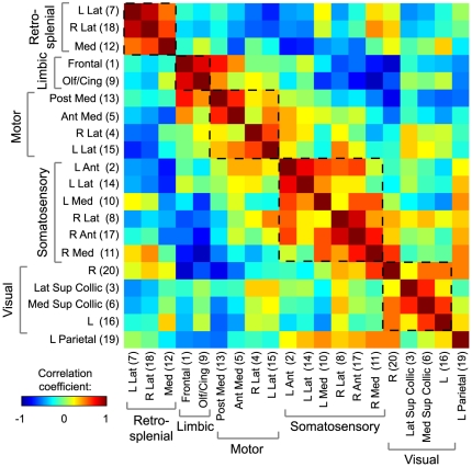
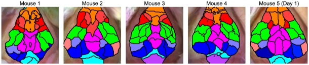
Functional neuroimaging (e.g., with fMRI) has been difficult to perform in mice, making it challenging to translate between human fMRI studies and molecular and genetic mechanisms. A method to easily perform large-scale functional neuroimaging in mice would enable the discovery of functional correlates of genetic manipulations and bridge with mouse models of disease. To satisfy this need, we combined resting-state functional connectivity mapping with optical intrinsic signal imaging (fcOIS). We demonstrate functional connectivity in mice through highly detailed fcOIS mapping of resting-state networks across most of the cerebral cortex. Synthesis of multiple network connectivity patterns through iterative parcellation and clustering provides a comprehensive map of the functional neuroarchitecture and demonstrates identification of the major functional regions of the mouse cerebral cortex. The method relies on simple and relatively inexpensive camera-based equipment, does not require exogenous contrast agents and involves only reflection of the scalp (the skull remains intact) making it minimally invasive. In principle, fcOIS allows new paradigms linking human neuroscience with the power of molecular/genetic manipulations in mouse models.
Overview
The claims and statistics of this paper haven’t been checked yet.
Run the check — free, no account needed. It reads the open-access text and checks the sources it cites.
Integrity signals
Registry
Cites
Statistics
Figures
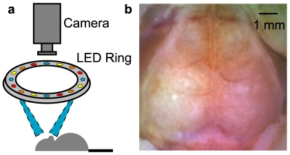
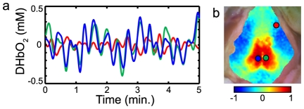
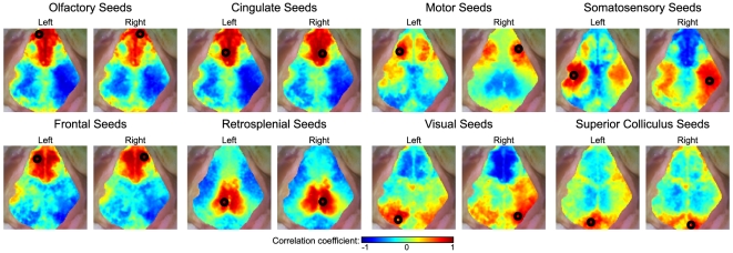
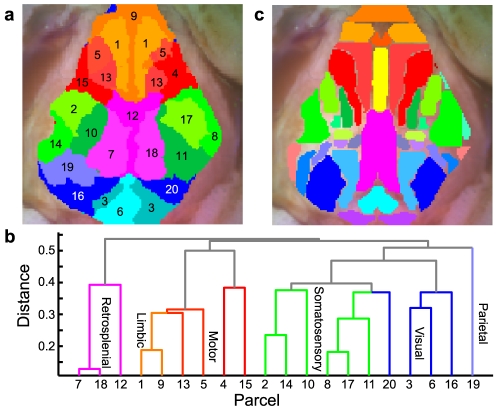
Verification
Claims the paper makes
The claims and statistics of this paper haven’t been checked yet.
Run the check — free, no account needed. It reads the open-access text and checks the sources it cites.
Claims other papers make about this one
Each is checked against this paper’s own text: does it say what the citing paper says it does?
The cited source says what the claim attributes to it.
In the citing paper
“We have shown previously that fcOIS is a sensitive imaging modality capable of measuring functional connectivity between brain regions in healthy mice (White et al., 2011;Bero et al., 2012)”
From Anti-ApoE Antibody Given after Plaque Onset Decreases Aβ Accumulation and Improves Brain Function in a Mouse Model of Aβ Amyloidosis (2014) · cites this paper as White et al., 2011
The cited source says what the claim attributes to it.
In the citing paper
“Compared with PBS-treated mice, bilateral functional connectivity in the HJ6.3 group appeared to be more globally restored and to resemble that of healthy, wild-type mice (Fig. 7C), as described previously (White et al., 2011;Bero et al., 2012).”
From Anti-ApoE Antibody Given after Plaque Onset Decreases Aβ Accumulation and Improves Brain Function in a Mouse Model of Aβ Amyloidosis (2014) · cites this paper as White et al., 2011
The check wasn’t confident either way, so it gives no verdict — never disputed.
In the citing paper
“and that there is functional disruption in APP/PS1 mice after A deposition (White et al., 2011;Bero et al., 2012).”
From Anti-ApoE Antibody Given after Plaque Onset Decreases Aβ Accumulation and Improves Brain Function in a Mouse Model of Aβ Amyloidosis (2014) · cites this paper as White et al., 2011
Experimental — accuracy not yet measured
Self-check
Percentages, sums and reported differences re-computed from the paper’s own numbers.
The self-check needs the paper’s full text.
Citations
Cites
The Mouse Brain in Stereotaxic Coordinates
Paxinos, G. (2001) · Cited by 12,859
Functional connectivity in the motor cortex of resting human brain using echo‐planar mri
Biswal, B., Zerrin Yetkin, F., Haughton, V. M., & Hyde, J. S. (1995) · Magnetic Resonance in Medicine · doi:10.1002/mrm.1910340409 · Cited by 9,812
The Brain's Default Network: Anatomy, Function, and Relevance to Disease
Buckner, R. L., Andrews‐Hanna, J. R., & Schacter, D. L. (2008) · Annals of the New York Academy of Sciences · doi:10.1196/annals.1440.011 · Cited by 9,761
The human brain is intrinsically organized into dynamic, anticorrelated functional networks
Fox, M. D., Snyder, A. Z., Vincent, J. L., Corbetta, M., Van Essen, D. C., & Raichle, M. E. (2005) · Proceedings of the National Academy of Sciences · doi:10.1073/pnas.0504136102 · Cited by 8,553
Self-citationSpontaneous fluctuations in brain activity observed with functional magnetic resonance imaging
Fox, M. D., & Raichle, M. E. (2007) · Nature Reviews Neuroscience · doi:10.1038/nrn2201 · Cited by 6,700
Functional connectivity in the resting brain: a network analysis of the default mode hypothesis
Greicius, M. D., Krasnow, B., Reiss, A. L., & Menon, V. (2003) · Proceedings of the National Academy of Sciences · doi:10.1073/pnas.0135058100 · Cited by 6,358
Talairach, J., Tournoux, P., & Rayport, M. (1988) · Thieme eBooks · Cited by 5,058
Buckner, R. L., Sepulcre, J., Talukdar, T., Krienen, F. M., Liu, H., Hedden, T., Andrews-Hanna, J. R., Sperling, R. A., & Johnson, K. A. (2009) · The Journal of Neuroscience · doi:10.1523/jneurosci.5062-08.2009 · Cited by 2,862
Neurodegenerative Diseases Target Large-Scale Human Brain Networks
Seeley, W. W., Crawford, R. K., Zhou, J., Miller, B. L., & Greicius, M. D. (2009) · Neuron · doi:10.1016/j.neuron.2009.03.024 · Cited by 2,350
Resting-State Functional Connectivity Reflects Structural Connectivity in the Default Mode Network
Greicius, M. D., Supekar, K., Menon, V., & Dougherty, R. F. (2009) · Cerebral Cortex · doi:10.1093/cercor/bhn059 · Cited by 2,175
The global signal and observed anticorrelated resting state brain networks
Fox, M. D., Zhang, D., Snyder, A. Z., & Raichle, M. E. (2009) · Journal of Neurophysiology · doi:10.1152/jn.90777.2008 · Cited by 1,877
Self-citationIntrinsic functional architecture in the anaesthetized monkey brain
Vincent, J. L., Patel, G. H., Fox, M. D., Snyder, A. Z., Baker, J. T., Van Essen, D. C., Zempel, J. M., Snyder, L. H., Corbetta, M., & Raichle, M. E. (2007) · Nature · doi:10.1038/nature05758 · Cited by 1,786
Self-citationRaichle, M. E., & Mintun, M. A. (2006) · Annual Review of Neuroscience · doi:10.1146/annurev.neuro.29.051605.112819 · Cited by 1,630
3D statistical neuroanatomical models from 305 MRI volumes
Evans, A. C., Collins, D. L., Mills, S., Brown, E. D., Kelly, R. L., & Peters, T. M. (2005) · IEEE Nuclear Science Symposium and Medical Imaging Conference (NSS/MIC) · doi:10.1109/nssmic.1993.373602 · Cited by 1,487
What does the retrosplenial cortex do?
Vann, S. D., Aggleton, J. P., & Maguire, E. A. (2009) · Nature Reviews Neuroscience · doi:10.1038/nrn2733 · Cited by 1,409
Neurophysiological Architecture of Functional Magnetic Resonance Images of Human Brain
Salvador, R. M., John Suckling, A., Coleman, M. R., Pickard, J. D., Menon, D. K., & Bullmore, E. T. (2005) · Cerebral Cortex · doi:10.1093/cercor/bhi016 · Cited by 1,334
Functional architecture of cortex revealed by optical imaging of intrinsic signals
Grinvald, A., Lieke, E. E., Frostig, R. D., Gilbert, C. D., & Wiesel, T. N. (1986) · Nature · doi:10.1038/324361a0 · Cited by 1,243
Spontaneously emerging cortical representations of visual attributes
Kenet, T., Bibitchkov, D., Tsodyks, M., Grinvald, A., & Arieli, A. (2003) · Nature · doi:10.1038/nature02078 · Cited by 924
Vanhaudenhuyse, A., Noirhomme, Q., Tshibanda, L. J.-F., Bruno, M.-A., Boveroux, P., Schnakers, C., Soddu, A., Perlbarg, V., Ledoux, D., Brichant, J.-F., et al. (2010) · Brain · doi:10.1093/brain/awp313 · Cited by 911 · cited 1×
“The discovery that functionally-related areas have correlated neural and hemodynamic activity even in the absence of tasks means that brain networks can be studied even in patients with brain-injury [5,6], those under anesthesia [7], or in subjects unable to perform detailed cognitive tasks, such as neonates [8,9].”
BackgroundIso-orientation domains in cat visual cortex are arranged in pinwheel-like patterns
Bonhoeffer, T., & Grinvald, A. (1991) · Nature · doi:10.1038/353429a0 · Cited by 873
Longitudinal Analysis of Neural Network Development in Preterm Infants
Smyser, C. D., Inder, T. E., Shimony, J. S., Hill, J. E., Degnan, A. J., Snyder, A. Z., & Neil, J. J. (2010) · Cerebral Cortex · doi:10.1093/cercor/bhq035 · Cited by 791
Self-citationBeckmann, M., Johansen-Berg, H., & Rushworth, M. F. S. (2009) · The Journal of Neuroscience · doi:10.1523/jneurosci.3328-08.2009 · Cited by 789
A network dysfunction perspective on neurodegenerative diseases
Palop, J. J., Chin, J., & Mucke, L. (2006) · Nature · doi:10.1038/nature05289 · Cited by 710
Resting-state networks in the infant brain
Fransson, P., Skiöld, B., Horsch, S., Nordell, A., Blennow, M., Lagercrantz, H., & Åden, U. (2007) · Proceedings of the National Academy of Sciences · doi:10.1073/pnas.0704380104 · Cited by 696
Arridge, S., Cope, M., & Delpy, D. T. (1992) · Physics in Medicine and Biology · doi:10.1088/0031-9155/37/7/005 · Cited by 597
Optical absorption of hemoglobin
Prahl, S. A. (1999) · Medical Entomology and Zoology · Cited by 579
New Paradigm for Optical Imaging
Kalatsky, V. A., & Stryker, M. P. (2003) · Neuron · doi:10.1016/s0896-6273(03)00286-1 · Cited by 571
Functional Organization of Primate Visual Cortex Revealed by High Resolution Optical Imaging
Ts’o, D. Y., Frostig, R. D., Lieke, E. E., & Grinvald, A. (1990) · Science · doi:10.1126/science.2165630 · Cited by 522
A review of systems and networks of the limbic forebrain/limbic midbrain
Morgane, P. J., Galler, J. R., & Mokler, D. J. (2005) · Progress in Neurobiology · doi:10.1016/j.pneurobio.2005.01.001 · Cited by 519
Synchronized delta oscillations correlate with the resting-state functional MRI signal
Lu, H., Zuo, Y., Gu, H., Waltz, J. A., Wang, Z., Scholl, C. A., Rea, W. P., Yang, Y., & Stein, E. A. (2007) · Proceedings of the National Academy of Sciences · doi:10.1073/pnas.0705791104 · Cited by 424
Culver, J. P., Durduran, T., Furuya, D., Cheung, C., Greenberg, J. H., & Yodh, A. G. (2003) · Journal of Cerebral Blood Flow & Metabolism · doi:10.1097/01.wcb.0000076703.71231.bb · Cited by 421
Self-citationDevor, A., Tian, P., Nishimura, N., Teng, I. C., Hillman, E. M. C., Narayanan, S. N., Ulbert, I., Boas, D. A., Kleinfeld, D., & Dale, A. M. (2007) · Journal of Neuroscience · doi:10.1523/jneurosci.0134-07.2007 · Cited by 393
Devor, A., Dunn, A. K., Andermann, M. L., Ulbert, I., Boas, D. A., & Dale, A. M. (2003) · Neuron · doi:10.1016/s0896-6273(03)00403-3 · Cited by 387
van Meer, M. P. A., van der Marel, K., Wang, K., Otte, W. M., el Bouazati, S., Roeling, T. A. P., Viergever, M. A., van der Sprenkel, J. W. B., & Dijkhuizen, R. M. (2010) · Journal of Neuroscience · doi:10.1523/jneurosci.5709-09.2010 · Cited by 352
Noninvasive Functional and Structural Connectivity Mapping of the Human Thalamocortical System
Zhang, D., Snyder, A. Z., Shimony, J. S., Fox, M., & Raichle, M. E. (2009) · Cerebral Cortex · doi:10.1093/cercor/bhp182 · Cited by 351
Self-citationPostsynaptic control of plasticity in developing somatosensory cortex
Schlaggar, B. L., Fox, K. D., & O'Leary, D. M. (1993) · Nature · doi:10.1038/364623a0 · Cited by 339
Self-citationDunn, A. K., Devor, A., Bolay, H., Andermann, M. L., Moskowitz, M. A., Dale, A. M., & Boas, D. A. (2003) · Optics Letters · doi:10.1364/ol.28.000028 · Cited by 333
Boly, M., Tshibanda, L., Vanhaudenhuyse, A., Noirhomme, Q., Schnakers, C., Ledoux, D., Boveroux, P., Garweg, C., Lambermont, B. C., Phillips, C. L. M., et al. (2009) · Human Brain Mapping · doi:10.1002/hbm.20672 · Cited by 333
Potential of Visual Cortex to Develop an Array of Functional Units Unique to Somatosensory Cortex
Schlaggar, B. L., & O’Leary, D. D. M. (1991) · Science · doi:10.1126/science.2047863 · Cited by 298
Self-citationResting-state functional connectivity in the human brain revealed with diffuse optical tomography
White, B. R., Snyder, A. Z., Cohen, A. L., Petersen, S. E., Raichle, M. E., Schlaggar, B. L., & Culver, J. P. (2009) · NeuroImage · doi:10.1016/j.neuroimage.2009.03.058 · Cited by 292
Self-citationHillman, E. M. C., Devor, A., Bouchard, M. B., Dunn, A. K., Krauss, G. W., Skoch, J. M., Bacskai, B. J., Dale, A. M., & Boas, D. A. (2007) · NeuroImage · doi:10.1016/j.neuroimage.2006.11.032 · Cited by 290
Dunn, A. K., Devor, A., Dale, A. M., & Boas, D. A. (2005) · NeuroImage · doi:10.1016/j.neuroimage.2005.04.024 · Cited by 285
Woolsey, T. A., Rovainen, C. M., Cox, S. B., Henegar, M. H., Liang, G. E., Liu, D., Moskalenko, Y. E., Sui, J., & Wei, L. (1996) · Cerebral Cortex · doi:10.1093/cercor/6.5.647 · Cited by 276
Investigating neural–hemodynamic coupling and the hemodynamic response function in the awake rat
Martin, C. J., Martindale, J., Berwick, J., & Mayhew, J. E. (2006) · NeuroImage · doi:10.1016/j.neuroimage.2006.02.021 · Cited by 255
A simple view of the brain through a frequency-specific functional connectivity measure
Salvador, R., Martínez, A., Pomarol-Clotet, E., Gomar, J., Vila, F., Sarró, S., Capdevila, A., & Bullmore, E. (2008) · NeuroImage · doi:10.1016/j.neuroimage.2007.08.018 · Cited by 225
Coupling of the cortical hemodynamic response to cortical and thalamic neuronal activity
Devor, A., Ulbert, I., Dunn, A. K., Narayanan, S. N., Jones, S. R., Andermann, M. L., Boas, D. A., & Dale, A. M. (2005) · Proceedings of the National Academy of Sciences · doi:10.1073/pnas.0407789102 · Cited by 222
Bouchard, M. B., Chen, B. R., Burgess, S. A., & Hillman, E. M. C. (2009) · Optics Express · doi:10.1364/oe.17.015670 · Cited by 221
Resting‐state functional connectivity of the rat brain
Pawela, C. P., Biswal, B. B., Cho, Y. R., Kao, D. S., Li, R., Jones, S. R., Schulte, M. L., Matloub, H. S., Hudetz, A. G., & Hyde, J. S. (2008) · Magnetic Resonance in Medicine · doi:10.1002/mrm.21524 · Cited by 200
Zhao, F., Zhao, T., Zhou, L., Qiu-lin, W., & Hu, X. P. (2007) · NeuroImage · doi:10.1016/j.neuroimage.2007.07.063 · Cited by 199
The effect of different anesthetics on neurovascular coupling
Franceschini, M. A., Radhakrishnan, H., Thakur, K. T., Wu, W., Ruvinskaya, S., Carp, S. A., & Boas, D. A. (2010) · NeuroImage · doi:10.1016/j.neuroimage.2010.03.060 · Cited by 174
Williams, K. A., Magnuson, M. E., Majeed, W., LaConte, S. M., Peltier, S., Hu, X. P., & Keilholz, S. D. (2010) · Magnetic Resonance Imaging · doi:10.1016/j.mri.2010.03.007 · Cited by 166
Hutchison, R. M., Mirsattari, S. M., Jones, C. K., Gati, J. S., & Leung, L.-W. S. (2010) · Journal of Neurophysiology · doi:10.1152/jn.00141.2010 · Cited by 165
Lindauer, U., Royl, G., Leithner, C., Kühl, M., Gold, L., Gethmann, J. M., Kohl‐Bareis, M., Villringer, A., & Dirnagl, U. (2001) · NeuroImage · doi:10.1006/nimg.2000.0709 · Cited by 154
Sheth, S. A., Nemoto, M., Guiou, M., Walker, M. A., Pouratian, N., Hageman, N. S., & Toga, A. W. (2004) · Journal of Neuroscience · doi:10.1523/jneurosci.4526-03.2004 · Cited by 137
Kannurpatti, S. S., Biswal, B. B., Kim, Y. R., & Rosen, B. R. (2007) · NeuroImage · doi:10.1016/j.neuroimage.2007.05.061 · Cited by 118
Pawela, C. P., Biswal, B. B., Hudetz, A. G., Li, R., Jones, S. R., Cho, Y. R., Matloub, H. S., & Hyde, J. S. (2009) · NeuroImage · doi:10.1016/j.neuroimage.2009.09.054 · Cited by 112
Culver, J. P., Siegel, A. M., Franceschini, M. A., Mandeville, J. B., & Boas, D. A. (2005) · NeuroImage · doi:10.1016/j.neuroimage.2005.05.052 · Cited by 80
Self-citationCortical area size dictates performance at modality-specific behaviors
Leingärtner, A., Thuret, S., Kroll, T. T., Chou, S., Leasure, J. L., Gage, F. H., & O’Leary, D. D. M. (2007) · Proceedings of the National Academy of Sciences · doi:10.1073/pnas.0611723104 · Cited by 55
Animal Models of Neurodegenerative Disease: Insights from In vivo Imaging Studies
Strome, E. M., & Doudet, D. J. (2007) · Molecular Imaging and Biology · doi:10.1007/s11307-007-0093-4 · Cited by 35
MRI of mouse models of neurological disorders
Anderson, S. A., & Frank, J. A. (2007) · NMR in Biomedicine · doi:10.1002/nbm.1167 · Cited by 20
Research works printed but not linked: 5
- A Devor, 2003, Coupling of total hemoglobin concentration, oxygenation, and neural activity in rat somatosensory cortex., Neuron, 39, 353, 10.1016/S0896-6273(03)00403-3 · DOI · Paper page
- A Devor, 2005, Coupling of the cortical hemodynamic response to cortical and thalamic neuronal activity., PNAS, 102, 3822, 10.1073/pnas.0407789102 · DOI
- MD Fox, 2007, Spontaneous fluctuations in brain activity observed with functional magnetic resonance imaging., Nature Reviews Neuroscience, 8, 700, 10.1038/nrn2201 · DOI · Paper page
- MD Fox, 2009, The Global Signal and Observed Anticorrelated Resting State Brain Networks., Journal of Neurophysiology, 101, 3270, 10.1152/jn.90777.2008 · DOI · Paper page
- AK Dunn, 2005, Spatial extent of oxygen metabolism and hemodynamic changes during functional activation of the rat somatosensory cortex., NeuroImage, 27, 279, 10.1016/j.neuroimage.2005.04.024 · DOI · Paper page
Cited by
Sorted by citations
Structure and function of complex brain networks
Sporns, O. (2013) · Dialogues in Clinical Neuroscience · doi:10.31887/dcns.2013.15.3/osporns · Cited by 964
Circadian clock proteins regulate neuronal redox homeostasis and neurodegeneration
Musiek, E. S., Lim, M. M., Yang, G., Bauer, A. Q., Qi, L., Lee, Y., Roh, J. H., Ortiz-Gonzalez, X., Dearborn, J. T., Culver, J. P., et al. (2013) · Journal of Clinical Investigation · doi:10.1172/jci70317 · Cited by 524
Self-citationLi, L. S., Zhu, L., Ma, C., Lin, L., Yao, J., Wang, L., Maslov, K., Zhang, R., Chen, W., Shi, J., et al. (2017) · Nature Biomedical Engineering · doi:10.1038/s41551-017-0071 · Cited by 476
Spontaneous cortical activity alternates between motifs defined by regional axonal projections
Mohajerani, M. H., Chan, A. W., Mohsenvand, M. ‘., LeDue, J. M., Liu, R., McVea, D. A., Boyd, J. D., Wang, Y. T., Reimers, M., & Murphy, T. H. (2013) · Nature Neuroscience · doi:10.1038/nn.3499 · Cited by 375
Grandjean, J., Schroeter, A., Batata, I., & Rudin, M. (2014) · NeuroImage · doi:10.1016/j.neuroimage.2014.08.043 · Cited by 339
Metadata
- Authors
- White, B. R., Bauer, A. Q., Snyder, A. Z., Schlaggar, B. L., Lee, J.-M., & Culver, J. P.
- Affiliations
- Washington University in St. Louis (US)
- Corresponding author
- Culver, Joseph P.
- ORCID
- Venue
- PLoS ONE
- Publisher
- Public Library of Science (PLoS)
- Published
- 2011-01-20
- Volume
- 6
- Issue
- 1
- Pages
- e16322
- ISSN
- 1932-6203 (ISSN-L)
- PMC
- PMC3024435
- OpenAlex
- W2065496603
- Cited by
- 253
- References
- 60
- Citations by year
- 2026: 7 · 2025: 15 · 2024: 18 · 2023: 25 · 2022: 16 · 2021: 15 · 2020: 15 · 2019: 22 · 2018: 15 · 2017: 30 · 2016: 16 · 2015: 9 · 2014: 23 · 2013: 16 · 2012: 7
- FWCI
- 7.96
- Type
- journal article
- Language
- en
- Open access
- gold
- doi.org
- Locations
- PLoS ONE (publishedVersion, cc-by)
- PubMed Central (publishedVersion, cc-by)
- PubMed (publishedVersion)
- DOAJ (DOAJ: Directory of Open Access Journals) (submittedVersion, cc-by-sa)
- Figshare (submittedVersion, cc-by)
- Funders
- National Institutes of Health
- National Institute on Drug Abuse (T90DA022871)
- National Institute of Neurological Disorders and Stroke (P01 NS32636, R01NS084028, P01NS032636)
- National Institute of Biomedical Imaging and Bioengineering (R01EB008085, R01EB009233)
- Topics
- Life Sciences › Neuroscience › Cognitive Neuroscience › Functional Brain Connectivity Studies
- Health Sciences › Medicine › Radiology, Nuclear Medicine and Imaging › Optical Imaging and Spectroscopy Techniques
- Health Sciences › Medicine › Radiology, Nuclear Medicine and Imaging › Advanced MRI Techniques and Applications
- Keywords
- functional connectivity, mouse brain, resting-state networks, intrinsic signal imaging, mouse models of human disease, non-invasive brain imaging, camera-based imaging
- MeSH terms
- Animals, Brain Mapping, Cerebral Cortex, Equipment Design, Magnetic Resonance Imaging, Methods, Neural Pathways, Mice, Optical Devices
About these checks
Registry, retraction, Cites and statistics results are computed, not judged, with one exception: a printed reference that no index lists is read once by a model before it counts as a citation error. Claims are extracted by a model and kept only when their quote appears verbatim in the text; each cited source is then checked, with a model’s help, for whether it says what the claim relies on. Those verdicts can be wrong — read the claim’s quote and the cited source before relying on one.
Statistics and arithmetic are recomputed, not judged.
Confidence bands are shown only for fields calibrated on our annotated test set. None is calibrated yet, so no band is shown.
What counts as a claim ›How we measure confidence ›How we check this ›
Are you an author, or do you see a problem with this page? Request a review
