

ApoE Receptor 2 Regulates Synapse and Dendritic Spine Formation
Open accessThe DOI matches its Crossref record; 45 of the 45 references it cites were found in a registry or an index.
Abstract
BACKGROUND: Apolipoprotein E receptor 2 (ApoEr2) is a postsynaptic protein involved in long-term potentiation (LTP), learning, and memory through unknown mechanisms. We examined the biological effects of ApoEr2 on synapse and dendritic spine formation-processes critical for learning and memory. METHODOLOGY/PRINCIPAL FINDINGS: In a heterologous co-culture synapse assay, overexpression of ApoEr2 in COS7 cells significantly increased colocalization with synaptophysin in primary hippocampal neurons, suggesting that ApoEr2 promotes interaction with presynaptic structures. In primary neuronal cultures, overexpression of ApoEr2 increased dendritic spine density. Consistent with our in vitro findings, ApoEr2 knockout mice had decreased dendritic spine density in cortical layers II/III at 1 month of age. We also tested whether the interaction between ApoEr2 and its cytoplasmic adaptor proteins, specifically X11α and PSD-95, affected synapse and dendritic spine formation. X11α decreased cell surface levels of ApoEr2 along with synapse and dendritic spine density. In contrast, PSD-95 increased cell surface levels of ApoEr2 as well as synapse and dendritic spine density. CONCLUSIONS/SIGNIFICANCE: These results suggest that ApoEr2 plays important roles in structure and function of CNS synapses and dendritic spines, and that these roles are modulated by cytoplasmic adaptor proteins X11α and PSD-95.
Overview
The claims and statistics of this paper haven’t been checked yet.
Free: it reads the open-access text and checks the first 5 claims that cite a source we can check. Create a free account to check every claim.
Integrity signals
Registry
Cites
Statistics
Figures




Verification
Claims the paper makes
The claims and statistics of this paper haven’t been checked yet.
Free: it reads the open-access text and checks the first 5 claims that cite a source we can check. Create a free account to check every claim.
Claims other papers make about this one
Each is checked against this paper’s own text: does it say what the citing paper says it does?
The source backs part of the claim, not all of it — or backs it for a different population, condition or comparison.
In the citing paper
“Our observation that IDOL bidirectionally regulate spine density through ApoER2 is consistent with previous studies showing that spine density is decreased in ApoER2 KO mice and increased in the setting of ApoER2 overexpression (Dumanis et al., 2011; Wasser et al., 2014).”
From The E3 ubiquitin ligase IDOL regulates synaptic ApoER2 levels and is important for plasticity and learning (2017) · cites this paper as Dumanis et al., 2011
Experimental — accuracy not yet measuredThe cited source says what the claim attributes to it.
In the citing paper
“suggesting a critical role of the receptor in promoting synaptogenesis (16).”
From Reelin: Neurodevelopmental Architect and Homeostatic Regulator of Excitatory Synapses (2017) · cites this paper as [16]
The cited source says what the claim attributes to it.
In the citing paper
“Our observation that IDOL bidirectionally regulate spine density through ApoER2 is consistent with previous studies showing that spine density is decreased in ApoER2 KO mice and increased in the setting of ApoER2 overexpression (Dumanis et al., 2011; Wasser et al., 2014).”
From The E3 ubiquitin ligase IDOL regulates synaptic ApoER2 levels and is important for plasticity and learning (2017) · cites this paper as Dumanis et al., 2011
In the citing paper
“Intriguingly, overexpression of the Reelin receptor ApoER2 in dissociated hippocampal neuron cultures can dramatically increase dendritic spine numbers in wild-type neuron cultures, suggesting a critical role of the receptor in promoting synaptogenesis (16).”
From Reelin: Neurodevelopmental Architect and Homeostatic Regulator of Excitatory Synapses (2017) · cites this paper as [16]
Self-check
Percentages, sums and reported differences re-computed from the paper’s own numbers.
The self-check needs the paper’s full text.
Citations
Cites
AMPA Receptor Trafficking and Synaptic Plasticity
Malinow, R., & Malenka, R. C. (2002) · Annual Review of Neuroscience · doi:10.1146/annurev.neuro.25.112701.142758 · Cited by 2,588 · cited 2×
“For example, GluA1 has been suggested to require activity-dependent stimulation for acute delivery, whereas GluA2 may undergo constitutive trafficking and turnover [28,45].”
BackgroundMatsuzaki, M., Ellis-Davies, G. C. R., Nemoto, T., Miyashita, Y., Iino, M., & Kasai, H. (2001) · Nature Neuroscience · doi:10.1038/nn736 · Cited by 1,802 · cited 1×
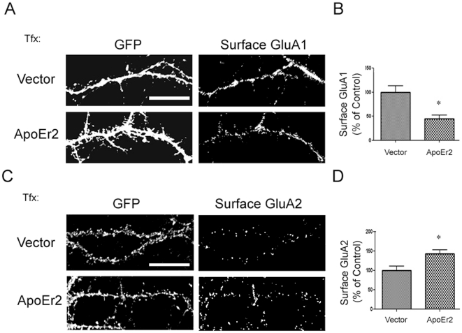
“Because AMPA receptors are important for synaptic plasticity and dendritic spine formation, we examined whether ApoEr2 affected total levels of AMPA receptor subunits by infecting neurons with ApoEr2 or GFP [28,29,30].”
BackgroundPDZ domain proteins of synapses
Kim, E., & Sheng, M. (2004) · Nature Reviews Neuroscience · doi:10.1038/nrn1517 · Cited by 1,577 · cited 1×
“Additionally, PSD-95 increased dendritic spine density by 21% compared to GFP – consistent with previous reports [31].”
ResultTrommsdorff, M., Gotthardt, M., Hiesberger, T., Shelton, J., Stockinger, W., Nimpf, J., Hammer, R. E., Richardson, J. A., & Herz, J. (1999) · Cell · doi:10.1016/s0092-8674(00)80782-5 · Cited by 1,261
Neuroligin Expressed in Nonneuronal Cells Triggers Presynaptic Development in Contacting Axons
Scheiffele, P., Fan, J., Choih, J., Fetter, R., & Serafini, T. (2000) · Cell · doi:10.1016/s0092-8674(00)80877-6 · Cited by 1,218
Regulation of distinct AMPA receptor phosphorylation sites during bidirectional synaptic plasticity
Lee, H.-K., Barbarosie, M., Kameyama, K., Bear, M. F., & Huganir, R. L. (2000) · Nature · doi:10.1038/35016089 · Cited by 1,116 · cited 5×
“These ApoE receptors are involved in neuronal migration during brain development [2], influx of calcium through NMDA channels [3], neurite outgrowth [4], LTP and memory [5].”
BackgroundInfluentialShi, S.-H., Hayashi, Y., Esteban, J. A., & Malinow, R. (2001) · Cell · doi:10.1016/s0092-8674(01)00321-x · Cited by 1,094 · cited 1×
“For example, GluA1 has been suggested to require activity-dependent stimulation for acute delivery, whereas GluA2 may undergo constitutive trafficking and turnover [28,45].”
BackgroundEvidence for multiple AMPA receptor complexes in hippocampal CA1/CA2 neurons
Wenthold, R., Petralia, R., Blahos J, II, & Niedzielski, A. (1996) · The Journal of Neuroscience · doi:10.1523/jneurosci.16-06-01982.1996 · Cited by 850 · cited 1×
“These results are somewhat surprising given that GluA1/A2 heteromers comprise the predominant AMPA receptor population in the hippocampus [41,42].”
Reelin Is a Ligand for Lipoprotein Receptors
D'Arcangelo, G., Homayouni, R., Keshvara, L., Rice, D. S., Sheldon, M., & Curran, T. (1999) · Neuron · doi:10.1016/s0896-6273(00)80860-0 · Cited by 808 · cited 1×
“One possible compensatory mechanism is modulation of extracellular ligands for ApoEr2, such as apolipoprotein E (apoE) or Reelin, which are known to be important for dendritic spine formation [22,37,38,39].”
BackgroundImportance of AMPA Receptors for Hippocampal Synaptic Plasticity But Not for Spatial Learning
Zamanillo, D., Sprengel, R., Hvalby, Ø., Jensen, V., Burnashev, N., Rozov, A., Kaiser, K. M. M., Köster, H. J., Borchardt, T., Worley, P., et al. (1999) · Science · doi:10.1126/science.284.5421.1805 · Cited by 791 · cited 1×
“While neither GluA1 nor GluA2 is absolutely required for normal dendritic spine formation or morphogenesis in vivo [43,44], GluA2 overexpression has been shown to enhance dendritic spine formation in hippocampal cultured neurons [30].”
Background
Research works printed but not linked: 2
- HS Hoe, 2006, Apolipoprotein E Receptor 2 Interactions with the N-Methyl-D-aspartate Receptor., J Biol Chem, 281, 3425, 10.1074/jbc.M509380200 · DOI · Paper page
- DT Pak, 2001, Regulation of dendritic spine morphology by SPAR, a PSD-95-associated RapGAP., Neuron, 31, 289, 10.1016/S0896-6273(01)00355-5 · DOI · Paper page
Cited by
Achariyar, T. M., Li, B., Peng, W., Verghese, P. B., Shi, Y., McConnell, E., Benraiss, A., Kasper, T., Song, W., Takano, T., et al. (2016) · Molecular Neurodegeneration · doi:10.1186/s13024-016-0138-8 · Cited by 252
What are lipoproteins doing in the brain?
Wang, H., & Eckel, R. H. (2014) · Trends in Endocrinology & Metabolism · doi:10.1016/j.tem.2013.10.003 · Cited by 234
Jossin, Y. (2020) · Biomolecules · doi:10.3390/biom10060964 · Cited by 202
Shaping Synapses by the Neural Extracellular Matrix
Ferrer-Ferrer, M., & Dityatev, A. (2018) · Frontiers in Neuroanatomy · doi:10.3389/fnana.2018.00040 · Cited by 169
“Significantly, it was shown that overexpression of ApoER2 in hippocampal neuron cultures increased dendritic spine density, suggesting a crucial role of this receptor in promoting spinogenesis (Dumanis et al., 2011).”
BackgroundKumar, A. (2011) · Frontiers in Aging Neuroscience · doi:10.3389/fnagi.2011.00007 · Cited by 165
Role of BACE1 in Alzheimer’s synaptic function
Das, B., & Yan, R. (2017) · Translational Neurodegeneration · doi:10.1186/s40035-017-0093-5 · Cited by 121
“In mouse models, ApoER2 increases the number of dendritic spines and synapses and stabilizes them by regulating the assembly of a complex of proteins involved in synaptic terminals across neurons, a process which is important for learning and memory [32].”
AMPA receptors and their minions: auxiliary proteins in AMPA receptor trafficking
Bissen, D., Foss, F., & Acker-Palmer, A. (2019) · Cellular and Molecular Life Sciences · doi:10.1007/s00018-019-03068-7 · Cited by 117
Reelin: Neurodevelopmental Architect and Homeostatic Regulator of Excitatory Synapses
Wasser, C. R., & Herz, J. (2017) · Journal of Biological Chemistry · doi:10.1074/jbc.r116.766782 · Cited by 111
“Loss of ApoER2 cleavage leads to increased ApoER2 expression and a concomitant enhancement of dendritic spine density (34), which is consistent with the published results of ApoER2 overexpression in wild-type neuron cultures (16).”
BackgroundResultFunctional Roles of the Interaction of APP and Lipoprotein Receptors
Pohlkamp, T., Wasser, C. R., & Herz, J. (2017) · Frontiers in Molecular Neuroscience · doi:10.3389/fnmol.2017.00054 · Cited by 92
“…Matsuki et al., 2008; Chai et al., 2009; Ventruti et al., 2011) as well as synapse formation and function (Glantz and Lewis, 2000; Sinagra et al., 2005; Groc et al., 2007; Qiu and Weeber, 2007; Niu et al., 2008; Campo et al., 2009; Dumanis et al., 2011; Hellwig et al., 2011; Bal et al., 2013).”
BackgroundECM receptors in neuronal structure, synaptic plasticity, and behavior
Kerrisk, M. E., Cingolani, L. A., & Koleske, A. J. (2014) · Progress in Brain Research · doi:10.1016/b978-0-444-63486-3.00005-0 · Cited by 96
“ApoER2 overexpression in primary neuronal cultures causes an increase in dendritic spine density, while ApoER2 knockout mice have a decrease in spine density (Dumanis et al., 2011; Trotter et al., 2011).”
Dab1 Is Required for Synaptic Plasticity and Associative Learning
Trotter, J., Lee, G. H., Kazdoba, T. M., Crowell, B., Domogauer, J., Mahoney, H. M., Franco, S. J., Müller, U., Weeber, E. J., & D'Arcangelo, G. (2013) · The Journal of Neuroscience · doi:10.1523/jneurosci.2010-13.2013 · Cited by 90
“Synaptic fractionation was performed on freshly dissected forebrains from 3-month-old C57BL6 mice as described previously (Dumanis et al., 2011).”
MethodSelf-citationThe benefit of docosahexaenoic acid for the adult brain in aging and dementia
Salem, N., Vandal, M., & Calon, F. (2015) · Prostaglandins, Leukotrienes and Essential Fatty Acids · doi:10.1016/j.plefa.2014.10.003 · Cited by 85
Santana, J., & Marzolo, M. (2017) · Biochemical Journal · doi:10.1042/bcj20160628 · Cited by 79
Olde Loohuis, N. F. M., Kole, K., Glennon, J. C., Karel, P., Van der Borg, G., Van Gemert, Y., Van den Bosch, D., Meinhardt, J., Kos, A., Shahabipour, F., et al. (2015) · Neurobiology of Disease · doi:10.1016/j.nbd.2015.05.006 · Cited by 54
Differential splicing and glycosylation of Apoer2 alters synaptic plasticity and fear learning
Wasser, C. R., Masiulis, I., Durakoglugil, M. S., Lane-Donovan, C., Xian, X., Beffert, U., Agarwala, A., Hammer, R. E., & Herz, J. (2014) · Science Signaling · doi:10.1126/scisignal.2005438 · Cited by 48
Ampuero, E., Jury, N., Härtel, S., Marzolo, M., & van Zundert, B. (2016) · Journal of Cellular Physiology · doi:10.1002/jcp.25605 · Cited by 49
“…the excitatory postsynaptic density, PSD95 drives the maturation of glutamatergic synapses and spines (El-Husseini et al., 2000; Losi et al., 2003; Ehrlich et al., 2007; Elias et al., 2008) and induces dendritic arbor refinement (Charych et al., 2006; Henriquez et al., 2013; Bustos et al., 2014).”
BackgroundLonskaya, I., Partridge, J., Lalchandani, R. R., Chung, A., Lee, T., Vicini, S., Hoe, H.-S., Lim, S. T., & Conant, K. (2013) · PLoS ONE · doi:10.1371/journal.pone.0069136 · Cited by 42
“Quantitation of surface signal and puncta were performed as previously described [36,37].”
MethodSelf-citationApoER2 processing by presenilin-1 modulates reelin expression
Balmaceda, V., Cuchillo‐Ibáñez, I., Pujadas, L., García‐Ayllón, M., Saura, C. A., Nimpf, J., Soriano, E., & Sáez‐Valero, J. (2014) · The FASEB Journal · doi:10.1096/fj.13-239350 · Cited by 40
Lane, R. F., Shineman, D. W., Steele, J., Lee, L. H., & Fillit, H. M. (2012) · Advances in pharmacology · doi:10.1016/b978-0-12-394816-8.00007-6 · Cited by 38
FE65 as a link between VLDLR and APP to regulate their trafficking and processing
Dumanis, S. B., Chamberlain, K. A., Jin Sohn, Y., Jin Lee, Y., Guénette, S. Y., Suzuki, T., Mathews, P. M., Pak, D. T., Rebeck, G. W., Suh, Y.-H., et al. (2012) · Molecular Neurodegeneration · doi:10.1186/1750-1326-7-9 · Cited by 34
“We and others have also demonstrated that ApoER2 and APP play an important role in dendritic spine formation [18,29,36,37,48,49].”
ResultSelf-citationGao, J., Marosi, M., Choi, J., Achiro, J. M., Kim, S., Li, S., Otis, K., Martin, K. C., Portera-Cailliau, C., & Tontonoz, P. (2017) · eLife · doi:10.7554/elife.29178 · Cited by 30
FoxP2 directly regulates the reelin receptor VLDLR developmentally and by singing
Adam, I., Mendoza, E., Kobalz, U., Wohlgemuth, S., & Scharff, C. (2016) · Molecular and Cellular Neuroscience · doi:10.1016/j.mcn.2016.04.002 · Cited by 29
ApoE4 Induces Synaptic and ERG Impairments in the Retina of Young Targeted Replacement ApoE4 Mice
Antes, R., Ezra-Elia, R., Weinberger, D., Solomon, A., Ofri, R., & Michaelson, D. M. (2013) · PLoS ONE · doi:10.1371/journal.pone.0064949 · Cited by 32
“Recent findings suggest that the apoE receptor apoER2 plays an important role in the maintenance of retinal synaptic connections and promotes presynaptic differentiation and dendritic spine formation [46,47].”
BackgroundDiBattista, A. M., Dumanis, S. B., Song, J. M., Bu, G., Weeber, E. J., Rebeck, G. W., & Hoe, H.-S. (2015) · Biochimica et Biophysica Acta (BBA) - Molecular Cell Research · doi:10.1016/j.bbamcr.2015.01.015 · Cited by 28
“After 24 hours, cell surface biotinlyation was conducted as previously described ([10]).”
BackgroundMethodSelf-citationLigand-induced Homotypic and Heterotypic Clustering of Apolipoprotein E Receptor 2
Divekar, S. D., Burrell, T. C., Lee, J. E., Weeber, E. J., & Rebeck, G. W. (2014) · Journal of Biological Chemistry · doi:10.1074/jbc.m113.537548 · Cited by 25
“They are neuronal proteins found in synaptic compartments (3, 4) that play an important role in neuronal migration during development (5, 6).”
BackgroundSelf-citationRamsden, C. E., Zamora, D., Horowitz, M. S., Jahanipour, J., Calzada, E., Li, X., Keyes, G. S., Murray, H. C., Curtis, M. A., Faull, R. L. M., et al. (2023) · Acta Neuropathologica Communications · doi:10.1186/s40478-023-01693-9 · Cited by 22
“2) [9, 22, 35, 50, 51, 53, 57, 72, 96, 98, 121, 124, 125, 128] (reviewed in [131]).”
BackgroundRamsden, C. E., Keyes, G. S., Calzada, E., Horowitz, M. S., Zamora, D., Jahanipour, J., Sedlock, A., Indig, F. E., Moaddel, R., Kapogiannis, D., et al. (2022) · Journal of Alzheimer's Disease · doi:10.3233/jad-220071 · Cited by 19
The apolipoprotein receptor LRP3 compromises APP levels
Cuchillo‐Ibáñez, I., Lennol, M. P., Escamilla, S., Mata‐Balaguer, T., Valverde-Vozmediano, L., López‐Font, I., Ferrer, I., & Sáez‐Valero, J. (2021) · Alzheimer s Research & Therapy · doi:10.1186/s13195-021-00921-5 · Cited by 16
ApoER2: Functional Tuning Through Splicing
Gallo, C. M., Ho, A., & Beffert, U. (2020) · Frontiers in Molecular Neuroscience · doi:10.3389/fnmol.2020.00144 · Cited by 16
Administration of Downstream ApoE Attenuates the Adverse Effect of Brain ABCA1 Deficiency on Stroke
Wang, X., Li, R., Zacharek, A., Landschoot‐Ward, J., Wang, F., Wu, K.-H., Chopp, M., Chen, J., & Cui, X. (2018) · International Journal of Molecular Sciences · doi:10.3390/ijms19113368 · Cited by 18
Gallo, C. M., Labadorf, A. T., Ho, A., & Beffert, U. (2022) · Genomics · doi:10.1016/j.ygeno.2022.110318 · Cited by 11
“It is possible that AS of the glycosylation domain, whether in tandem with the EGF precursor-like repeat before it or by itself, emerged in mice and primates as a regulatory mechanism of Apoer2 surface levels, which are critical for maintaining the proper balance of dendritic spines [35].”
BackgroundNeurotrophins regulate ApoER2 proteolysis through activation of the Trk signaling pathway
Larios, J. A., Jausoro, I., Benitez, M.-L., Bronfman, F. C., & Marzolo, M.-P. (2014) · BMC Neuroscience · doi:10.1186/1471-2202-15-108 · Cited by 12
“aptic plasticity [47,48], dendritic branching [49], actin remodeling [50], growth cone motility, filopodia formation”
Neuronal PCSK9 regulates cognitive performances via the modulation of ApoER2 synaptic localization
Pelucchi, S., Da Dalt, L., De Cesare, G., Stringhi, R., D’Andrea, L., La Greca, F., Cambria, C., Vandermeulen, L., Zianni, E., Musardo, S., et al. (2025) · Pharmacological Research · doi:10.1016/j.phrs.2025.107652 · Cited by 12
Regulation of the hippocampal translatome by Apoer2-ICD release
Wasser, C. R., Werthmann, G. C., Hall, E. M., Kuhbandner, K., Wong, C. H. Y., Durakoglugil, M. S., & Herz, J. (2023) · Molecular Neurodegeneration · doi:10.1186/s13024-023-00652-1 · Cited by 12
“We know that the abundance of Apoer2 can pro-portionately regulate synapse number [26, 86], and its cleavage can block extracellular signaling as well as transcription by the release of the ICD [1, 48, 81].”
BackgroundInfluentialLee, N. J., Song, J. M., Cho, H.-J., Sung, Y. M., Lee, T., Chung, A., Hong, S., Cifelli, J. L., Rubinshtein, M., Habib, L. K., et al. (2015) · Biochimica et Biophysica Acta (BBA) - Molecular Basis of Disease · doi:10.1016/j.bbadis.2015.12.007 · Cited by 11
“The cell surface biotinylation was conducted as previously described [21].”
MethodSelf-citationAlzheimer's disease: the large gene instability hypothesisPreprint
Soheili‐Nezhad, S. (2017) · bioRxiv (Cold Spring Harbor Laboratory) · doi:10.1101/189712 · Cited by 5
Contributing Factors of Neurodegeneration in Alzheimer’s Disease
Tailor, B. A., Pilozzi, A. R., & Huang, X. (2019) · Codon Publications eBooks · doi:10.15586/alzheimersdisease.2019.ch5 · Cited by 6
“Apolipoprotein E (ApoE) regulates synaptic function, promotes plasticity, increases the number of dendritic spines, and regulates protein trafficking across neurons (54).”
BackgroundApolipoprotein E acts at pre‐synaptic sites… among others
Barger, S. W. (2012) · Journal of Neurochemistry · doi:10.1111/j.1471-4159.2012.07935.x · Cited by 3
Fyn Tyrosine Kinase Increases Apolipoprotein E Receptor 2 Levels and Phosphorylation
Burrell, T. C., Divekar, S. D., Weeber, E. J., & Rebeck, G. W. (2014) · PLoS ONE · doi:10.1371/journal.pone.0110845 · Cited by 2
“However, the members of the Reelin pathway also have a role in dendritic spine density as ApoER2 knockout mice and Fyn knockout mice have reduced spine density in the cortex [53–54].”
Self-citationRamsden, C. E., Zamora, D., Horowitz, M. S., Jahanipour, J., Keyes, G. S., Li, X., Murray, H. C., Curtis, M. A., Faull, R. L. M., Sedlock, A. B., et al. (2023) · medRxiv · doi:10.21203/rs.3.rs-2968020/v1 · Cited by 2
Apoer2-ICD-Dependent Regulation of the Hippocampal TranslatomePreprint
Wasser, C. R., Hall, E. M., Kuhbandner, K., Durakoglugil, M. S., Wong, C. H. Y., Werthmann, G. C., & Herz, J. (2022) · SSRN Electronic Journal · doi:10.2139/ssrn.4124630 · Cited by 1
Role of LSR in the regulation of cholesterol homeostasis in the central nervous system
Hajj, A. E. (2019) · theses.fr (ABES) · doi:10.70675/5772a646z01eez4001z9947z40d5d34bcfbb
Molecular mechanisms underlying the surface organization of the NMDA receptors during development
Bénac, N. (2024) · theses.fr (ABES) · doi:10.70675/e3508a2ezb62ez4f99zb27bz156201271974
The effect of human-specific genetic variants on neuronal spinogenesis
Rosas, N. M., Szombathy, A. M., & Szigeti, K. (2026) · Frontiers in Genetics · doi:10.3389/fgene.2026.1786287
“The apolipoprotein E receptor (ApoE) through the APOE2 , APOE3 and APOE4 variants modify the recycling of GluA1-4 subunits of AMPA receptors and regulate spine density and maturation (Dumanis et al., 2011; Taxier et al., 2022; Dumanis et al., 2009; Neustadtl et al., 2017).”
Metadata
- Authors
- Dumanis, S. B., Cha, H.-J., Song, J. M., Trotter, J. H., Spitzer, M., Lee, J.-Y., Weeber, E. J., Turner, R. S., Pak, D. T. S., Rebeck, G. W., & Hoe, H.-S.
- Affiliations
- Georgetown University Medical Center (US)
- University of South Florida (US)
- Corresponding author
- Hoe, Hyang-Sook
- ORCID
- Venue
- PLoS ONE
- Publisher
- Public Library of Science (PLoS)
- Published
- 2011-02-15
- Volume
- 6
- Issue
- 2
- Pages
- e17203
- ISSN
- 1932-6203 (ISSN-L)
- PMC
- PMC3039666
- OpenAlex
- W2045877537
- Cited by
- 45
- References
- 45
- Citations by year
- 2026: 1 · 2025: 1 · 2024: 1 · 2023: 3 · 2022: 3 · 2021: 1 · 2020: 2 · 2019: 4 · 2018: 2 · 2017: 5 · 2016: 4 · 2015: 3 · 2014: 6 · 2013: 5 · 2012: 3
- FWCI
- 1.99
- Type
- journal article
- Language
- en
- Open access
- gold
- doi.org
- Locations
- PLoS ONE (publishedVersion, cc-by)
- PubMed Central (publishedVersion, cc-by)
- PubMed (publishedVersion)
- DOAJ (DOAJ: Directory of Open Access Journals) (submittedVersion, cc-by-sa)
- Figshare (submittedVersion, cc-by)
- Funders
- National Science Foundation
- Department of Transportation of Sichuan Province
- National Institutes of Health
- National Institute on Aging (R01AG026478, P01AG030128, AG032330-02S1, R03AG034253, R03AG032330, AG032330, AG034253, AG026478)
- National Institute of Neurological Disorders and Stroke (R01NS048085, NS048085)
- Topics
- Life Sciences › Neuroscience › Cellular and Molecular Neuroscience › Neuroscience and Neuropharmacology Research
- Life Sciences › Biochemistry, Genetics and Molecular Biology › Genetics › Genetics and Neurodevelopmental Disorders
- Life Sciences › Neuroscience › Cellular and Molecular Neuroscience › Alzheimer's disease research and treatments
- Keywords
- synaptogenesis, dendritic spine formation, PSD-95, synaptophysin, hippocampal neurons, ApoE-deficient mice, learning and memory, long-term potentiation
- MeSH terms
- Disks Large Homolog 4 Protein, Animals, Chlorocebus aethiops, Cytoplasm, Extracellular Space, Hippocampus, Humans, Membrane Proteins, Nerve Tissue Proteins, Synapses, Protein Structure, Tertiary, Receptors, AMPA, Coculture Techniques, COS Cells, LDL-Receptor Related Proteins, Adaptor Proteins, Signal Transducing, Dendritic Spines, Mice, Guanylate Kinases
About these checks
Registry, retraction, Cites and statistics results are computed, not judged, with one exception: a printed reference that no index lists is read once by a model before it counts as a citation error. Claims are extracted by a model and kept only when their quote appears verbatim in the text; each cited source is then checked, with a model’s help, for whether it says what the claim relies on. Those verdicts can be wrong — read the claim’s quote and the cited source before relying on one.
Statistics and arithmetic are recomputed, not judged.
Confidence bands are shown only for fields calibrated on our annotated test set. None is calibrated yet, so no band is shown.
What counts as a claim ›How we measure confidence ›How we check this ›
Are you an author, or do you see a problem with this page? Request a review
Data sources
Bibliographic data: Semantic Scholar, OpenAlex, Crossref.
Contains information from the Semantic Scholar Academic Graph Datasets, made available under the ODC Attribution License (ODC-BY 1.0).
Semantic Scholar’s data is described in: Kinney, R., et al. (2023). The Semantic Scholar Open Data Platform. arXiv:2301.10140.
