

The DOI matches its Crossref record; 78 of the 82 references it cites were found in a registry or an index.
The Regulation of Skeletal Muscle Protein Turnover during the Progression of Cancer Cachexia in the ApcMin/+ Mouse
Open accessAbstract
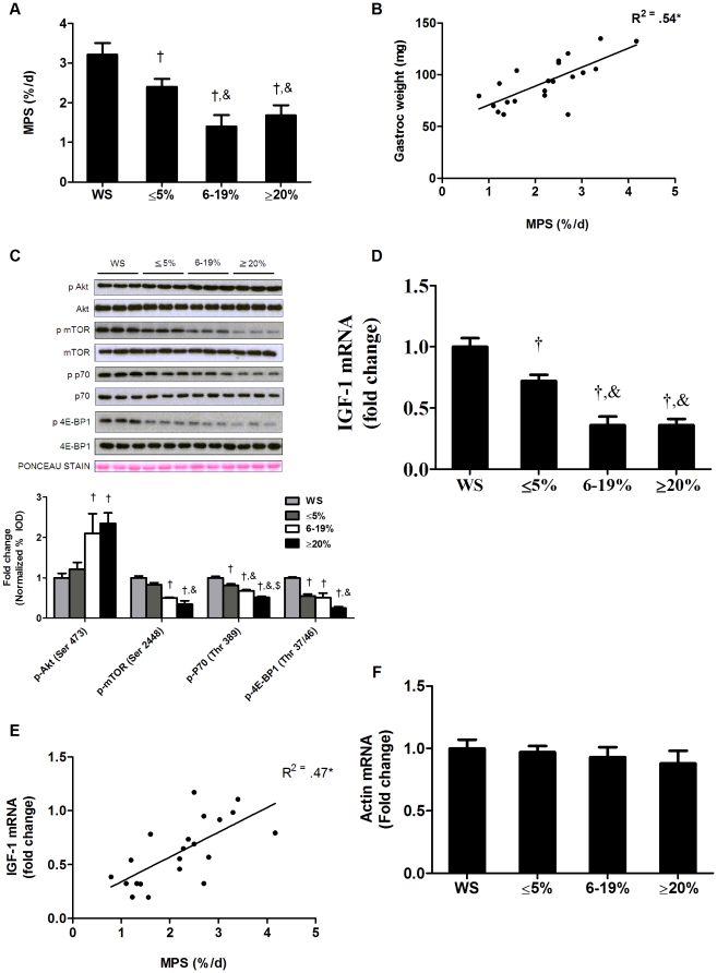
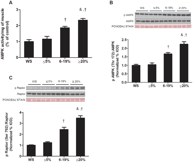
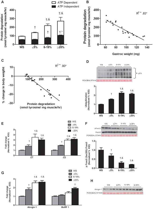
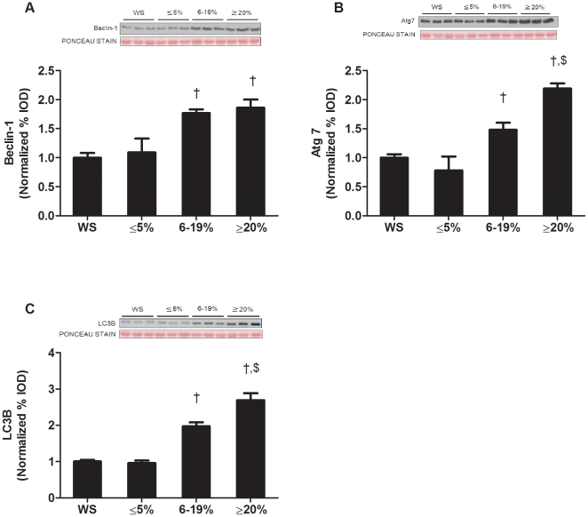
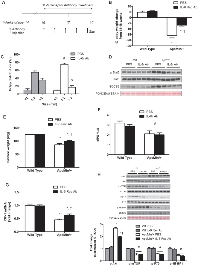
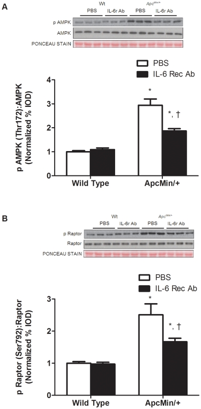
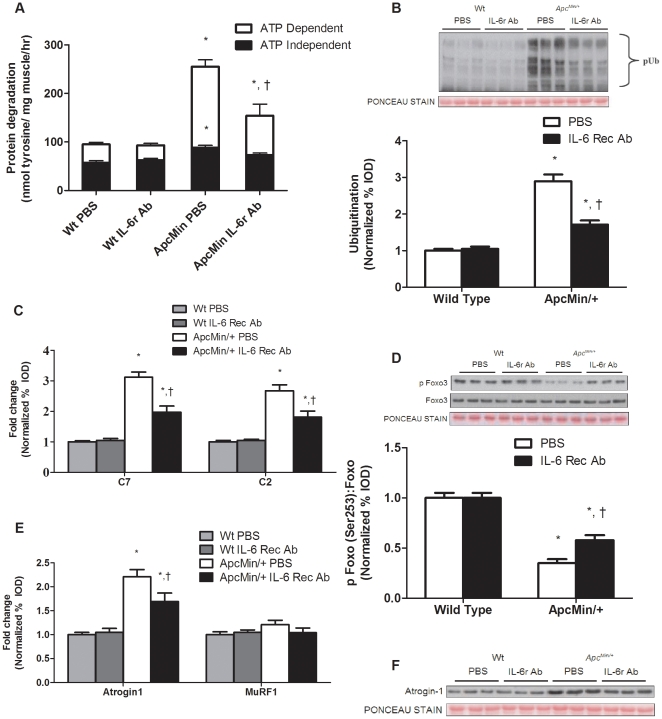
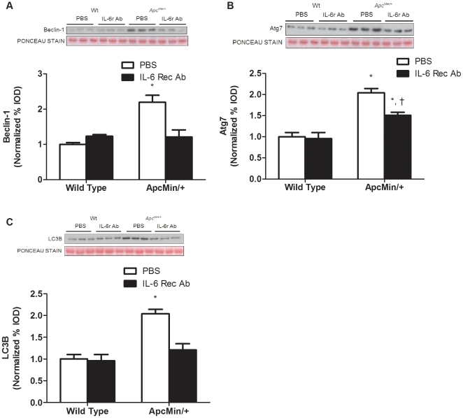
Muscle wasting that occurs with cancer cachexia is caused by an imbalance in the rates of muscle protein synthesis and degradation. The Apc(Min/+) mouse is a model of colorectal cancer that develops cachexia that is dependent on circulating IL-6. However, the IL-6 regulation of muscle protein turnover during the initiation and progression of cachexia in the Apc(Min/+) mouse is not known. Cachexia progression was studied in Apc(Min/+) mice that were either weight stable (WS) or had initial (≤5%), intermediate (6-19%), or extreme (≥20%) body weight loss. The initiation of cachexia reduced %MPS 19% and a further ∼50% with additional weight loss. Muscle IGF-1 mRNA expression and mTOR targets were suppressed with the progression of body weight loss, while muscle AMPK phosphorylation (Thr 172), AMPK activity, and raptor phosphorylation (Ser 792) were not increased with the initiation of weight loss, but were induced as cachexia progressed. ATP dependent protein degradation increased during the initiation and progression of cachexia. However, ATP independent protein degradation was not increased until cachexia had progressed beyond the initial phase. IL-6 receptor antibody administration prevented body weight loss and suppressed muscle protein degradation, without any effect on muscle %MPS or IGF-1 associated signaling. In summary, the %MPS reduction during the initiation of cachexia is associated with IGF-1/mTOR signaling repression, while muscle AMPK activation and activation of ATP independent protein degradation occur later in the progression of cachexia. IL-6 receptor antibody treatment blocked cachexia progression through the suppression of muscle protein degradation, while not rescuing the suppression of muscle protein synthesis. Attenuation of IL-6 signaling was effective in blocking the progression of cachexia, but not sufficient to reverse the process.
Overview
The claims and statistics of this paper haven’t been checked yet.
Run the check — free, no account needed. It reads the open-access text and checks the sources it cites.
Integrity signals
Registry
Cites
Statistics
Figures
Verification
Claims the paper makes
The claims and statistics of this paper haven’t been checked yet.
Run the check — free, no account needed. It reads the open-access text and checks the sources it cites.
Claims other papers make about this one
Each is checked against this paper’s own text: does it say what the citing paper says it does?
The source backs part of the claim, not all of it — or backs it for a different population, condition or comparison.
In the citing paper
“The link between IL-6/STAT3 signaling and muscle and adipose tissue wasting is well established in mouse models of cachexia (White et al., 2011; Silva et al., 2015) and has been reinforced preliminarily in clinical studies.”
From Targeting Janus Kinases and Signal Transducer and Activator of Transcription 3 To Treat Inflammation, Fibrosis, and Cancer: Rationale, Progress, and Caution (2020) · cites this paper as White et al., 2011
Experimental — accuracy not yet measured
Self-check
Percentages, sums and reported differences re-computed from the paper’s own numbers.
The self-check needs the paper’s full text.
Citations
Cites
Bradford, M. M. (1976) · Analytical Biochemistry · doi:10.1016/0003-2697(76)90527-3 · Cited by 255,302 · cited 1×
“Briefly, frozen gastrocnemius muscle was homogenized in Mueller buffer and protein concentration determined by the Bradford method [49].”
TSC2 Mediates Cellular Energy Response to Control Cell Growth and Survival
Inoki, K., Zhu, T., & Guan, K.-L. (2003) · Cell · doi:10.1016/s0092-8674(03)00929-2 · Cited by 3,914 · cited 1×
“AMPK activation can reduce muscle mTOR signaling and protein synthesis [60,61,62] through multiple mechanisms, including activation of TSC2 [23,63], direct inhibition of mTOR itself [64], or inhibition of raptor [23].”
AMPK Phosphorylation of Raptor Mediates a Metabolic Checkpoint
Gwinn, D. M., Shackelford, D. B., Egan, D. F., Mihaylova, M. M., Mery, A., Vasquez, D. S., Turk, B. E., & Shaw, R. J. (2008) · Molecular Cell · doi:10.1016/j.molcel.2008.03.003 · Cited by 3,849 · cited 4×
“AMPK can inhibit mTOR signaling through several mechanisms, including phosphorylation of raptor at Ser792 [23], which prevent binding and subsequent phosphorylation of p70S6K and 4EBP1.”
InfluentialKim, D., Sarbassov, D. D., Ali, S. M., King, J. E., Latek, R. R., Erdjument‐Bromage, H., Tempst, P., & Sabatini, D. M. (2002) · Cell · doi:10.1016/s0092-8674(02)00808-5 · Cited by 3,051 · cited 1×
“Raptor acts as a scaffolding protein to recruit downstream targets of mTOR, S6K and 4EBP1 [65,66].”
Sandri, M., Sandri, C., Gilbert, A., Skurk, C., Calabria, E., Picard, A., Walsh, K., Schiaffino, S., Lecker, S. H., & Goldberg, A. L. (2004) · Cell · doi:10.1016/s0092-8674(04)00400-3 · Cited by 3,012 · cited 1×
“IGF-1 signaling can also regulate muscle protein degradation through the suppression of forkhead box O (FOXO), whose transcriptional targets include muscle atrogin-1/MAFbx, Muscle RING Finger-1 (MuRF1) and autophagy-related genes [16,17,18].”
Evans, W., Morley, J., Argilés, J., Bales, C., Baracos, V., Guttridge, D., Jatoi, A., Kalantar-zadeh, K., Lochs, H., Mantovani, G., et al. (2008) · Clinical Nutrition · doi:10.1016/j.clnu.2008.06.013 · Cited by 2,341 · cited 1×
“Skeletal muscle mass loss is a hallmark of cachexia, and muscle mass preservation is critical for the survival of many cancer patients [1].”
Raptor, a Binding Partner of Target of Rapamycin (TOR), Mediates TOR Action
Hara, K., Maruki, Y., Long, X., Yoshino, K.-I., Oshiro, N., Sujuti, H., Tokunaga, C., Avruch, J., & Yonezawa, K. (2002) · Cell · doi:10.1016/s0092-8674(02)00833-4 · Cited by 1,810 · cited 1×
“Raptor acts as a scaffolding protein to recruit downstream targets of mTOR, S6K and 4EBP1 [65,66].”
Muscaritoli, M., Anker, S. D., Argilés, J., Aversa, Z., Bauer, J. M., Biolo, G., Boirie, Y., Bosaeus, I., Cederholm, T., Costelli, P., et al. (2010) · Clinical Nutrition · doi:10.1016/j.clnu.2009.12.004 · Cited by 1,732 · cited 1×
“accelerates during the progression of cachexia [5].”
Multiple types of skeletal muscle atrophy involve a common program of changes in gene expression
Lecker, S. H., Jagoe, R. T., Gilbert, A., Gomes, M., Baracos, V., Bailey, J., Price, S. R., Mitch, W. E., & Goldberg, A. L. (2004) · The FASEB Journal · doi:10.1096/fj.03-0610com · Cited by 1,539 · cited 3×
“expression of autophagy [26,31] and lysosomal [31] related”
Zhao, J., Brault, J. J., Schild, A., Cao, P., Sandri, M., Schiaffino, S., Lecker, S. H., & Goldberg, A. L. (2007) · Cell Metabolism · doi:10.1016/j.cmet.2007.11.004 · Cited by 1,489 · cited 2×
“IGF-1 signaling can also regulate muscle protein degradation through the suppression of forkhead box O (FOXO), whose transcriptional targets include muscle atrogin-1/MAFbx, Muscle RING Finger-1 (MuRF1) and autophagy-related genes [16,17,18].”
Autophagy is required to maintain muscle mass.
Masiero, E., Agatea, L., Mammucari, C., Blaauw, B., Loro, E., Komatsu, M., Metzger, D., Reggiani, C., Schiaffino, S., & Sandri, M. (2009) · Cell Metabolism · doi:10.1016/j.cmet.2009.10.008 · Cited by 1,282 · cited 1×
“Deletion of Atg7, a critical gene involved in autophagy, results in skeletal muscle atrophy, abnormal mitochondria and disorganization of sarcomeres [29].”
Tisdale, M. J. (2009) · Physiological Reviews · doi:10.1152/physrev.00016.2008 · Cited by 1,216 · cited 2×
“Muscle proteolysis, primarily through ubiquitin dependent mechanisms, is increased during late stage cachexia [6], while a single report has reported no difference in muscle proteolysis during the initial stages of cachexia in tumor bearing mice [7].”
AMP-activated protein kinase signaling in metabolic regulation
Long, Y. C. (2006) · Journal of Clinical Investigation · doi:10.1172/jci29044 · Cited by 1,125 · cited 1×
“AMPK is a negative regulator of cell growth, activated under circumstances of nutrient deprivation, energy depletion and cell stress [59].”
Carey, A. L., Steinberg, G. R., Macaulay, S. L., Thomas, W. G., Holmes, A. G., Ramm, G., Prelovsek, O., Hohnen-Behrens, C., Watt, M. J., James, D. E., et al. (2006) · Diabetes · doi:10.2337/db05-1404 · Cited by 898 · cited 1×
“The increase in muscle AMPK activity that corresponds with the acceleration of body weight loss could be due to surging IL-6 levels [67] and/or the reduction in muscle oxidative capacity found in severely cachectic Apc mice [40].”
Bolster, D. R., Crozier, S. J., Kimball, S. R., & Jefferson, L. S. (2002) · Journal of Biological Chemistry · doi:10.1074/jbc.c200171200 · Cited by 851 · cited 1×
“Muscle 59adenosine monophosphate-activated protein kinase (AMPK), a sensor of cellular energy status, also regulates protein synthesis [19,20,21].”
Autophagy in metazoans: cell survival in the land of plenty
Lum, J. J., DeBerardinis, R. J., & Thompson, C. B. (2005) · Nature Reviews Molecular Cell Biology · doi:10.1038/nrm1660 · Cited by 774 · cited 1×
“mitochondria and portions of the cytoplasm [28].”
Garofalo, R. S., Orena, S. J., Rafidi, K., Torchia, A. J., Stock, J. L., Hildebrandt, A. L., Coskran, T., Black, S. C., Brees, D. J., Wicks, J. R., et al. (2003) · Journal of Clinical Investigation · doi:10.1172/jci16885 · Cited by 726 · cited 1×
“An explanation worth further examination could be related to Akt phosphorylation on Akt2, which is involved in glucose metabolism [57].”
Bentzinger, C. F., Romanino, K., Cloëtta, D., Lin, S., Mascarenhas, J. B., Oliveri, F., Xia, J., Casanova, E., Costa, C. F., Brink, M., et al. (2008) · Cell Metabolism · doi:10.1016/j.cmet.2008.10.002 · Cited by 640 · cited 2×
“In skeletal muscle, raptor KO mice have a marked reduction in phosphorylation of both S6K and 4EBP1 [58].”
Mitochondrial fission and remodelling contributes to muscle atrophy
Romanello, V., Guadagnin, E., Gomes, L., Roder, I., Sandri, C., Petersen, Y., Milan, G., Masiero, E., Del Piccolo, P., Foretz, M., et al. (2010) · The EMBO Journal · doi:10.1038/emboj.2010.60 · Cited by 596 · cited 1×
“both atrogin1 and MuRF1 expression in muscle [77,78].”
Cancer cachexia is regulated by selective targeting of skeletal muscle gene products
Acharyya, S., Ladner, K. J., Nelsen, L., Damrauer, J. S., Reiser, P. J., Swoap, S. J., & D, D. C. G. (2004) · Journal of Clinical Investigation · doi:10.1172/jci20174 · Cited by 541 · cited 1×
“In both humans [68,69,70] and rodents cachexia [7,31,71,72,73] induces muscle proteolysis , and the ubiquitin dependent proteasomal system has been reported to be the dominant proteolytic pathway during cachexia [6].”
Latres, E., Amini, A. R., Amini, A. A., Griffiths, J., Martin, F. J., Wei, Y., Lin, H. C., Yancopoulos, G. D., & Glass, D. J. (2004) · Journal of Biological Chemistry · doi:10.1074/jbc.m407517200 · Cited by 538 · cited 1×
“IGF-1 signaling can also regulate muscle protein degradation through the suppression of forkhead box O (FOXO), whose transcriptional targets include muscle atrogin-1/MAFbx, Muscle RING Finger-1 (MuRF1) and autophagy-related genes [16,17,18].”
Horman, S., Browne, G. J., Krause, U., Patel, J. V., Vertommen, D., Bertrand, L., Lavoinne, A., Hue, L., Proud, C. G., & Rider, M. H. (2002) · Current Biology · doi:10.1016/s0960-9822(02)01077-1 · Cited by 467 · cited 1×
“Muscle 59adenosine monophosphate-activated protein kinase (AMPK), a sensor of cellular energy status, also regulates protein synthesis [19,20,21].”
Tsujinaka, T., Fujita, J., Ebisui, C., Yano, M., Kominami, E., Suzuki, K., Tanaka, K., Katsume, A., Ohsugi, Y., Shiozaki, H., et al. (1996) · Journal of Clinical Investigation · doi:10.1172/jci118398 · Cited by 421 · cited 1×
“Transgenic mice over-expressing IL-6 have muscle atrophy associated with increased expression of lysosomal and ubiquitin-related mRNA and proteins [34,35].”
PI3 Kinase Regulation of Skeletal Muscle Hypertrophy and Atrophy
Glass, D. J. (2010) · Current topics in microbiology and immunology · doi:10.1007/82_2010_78 · Cited by 343
Cheng, S., Fryer, L. G. D., Carling, D., & Shepherd, P. R. (2004) · Journal of Biological Chemistry · doi:10.1074/jbc.c300534200 · Cited by 340 · cited 1×
“AMPK activation can reduce muscle mTOR signaling and protein synthesis [60,61,62] through multiple mechanisms, including activation of TSC2 [23,63], direct inhibition of mTOR itself [64], or inhibition of raptor [23].”
Chan, A. Y. M., Soltys, C.-L. M., Young, M. E., Proud, C. G., & Dyck, J. R. B. (2004) · Journal of Biological Chemistry · doi:10.1074/jbc.m403528200 · Cited by 331 · cited 1×
“Muscle 59adenosine monophosphate-activated protein kinase (AMPK), a sensor of cellular energy status, also regulates protein synthesis [19,20,21].”
Interleukin-6 and cachexia in Apc Min/+ mice
Baltgalvis, K. A., Berger, F. G., Peña, M. M. O., Davis, J. M., Muga, S. J., & Carson, J. A. (2007) · American Journal of Physiology-Regulatory, Integrative and Comparative Physiology · doi:10.1152/ajpregu.00716.2007 · Cited by 283 · cited 2×
“In contrast, cachexia in the Apc mouse develops over a much longer time period [38].”
Self-citationP38 MAPK Links Oxidative Stress to Autophagy-Related Gene Expression in Cachectic Muscle Wasting
McClung, J. M., Judge, A. R., Powers, S. K., & Yan, Z. (2010) · American Journal of Physiology-Cell Physiology · doi:10.1152/ajpcell.00192.2009 · Cited by 277
Baracos, V., DeVivo, C., Hoyle, D., & Goldberg, A. (1995) · American Journal of Physiology · doi:10.1152/ajpendo.1995.268.5.e996 · Cited by 275 · cited 1×
“In both humans [68,69,70] and rodents cachexia [7,31,71,72,73] induces muscle proteolysis , and the ubiquitin dependent proteasomal system has been reported to be the dominant proteolytic pathway during cachexia [6].”
Changes in nutritional status associated with unresectable pancreatic cancer.
Wigmore, S., Plester, C., Richardson, R., & Fearon, K. (1997) · British Journal of Cancer · doi:10.1038/bjc.1997.17 · Cited by 252 · cited 1×
“Often cancer patients are not diagnosed until significant body weight loss has occurred [2], which limits treatment options in cachectic patients [3].”
Prevention and treatment of cancer cachexia: New insights into an old problem
Muscaritoli, M., Bossola, M., Aversa, Z., Bellantone, R. D. A., & Fanelli, F. R. (2005) · European Journal of Cancer · doi:10.1016/j.ejca.2005.07.026 · Cited by 241 · cited 1×
“Understanding the regulation of muscle wasting throughout the progression of cachexia is critical for developing both prevention and intervention strategies for treatment of cachexia [4].”
Interleukin-6 as a Key Regulator of Muscle Mass during Cachexia
Carson, J., & Baltgalvis, K. A. (2010) · Exercise and sport sciences reviews · doi:10.1097/jes.0b013e3181f44f11 · Cited by 217 · cited 1×
“Pro-inflammatory cytokine IL-6 has been implicated in the regulation of muscle wasting during cachexia in both humans and rodents [33].”
Self-citationNakashima, K., & Yakabe, Y. (2007) · Bioscience Biotechnology and Biochemistry · doi:10.1271/bbb.70057 · Cited by 208 · cited 13×
“In addition, AMPK activation has shown to increase protein degradation in myotubes, being associated with an increase in FOXO transcriptional targets atrogin1 and MuRF1 [24].”
InfluentialFOXO signaling is required for disuse muscle atrophy and is directly regulated by Hsp70
Judge, S. M., DODD, S. L., & Judge, A. R. (2009) · American Journal of Physiology-Cell Physiology · doi:10.1152/ajpcell.00315.2009 · Cited by 202 · cited 1×
“It has been proposed Foxo-induced transcription of atrogin1 and MuRF1 are differently regulated and MuRF1 transcription may involve additional co-factors [76].”
Asp, M. L., Tian, M., Wendel, A. A., & Belury, M. A. (2009) · International Journal of Cancer · doi:10.1002/ijc.24784 · Cited by 190
Increased Muscle Proteasome Activity Correlates With Disease Severity in Gastric Cancer Patients
Bossola, M., Muscaritoli, M., Costelli, P., Grieco, G., Bonelli, G., Pacelli, F., Fanelli, F., Doglietto, G., & Baccino, F. (2003) · Annals of Surgery · doi:10.1097/01.sla.0000055225.96357.71 · Cited by 189 · cited 2×
“In both humans [68,69,70] and rodents cachexia [7,31,71,72,73] induces muscle proteolysis , and the ubiquitin dependent proteasomal system has been reported to be the dominant proteolytic pathway during cachexia [6].”
Protein synthesis in muscle measured in vivo in cachectic patients with cancer.
Emery, P., Edwards, R., Rennie, M., Souhami, R., & Halliday, D. (1984) · British medical journal · doi:10.1136/bmj.289.6445.584 · Cited by 187 · cited 1×
“There is an overall trend in the literature to support that protein synthesis is reduced during the later stages of cachexia in both humans [8,52,53] and rodents [7,54].”
Effect of Branched-Chain Amino Acids on Muscle Atrophy in Cancer Cachexia
Eley, H. L., Russell, S. T., & Tisdale, M. J. (2007) · Biochemical Journal · doi:10.1042/bj20070651 · Cited by 184 · cited 3×
“The phosphorylation of mTOR target substrates, p70S6K and 4EBP1, corresponded with the reduction in myofibrillar protein synthesis throughout the progression of cachexia and are consistent with observations in other models of cancer cachexia [14,15].”
IGF-1 is downregulated in experimental cancer cachexia
Costelli, P., Muscaritoli, M., Bossola, M., Penna, F., Reffo, P., Bonetto, A., Busquets, S., Bonelli, G., López-Soriano, F. J., Doglietto, G. B., et al. (2006) · American Journal of Physiology-Regulatory, Integrative and Comparative Physiology · doi:10.1152/ajpregu.00104.2006 · Cited by 182 · cited 2×
“Circulating IGF-1 and muscle IGF-1 gene expression are generally decreased with wasting conditions [11] including cachexia [12,13].”
Muscle oxidative capacity during IL-6-dependent cancer cachexia
White, J. P., Baltgalvis, K. A., Puppa, M., Sato, S., Baynes, J., & Carson, J. A. (2010) · American Journal of Physiology-Regulatory, Integrative and Comparative Physiology · doi:10.1152/ajpregu.00300.2010 · Cited by 174 · cited 3×
“In support of these data, we have also previously published muscle myosin heavy chain IIa and IIb mRNA expression was not affected by cachexia in the Apc mouse [40].”
Self-citationIncreased muscle ubiquitin mRNA levels in gastric cancer patients.
Bossola, M., Muscaritoli, M., Costelli, P., Bellantone, R., Pacelli, F., Busquets, S., Argilès, J., López-Soriano, F., Civello, I. M., Baccino, F., et al. (2001) · American Journal of Physiology. Regulatory Integrative and Comparative Physiology · doi:10.1152/ajpregu.2001.280.5.r1518 · Cited by 164 · cited 1×
“Gastric cancer patients showed an increase in ubiquitin gene expression with only 5% body weight loss [75].”
Tsujinaka, T., Ebisui, C., Fujita, J., Kishibuchi, M., Morimoto, T., Ogawa, A., Katsume, A., Ohsugi, Y., Kominami, E., & Monden, M. (1995) · Biochemical and Biophysical Research Communications - BBRC · doi:10.1006/bbrc.1995.1168 · Cited by 159 · cited 1×
“Transgenic mice over-expressing IL-6 have muscle atrophy associated with increased expression of lysosomal and ubiquitin-related mRNA and proteins [34,35].”
Fan, J., Molina, P. E., Gelato, M. C., & Lang, C. H. (1994) · Endocrinology · doi:10.1210/endo.134.4.7511091 · Cited by 154 · cited 2×
“Circulating IGF-1 and muscle IGF-1 gene expression are generally decreased with wasting conditions [11] including cachexia [12,13].”
Thomson, D. M., Fick, C. A., & Gordon, S. E. (2008) · Journal of Applied Physiology · doi:10.1152/japplphysiol.00915.2007 · Cited by 153 · cited 1×
“AMPK activation can reduce muscle mTOR signaling and protein synthesis [60,61,62] through multiple mechanisms, including activation of TSC2 [23,63], direct inhibition of mTOR itself [64], or inhibition of raptor [23].”
Smith, K., & Tisdale, M. (1993) · British Journal of Cancer · doi:10.1038/bjc.1993.126 · Cited by 149 · cited 8×
“Muscle proteolysis, primarily through ubiquitin dependent mechanisms, is increased during late stage cachexia [6], while a single report has reported no difference in muscle proteolysis during the initial stages of cachexia in tumor bearing mice [7].”
InfluentialEnergy sensing by the AMP-activated protein kinase and its effects on muscle metabolism
Hardie, D. G. (2011) · Proceedings of the Nutrition Society · doi:10.1017/s0029665110003915 · Cited by 146 · cited 1×
“AMPK is activated by low cellular energy status and also muscle contraction [22].”
Increased expression of proteasome subunits in skeletal muscle of cancer patients with weight loss
Khal, J., Hine, A. V., Fearon, K. C. H., Dejong, C. H. C., & Tisdale, M. J. (2004) · The International Journal of Biochemistry & Cell Biology · doi:10.1016/j.biocel.2004.10.017 · Cited by 141 · cited 2×
“In both humans [68,69,70] and rodents cachexia [7,31,71,72,73] induces muscle proteolysis , and the ubiquitin dependent proteasomal system has been reported to be the dominant proteolytic pathway during cachexia [6].”
Muscle wasting and interleukin-6-induced atrogin-I expression in the cachectic Apc Min/+ mouse
Baltgalvis, K. A., Berger, F. G., Peña, M. M. O., Davis, J. M., White, J. P., & Carson, J. A. (2008) · Pflügers Archiv - European Journal of Physiology · doi:10.1007/s00424-008-0574-6 · Cited by 129 · cited 5×
“Apc Min/ + mice with extreme body weigh loss in our current study had significantly more pronounced weight loss and higher circulating IL-6 levels than that seen in our earlier study by Baltgalvis et al [39].”
InfluentialSelf-citationKrawiec, B. J., Nystrom, G. J., Frost, R. A., Jefferson, L. S., & Lang, C. H. (2007) · American Journal of Physiology-Endocrinology and Metabolism · doi:10.1152/ajpendo.00622.2006 · Cited by 125 · cited 1×
“both atrogin1 and MuRF1 expression in muscle [77,78].”
Kimball, S., & Jefferson, L. (2010) · Journal of Biological Chemistry · doi:10.1074/jbc.r110.137208 · Cited by 122 · cited 1×
“The muscle’s capacity to synthesize protein is responsive to many stimuli including energy status, anabolic hormones, catabolic hormones, and loading [9].”
van Hall, G., Steensberg, A., Fischer, C., Keller, C., Møller, K., Moseley, P., & Pedersen, B. (2008) · Journal of Clinical Endocrinology and Metabolism · doi:10.1210/jc.2007-2223 · Cited by 116 · cited 2×
“The administration of an IL-6 receptor antibody has effectively countered muscle wasting in tumor bearing mice [36,37].”
Muscle atrophy in experimental cancer cachexia: Is the IGF‐1 signaling pathway involved?
Penna, F., Bonetto, A., Muscaritoli, M., Costamagna, D., Minero, V. G., Bonelli, G., Fanelli, F. R., Baccino, F. M., & Costelli, P. (2009) · International Journal of Cancer · doi:10.1002/ijc.25146 · Cited by 109 · cited 4×
“Previous studies have shown Akt activity during cachexia in tumor bearing rodents to be unchanged [25] or decreased [26].”
InfluentialDepressed protein synthesis is the dominant characteristic of muscle wasting and cachexia.
Rennie, M., Edwards, R., Emery, P., Halliday, D., Lundholm, K., & Millward, D. (1983) · Clinical Physiology · doi:10.1111/j.1475-097x.1983.tb00847.x · Cited by 106 · cited 1×
“There is an overall trend in the literature to support that protein synthesis is reduced during the later stages of cachexia in both humans [8,52,53] and rodents [7,54].”
Williamson, D. L., Bolster, D. R., Kimball, S. R., & Jefferson, L. S. (2006) · American Journal of Physiology-Endocrinology and Metabolism · doi:10.1152/ajpendo.00566.2005 · Cited by 100 · cited 1×
“AMPK activation can reduce muscle mTOR signaling and protein synthesis [60,61,62] through multiple mechanisms, including activation of TSC2 [23,63], direct inhibition of mTOR itself [64], or inhibition of raptor [23].”
Decreased intestinal polyp multiplicity is related to exercise mode and gender in ApcMin/+ mice
Mehl, K. A., Davis, J. M., Clements, J. M., Berger, F. G., Pena, M. M., & Carson, J. A. (2005) · Journal of Applied Physiology · doi:10.1152/japplphysiol.00975.2004 · Cited by 99 · cited 1×
“Intestinal tissue collection was performed as described previously with a slight modification [39,42,43,44].”
Self-citationMyofibrillar protein synthesis in myostatin-deficient mice
Welle, S. L., Bhatt, K. S., & Pinkert, C. A. (2005) · American Journal of Physiology-Endocrinology and Metabolism · doi:10.1152/ajpendo.00433.2005 · Cited by 94 · cited 1×
“Thirty minutes prior to sacrifice, all mice were given an intraperitoneal injection of 150 mM 2H5-phenylalanine (Cambridge Isotope Laboratories) in a 75 mM NaCl solution at a dose of 2 ml/100 g body weight [41].”
Self-citationExpression of the ubiquitin-proteasome pathway and muscle loss in experimental cancer cachexia
Khal, J., Wyke, S. M., Russell, S. T., Hine, A. V., & Tisdale, M. J. (2005) · British Journal of Cancer · doi:10.1038/sj.bjc.6602780 · Cited by 94 · cited 1×
“Recent studies have supported these findings demonstrating an induction of the proteasomal system during later stages of cachexia, but not during initial stages of weight loss [69,70,74].”
Costelli, P., Bossola, M., Muscaritoli, M., Grieco, G., Bonelli, G., Bellantone, R., Doglietto, G., Baccino, F., & Fanelli, F. (2002) · Cytokine · doi:10.1006/cyto.2002.1036 · Cited by 93 · cited 1×
“However, tumor bearing rats treated with suramin, a global cytokine inhibitor, demonstrated a suppression of muscle proteasomal activity [80].”
Llovera, M., García-Martínez, C., López-Soriano, J., Agell, N., López-Soriano, F., Garcia, I., & Argilés, J. (1998) · Cancer Letters · doi:10.1016/s0304-3835(98)00137-2 · Cited by 87 · cited 1×
“While the importance of activated muscle protein degradation during wasting has been well established, the relationship of muscle protein synthesis regulation to muscle mass loss during cachexia has demonstrated equivocal results [50,51].”
Clark, A. S., & Mitch, W. E. (1983) · Journal of Clinical Investigation · doi:10.1172/jci111054 · Cited by 84 · cited 1×
“The reaction was stopped with trichloroacetic acid, precipitated proteins were removed by centrifugation, and free tyrosine was measured fluorometrically (450 nm excitation/550 nm emission) to calculate the rate of protein degradation [46].”
Strelkov, A. B., Fields, A., & Baracos, V. (1989) · American Journal of Physiology · doi:10.1152/ajpcell.1989.257.2.c261 · Cited by 78 · cited 1×
“In both humans [68,69,70] and rodents cachexia [7,31,71,72,73] induces muscle proteolysis , and the ubiquitin dependent proteasomal system has been reported to be the dominant proteolytic pathway during cachexia [6].”
Combaret, L., Rallière, C., Taillandier, D., Tanaka, K., & Attaix, D. (1999) · Molecular Biology Reports · doi:10.1023/a:1006955832323 · Cited by 77 · cited 1×
“While the importance of activated muscle protein degradation during wasting has been well established, the relationship of muscle protein synthesis regulation to muscle mass loss during cachexia has demonstrated equivocal results [50,51].”
Maintenance of epithelial cell differentiation: The mode of action of vitamin A
Luca, L. D., Maestri, N. E., Bonanni, F., & Nelson, D. (1972) · Cancer · doi:10.1002/1097-0142(197211)30:5 · Cited by 77
Keller, G., Grimm, V., Vogelsang, H., Bischoff, P., Mueller, J., Siewert, J. R., & Höfler, H. (1996) · International Journal of Cancer · doi:10.1002/(sici)1097-0215(19961127)68:5 · Cited by 76
Baltgalvis, K. A., Berger, F., Peña, M., Davis, J., & Carson, J. (2009) · Cancer Prevention Research · doi:10.1158/1940-6207.capr-09-0017 · Cited by 73 · cited 1×
“Intestinal tissue collection was performed as described previously with a slight modification [39,42,43,44].”
Self-citationUbiquitin and proteasome gene expression is increased in skeletal muscle of slim AIDS patients.
Llovera, M., García-Martínez, C., Agell, N., López-Soriano, F., Authier, F., Ghérardi, R., & Argilés, J. (1998) · International Journal of Molecular Medicine · doi:10.3892/ijmm.2.1.69 · Cited by 68 · cited 1×
“In both humans [68,69,70] and rodents cachexia [7,31,71,72,73] induces muscle proteolysis , and the ubiquitin dependent proteasomal system has been reported to be the dominant proteolytic pathway during cachexia [6].”
Protein synthesis measured in vivo in muscle and liver of cachectic tumor-bearing mice.
Emery, P., Lovell, L., & Rennie, M. J. (1984) · Cancer Research · doi:10.1016/0278-2391(85)90177-6 · Cited by 66 · cited 1×
“There is an overall trend in the literature to support that protein synthesis is reduced during the later stages of cachexia in both humans [8,52,53] and rodents [7,54].”
Elevated levels of telomerase activity in malignant pheochromocytoma
Kubota, Y., Nakada, T., Sasagawa, I., Yanai, H., & Itoh, K. (1998) · Cancer · doi:10.1002/(sici)1097-0142(19980101)82:1 · Cited by 65
Effect of nandrolone decanoate administration on recovery from bupivacaine-induced muscle injury.
White, J., Baltgalvis, K. A., Sato, S., Wilson, L., & Carson, J. A. (2009) · Journal of applied physiology · doi:10.1152/japplphysiol.00668.2009 · Cited by 62 · cited 1×
“Western blot analysis was performed as previously described [48].”
Self-citationAbnormal expression of lysosomal cysteine proteinases in muscle wasting diseases.
Katunuma, N., & Kominami, E. (1987) · Reviews of Physiology, Biochemistry and Pharmacology · doi:10.1007/bfb0034070 · Cited by 62 · cited 1×
“There have been reports that show autophagy-related genes increase during muscle dystrophy [30], diabetes [31], sepsis-induced wasting [27] and cancer related cachexia [31,32].”
Wang, H., Casaburi, R., Taylor, W. E., Aboellail, H., Storer, T., & Kopple, J. (2005) · Kidney International · doi:10.1111/j.1523-1755.2005.00409.x · Cited by 59 · cited 1×
“The reduction in both circulating and muscle expression of IGF-1 has been shown in various wasting condition [11,12,13,25,55].”
Pruznak, A. M., Kazi, A. A., Frost, R. A., Vary, T. C., & Lang, C. H. (2008) · Journal of Nutrition · doi:10.1093/jn/138.10.1887 · Cited by 58
Effect of exercise on biological pathways in ApcMin/+ mouse intestinal polyps.
Baltgalvis, K. A., Berger, F. G., Peña, M. M. O., Davis, J. M., & Carson, J. A. (2008) · Journal of applied physiology · doi:10.1152/japplphysiol.00955.2007 · Cited by 54 · cited 1×
“Intestinal tissue collection was performed as described previously with a slight modification [39,42,43,44].”
Self-citationWhite, J. P., Reecy, J. M., Washington, T. A., Sato, S., Le, M. E., Davis, J. M., Wilson, L. B., & Carson, J. A. (2009) · Acta Physiologica · doi:10.1111/j.1748-1716.2009.02029.x · Cited by 53 · cited 1×
“RNA isolation, cDNA synthesis, and real-time PCR were performed as previously described [47], using reagents from Applied Biosystems (Foster City, CA).”
Self-citationHu, J., Klein, J., Du, J., & Wang, X.-N. H. (2008) · Endocrinology · doi:10.1210/en.2008-0132 · Cited by 51 · cited 1×
“To assay the degradation of soluble muscle protein, a modified proteasomal proteolysis assay reported by Hu et al [45] was be”
Clinical anticachexia treatments.
Tisdale, M. (2006) · Nutrition in clinical practice · doi:10.1177/0115426506021002168 · Cited by 41 · cited 1×
“Often cancer patients are not diagnosed until significant body weight loss has occurred [2], which limits treatment options in cachectic patients [3].”
Changes in protein turnover, IGF‐I and IGF binding proteins in children with cancer
Attard-Montalto, S., Camacho-Hübner, C., Cotterill, A., D’Souza-Li, L., Daley, S., Bartlett, K., Halliday, D., & Eden, O. (1998) · Acta paediatrica · doi:10.1111/j.1651-2227.1998.tb01386.x · Cited by 37 · cited 2×
“Circulating IGF-1 and muscle IGF-1 gene expression are generally decreased with wasting conditions [11] including cachexia [12,13].”
Attenuation of skeletal muscle atrophy in cancer cachexia by d-myo-inositol 1,2,6-triphosphate
Russell, S., Siren, P. M., Sirén, M., & Tisdale, M. J. (2009) · Cancer Chemotherapy and Pharmacology · doi:10.1007/s00280-008-0899-z · Cited by 30 · cited 2×
“The phosphorylation of mTOR target substrates, p70S6K and 4EBP1, corresponded with the reduction in myofibrillar protein synthesis throughout the progression of cachexia and are consistent with observations in other models of cancer cachexia [14,15].”
Research works printed but not linked: 2
- ML Asp, 2010, Evidence for the contribution of insulin resistance to the development of cachexia in tumor-bearing mice., Int J Cancer, 126, 756, 10.1002/ijc.24784 · DOI
- RA Frost, 2007, Protein kinase B/Akt: a nexus of growth factor and cytokine signaling in determining muscle mass., J Appl Physiol, 103, 378, 10.1152/japplphysiol.00089.2007 · DOI · Paper page
Cited by
Sorted by citations
Cancer Cachexia: Mediators, Signaling, and Metabolic Pathways
Fearon, K. C. H., Glass, D. J., & Guttridge, D. C. (2012) · Cell Metabolism · doi:10.1016/j.cmet.2012.06.011 · Cited by 1,244
“IL-6 receptor antibody blocked cachexia progression via suppression of muscle protein degradation, while not rescuing the suppression of synthesis (White et al., 2011).”
BackgroundGreen justice or just green? Provision of urban green spaces in Berlin, Germany
Kabisch, N., & Haase, D. (2013) · Landscape and Urban Planning · doi:10.1016/j.landurbplan.2013.11.016 · Cited by 953
A PGC-1α isoform induced by resistance training regulates skeletal muscle hypertrophy
Ruas, J. L., White, J. P., Rao, R. R., Kleiner, S., Brannan, K. T., Harrison, B. C., Greene, N. P., Wu, J., Estall, J. L., Irving, B. A., et al. (2012) · Cell · doi:10.1016/j.cell.2012.10.050 · Cited by 677
“Cancer-induced cachexia has been shown to occur concomitantly with dysregulation of myostatin and IGF1 expression in muscle (Busquets et al., 2012; Fearon et al., 2012; White et al., 2011).”
Self-citationMechanisms of IGF-1-Mediated Regulation of Skeletal Muscle Hypertrophy and Atrophy
Yoshida, T., & Delafontaine, P. (2020) · Cells · doi:10.3390/cells9091970 · Cited by 772
Bonetto, A., Aydogdu, T., Jin, X., Zhang, Z., Zhan, R., Puzis, L., Koniaris, L. G., & Zimmers, T. A. (2012) · American Journal of Physiology-Endocrinology and Metabolism · doi:10.1152/ajpendo.00039.2012 · Cited by 463
Metadata
- Authors
- White, J. P., Baynes, J. W., Welle, S. L., Kostek, M. C., Matesic, L. E., Sato, S., & Carson, J. A.
- Affiliations
- University of South Carolina (US)
- University of Rochester Medicine (US)
- Corresponding author
- Carson, James A.
- Venue
- PLoS ONE
- Publisher
- Public Library of Science (PLoS)
- Published
- 2011-09-19
- Volume
- 6
- Issue
- 9
- Pages
- e24650
- ISSN
- 1932-6203 (ISSN-L)
- PMC
- PMC3176277
- OpenAlex
- W2151316758
- Cited by
- 260
- References
- 82
- Citations by year
- 2026: 8 · 2025: 16 · 2024: 21 · 2023: 22 · 2022: 20 · 2021: 29 · 2020: 24 · 2019: 18 · 2018: 18 · 2017: 19 · 2016: 11 · 2015: 11 · 2014: 12 · 2013: 17 · 2012: 12
- FWCI
- 5.59
- Type
- journal article
- Language
- en
- Open access
- gold
- doi.org
- Locations
- PLoS ONE (publishedVersion, cc-by)
- Europe PMC (PubMed Central) (publishedVersion, cc-by)
- PubMed Central (publishedVersion, cc-by)
- PubMed (publishedVersion)
- Scholar Commons (University of South Carolina) (submittedVersion)
- DOAJ (DOAJ: Directory of Open Access Journals) (submittedVersion)
- Figshare (submittedVersion, cc-by)
- Funders
- National Institutes of Health
- Chugai Pharmaceutical
- National Cancer Institute (R01CA121249)
- National Center for Research Resources (P20RR017698)
- Topics
- Life Sciences › Biochemistry, Genetics and Molecular Biology › Molecular Biology › Muscle Physiology and Disorders
- Health Sciences › Medicine › Physiology (medical) › Nutrition and Health in Aging
- Health Sciences › Nursing › Nutrition and Dietetics › Muscle metabolism and nutrition
- Keywords
- cancer cachexia, skeletal muscle protein turnover, muscle protein synthesis, muscle protein degradation, colorectal cancer, IL-6 signaling, AMPK, Raptor phosphorylation, muscle atrophy, weight loss, IL-6 receptor antibody
- MeSH terms
- Adenosine Triphosphate, Animals, Body Weight, Cachexia, Enzyme Activation, Inflammation, Insulin-Like Growth Factor I, Mice, Inbred C57BL, Mice, Mutant Strains, Muscle Proteins, Myofibrils, Neoplasms, Organ Size, Phosphorylation, Protein Biosynthesis, Signal Transduction, Interleukin-6, Disease Progression, Muscle, Skeletal, Adiposity, Mice, AMP-Activated Protein Kinases, TOR Serine-Threonine Kinases, Proteolysis
About these checks
Registry, retraction, Cites and statistics results are computed, not judged, with one exception: a printed reference that no index lists is read once by a model before it counts as a citation error. Claims are extracted by a model and kept only when their quote appears verbatim in the text; each cited source is then checked, with a model’s help, for whether it says what the claim relies on. Those verdicts can be wrong — read the claim’s quote and the cited source before relying on one.
Statistics and arithmetic are recomputed, not judged.
Confidence bands are shown only for fields calibrated on our annotated test set. None is calibrated yet, so no band is shown.
What counts as a claim ›How we measure confidence ›How we check this ›
Are you an author, or do you see a problem with this page? Request a review
