The DOI matches its Crossref record; 47 of the 49 references it cites were found in a registry or an index.
Comparative Analysis of the Subventricular Zone in Rat, Ferret and Macaque: Evidence for an Outer Subventricular Zone in Rodents
Open accessAbstract
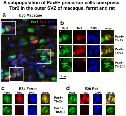
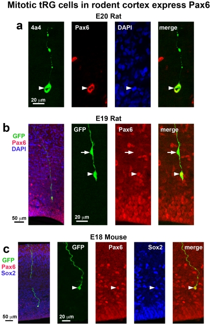
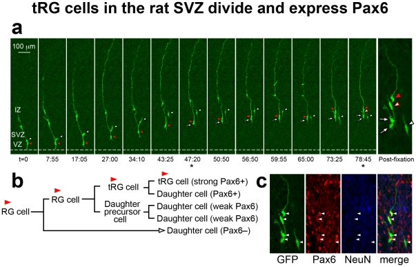
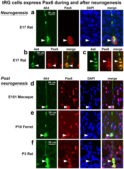
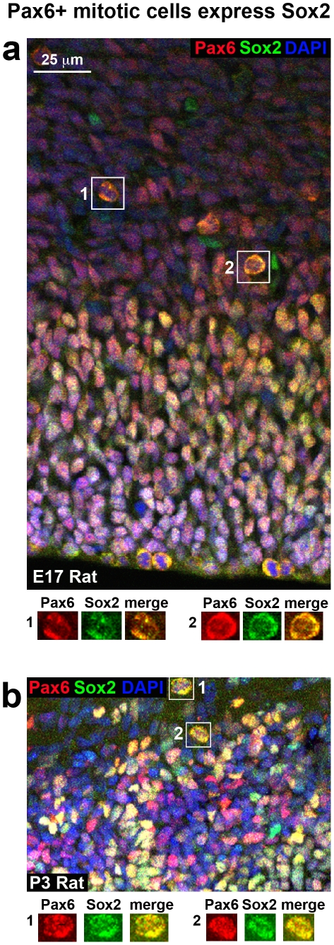
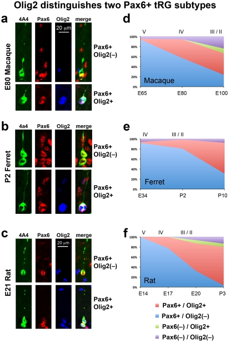
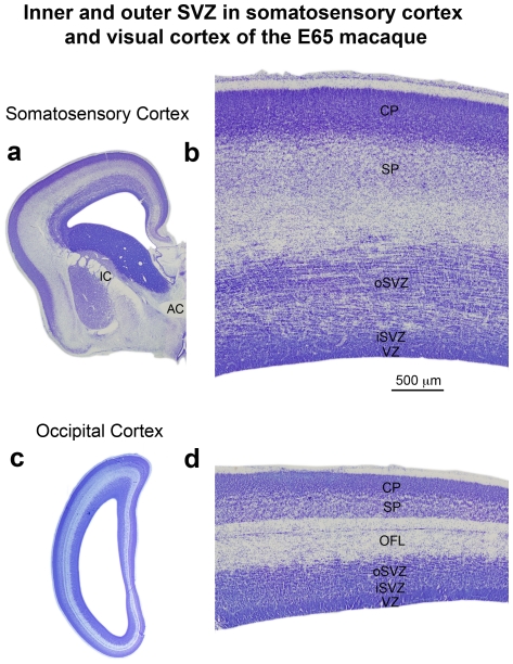
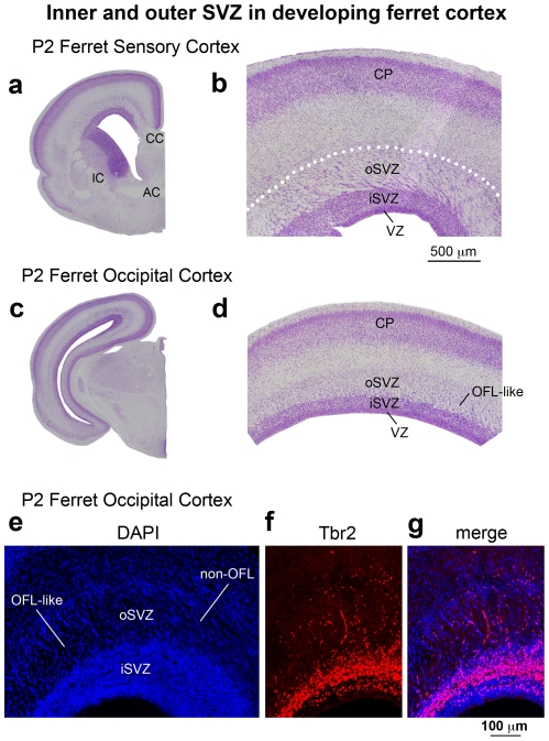
The mammalian cerebral cortex arises from precursor cells that reside in a proliferative region surrounding the lateral ventricles of the developing brain. Recent work has shown that precursor cells in the subventricular zone (SVZ) provide a major contribution to prenatal cortical neurogenesis, and that the SVZ is significantly thicker in gyrencephalic mammals such as primates than it is in lissencephalic mammals including rodents. Identifying characteristics that are shared by or that distinguish cortical precursor cells across mammalian species will shed light on factors that regulate cortical neurogenesis and may point toward mechanisms that underlie the evolutionary expansion of the neocortex in gyrencephalic mammals. We immunostained sections of the developing cerebral cortex from lissencephalic rats, and from gyrencephalic ferrets and macaques to compare the distribution of precursor cell types in each species. We also performed time-lapse imaging of precursor cells in the developing rat neocortex. We show that the distribution of Pax6+ and Tbr2+ precursor cells is similar in lissencephalic rat and gyrencephalic ferret, and different in the gyrencephalic cortex of macaque. We show that mitotic Pax6+ translocating radial glial cells (tRG) are present in the cerebral cortex of each species during and after neurogenesis, demonstrating that the function of Pax6+ tRG cells is not restricted to neurogenesis. Furthermore, we show that Olig2 expression distinguishes two distinct subtypes of Pax6+ tRG cells. Finally we present a novel method for discriminating the inner and outer SVZ across mammalian species and show that the key cytoarchitectural features and cell types that define the outer SVZ in developing primates are present in the developing rat neocortex. Our data demonstrate that the developing rat cerebral cortex possesses an outer subventricular zone during late stages of cortical neurogenesis and that the developing rodent cortex shares important features with that of primates.
Overview
The claims and statistics of this paper haven’t been checked yet.
Run the check — free, no account needed. It reads the open-access text and checks the sources it cites.
Integrity signals
Registry
Cites
Statistics
Figures
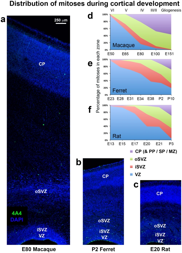
![Figure 1 The distribution of mitoses during cortical development is similar in ferrets and rats, but different in macaques. (a–c) Coronal sections of somatosensory cortex from E80 macaque, P2 ferret and E20 rat immunostained for phosphorylated vimentin (4A4, green) to label dividing cells and counterstained with DAPI (blue). Images are displayed at the same scale. There are a significant number of abventricular mitoses in each species. (d–f) Graphs showing changes in the distribution of mitoses during development of the neocortex. The stage of development is shown at the bottom of the graphs. The approximate cortical layer generated at each stage of development is indicated along the top of the macaque graph, and applies to each graph. (d) Early in macaque neurogenesis the majority of mitoses shifted from the ventricular zone (VZ) to abventricular locations. By E80, when layer 4 neurons are being generated [20], less than 20% of mitoses remained in the VZ and 57% were located in the outer subventricular zone (oSVZ). (e) The distribution of mitoses in the gyrencephalic ferret did not shift away from the VZ during early stages of neurogenesis as in macaque. At E34 during generation of layer 4 neurons [19], greater than half of all mitoses were still located in the VZ. (f) The distribution of mitoses in the lissencephalic rat cortex was similar to that in the gyrencephalic ferret cortex. At E21, which represents the genesis of layer 2 neurons in somatosensory cortex [21], 35% of mitoses were located superficial to the SVZ in a zone that we term the oSVZ. Legend indicates histological zones: VZ: blue; inner SVZ (iSVZ): red; oSVZ: green; cortical plate (CP)/preplate (PP)/subplate (SP)/marginal zone (MZ): purple. Scale bar in (a) applies to (a–c).](/paper-figures/c02a1d51cf384a49baab2b18f393f123/comparative-analysis-of-the-subventricular-zone-in-rat-ferret-and-macaque-figure-1.jpg)
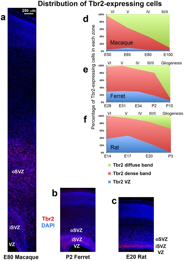
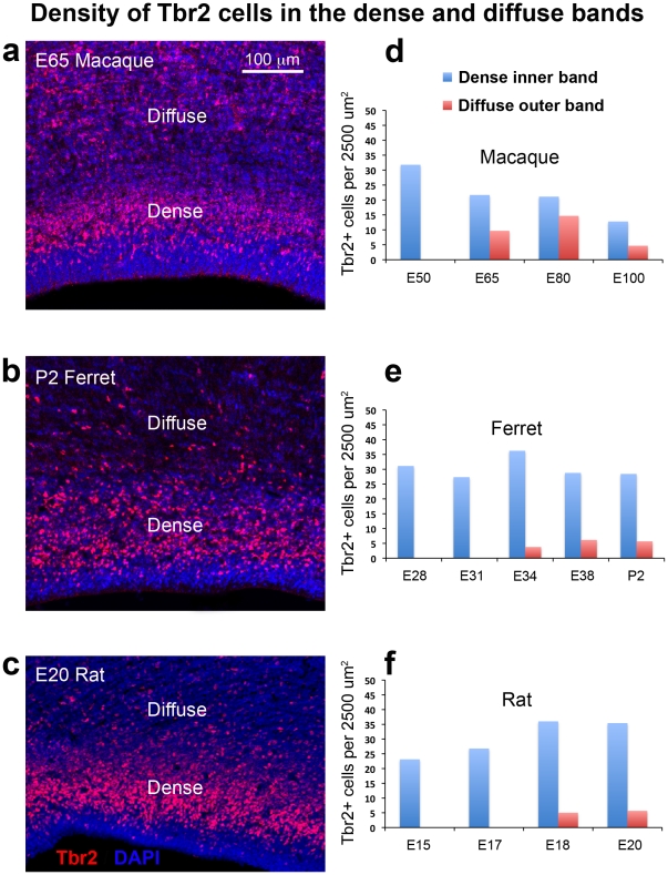
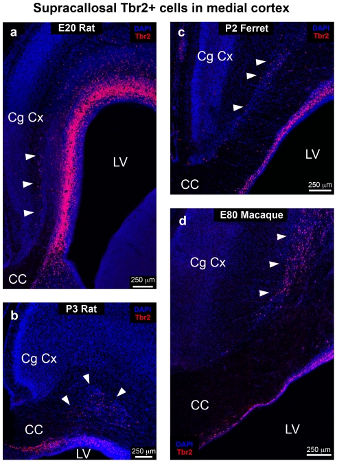
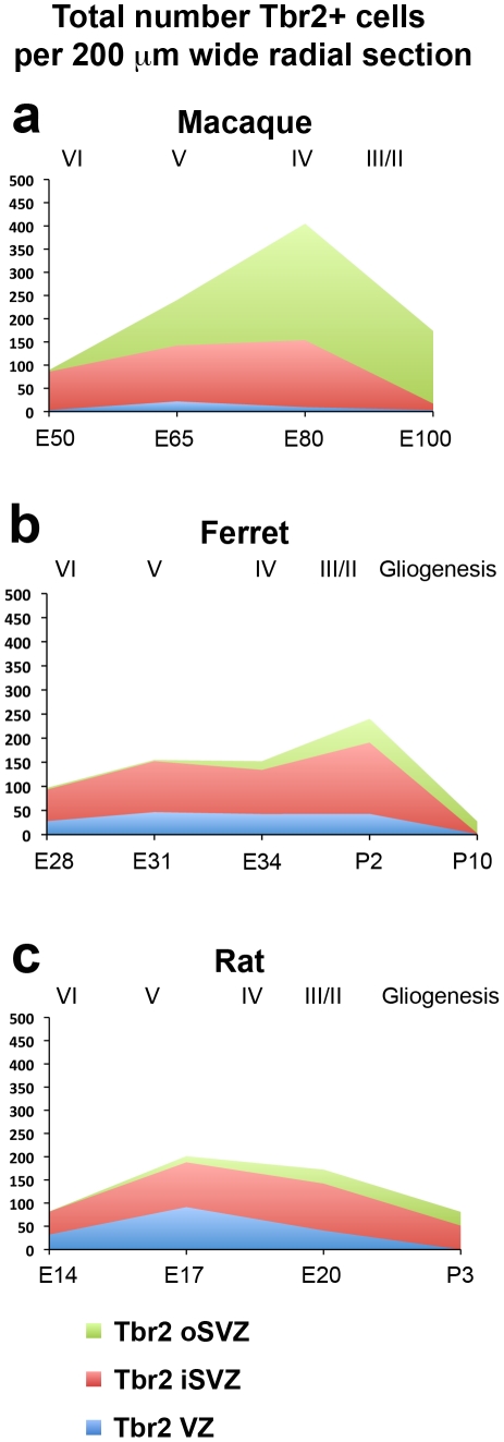
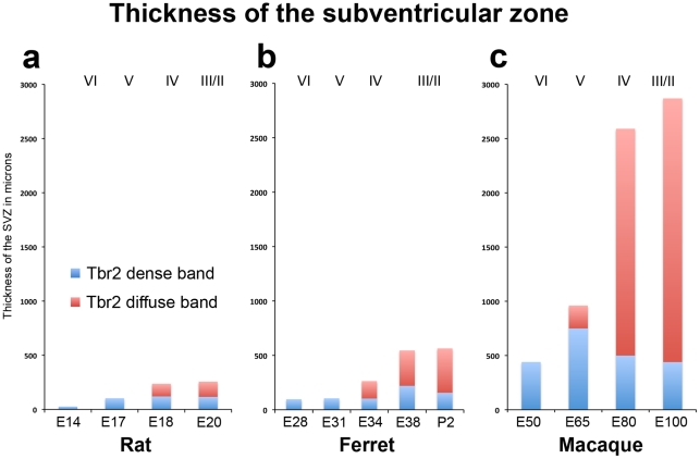
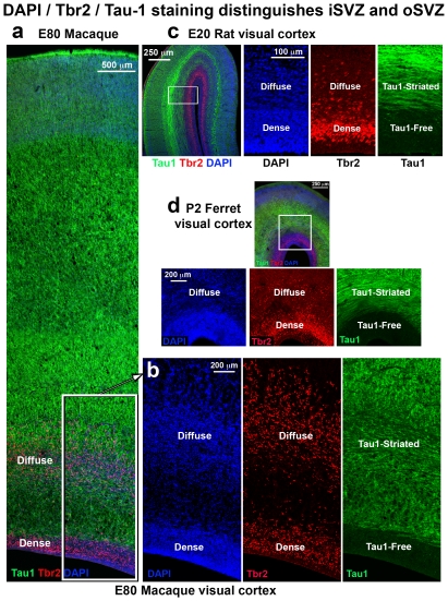
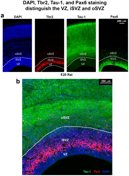
![Figure 2 The distribution of Tbr2+ cells during cortical development is similar in ferrets and rats, but different in macaques. (a–c) Coronal sections of somatosensory cortex from E80 macaque, P2 ferret and E20 rat immunostained for Tbr2 (red) and counterstained with DAPI (blue). Images are displayed at the same scale. In each species a dense inner band of Tbr2+ cells was located within the cell dense zone surrounding the lateral ventricle visualized with DAPI. The diffuse outer band of Tbr2 expression was located within a striated zone marked by streams of cells organized in tangential clusters. (d–f) Graphs showing changes in the distribution of Tbr2+ cells during development of the cerebral cortex. The stage of development is shown at the bottom of the graphs, and the approximate cortical layer generated at each stage of development is indicated along the top of each graph. (d) In macaque the distribution of Tbr2+ cells progressively shifted to the outer subventricular zone (oSVZ) during development. By E80, during genesis of layer 4 neurons [20], 59% of Tbr2+ cells were located in the oSVZ. (e) The distribution of Tbr2+ cells in the gyrencephalic ferret shifted to the oSVZ more slowly in comparison to macaque. At P2 during genesis of layer 2 neurons [19], 62% of Tbr2+ cells remained in the inner SVZ (iSVZ). (f) The distribution of Tbr2+ cells in the lissencephalic rat was similar to that of the gyrencephalic ferret. At E20 during genesis of upper layer neurons [21], the majority of Tbr2+ cells (59%) were located in the iSVZ, and 17% were located in the oSVZ. Legend indicates histological zones: VZ: blue; iSVZ: red; oSVZ: green. Scale bar in (a) applies to (a–c).](/paper-figures/c9b7abfae4d645b89d7fb6a55b04e0dc/comparative-analysis-of-the-subventricular-zone-in-rat-ferret-and-macaque-figure-2.jpg)


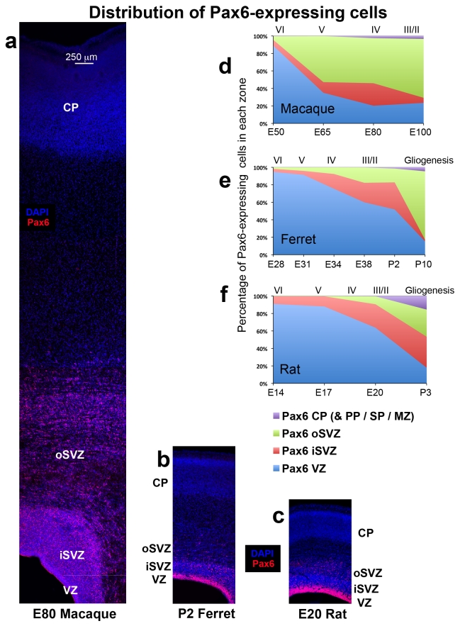
![Figure 9 The distribution of Pax6+ cells during cortical development is similar in ferrets and rats, but different in macaques. (a–c) Coronal sections of somatosensory cortex from E80 macaque, P2 ferret and E20 rat immunostained for Pax6 (red) and counterstained with DAPI (blue). Images are displayed at the same scale. In each species a dense inner band of Pax6+ cells was colocalized with the ventricular zone (VZ), and a diffuse band of Pax6-expressing cells extended outward through the inner subventricular zone (iSVZ) and the outer SVZ (oSVZ). The iSVZ and oSVZ were identified based on the pattern of DAPI staining as described above. (d–f) Graphs showing changes in the distribution of Pax6+ cells during development of the somatosensory cortex. The stage of development is shown at the bottom of the graphs, and the approximate cortical layer generated at each stage of development is indicated along the top of each graph. (d) In macaque the distribution of Pax6+ cells rapidly shifted to the oSVZ. At E50 nearly 90% of Pax6+ cells were located in the VZ. But by E65 during production of layer 5 neurons [20], the majority of Pax6+ cells (60%) were located in the oSVZ. (e) The distribution of Pax6+ cells in the gyrencephalic ferret shifted to the oSVZ much more slowly than in macaque. At P2 during genesis of layer 2 neurons [19], 52% of Pax6+ cells remained in the VZ. (f) The distribution of Pax6+ cells in the lissencephalic rat was similar to that of the gyrencephalic ferret. At the end of neurogenesis on E20 during production of layer 2 neurons [21], the majority of Pax6+ cells (64%) were still located in the VZ. Legend indicates histological zones: VZ: blue; iSVZ: red; oSVZ: green. Cortical plate (CP)/preplate (PP)/subplate (SP)/marginal zone (MZ): purple. Scale bar in (a) applies to (a–c).](/paper-figures/17f58376f6c04a0aa1f87b353c62b018/comparative-analysis-of-the-subventricular-zone-in-rat-ferret-and-macaque-figure-9.jpg)

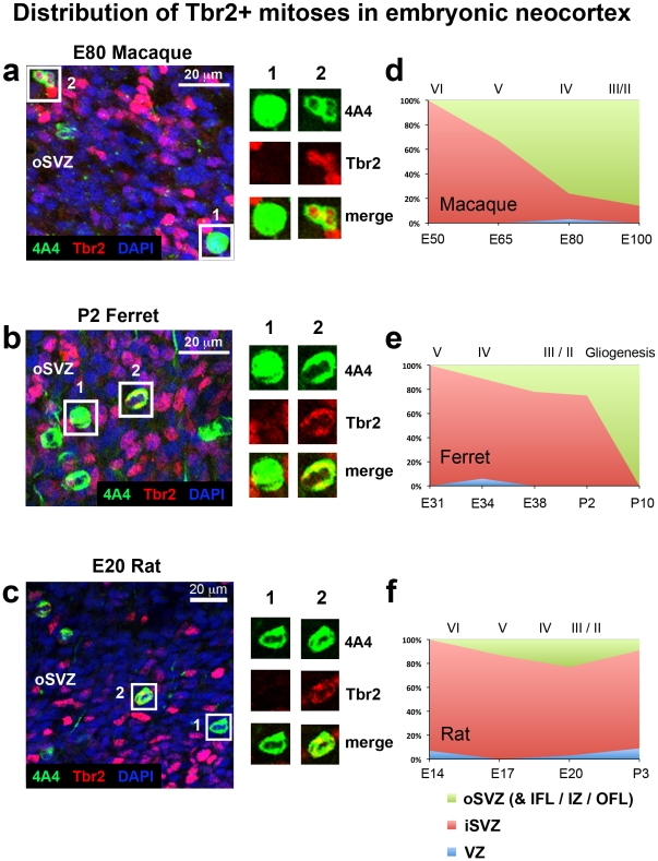
![Figure 11 The distribution of Tbr2+ mitoses shifts to the outer subventricular zone (oSVZ) during early stages of cortical development in macaque, but not in ferret or rat. (a–c) Images taken from coronal sections of the E80 macaque, P2 ferret, and E20 rat oSVZ. Tissue was immunostained for 4A4 (green), Tbr2 (red) and counterstained with DAPI (blue). Boxes to the right highlight examples of 4A4+ mitotic cells that either do not express Tbr2 (#1) or that do express Tbr2 (#2). (d–f) Graphs showing changes in the distribution of Tbr2+ mitoses during development of the somatosensory cortex. The stage of development is shown at the bottom of the graphs, and the approximate cortical layer generated at each stage of development is indicated along the top of each graph. (d) At E50 in the macaque all Tbr2+ mitoses were located in the inner SVZ (iSVZ). There was a steady decrease in the proportion of Tbr2+ divisions occurring in the iSVZ. By E80 the majority of Tbr2+ mitoses (76%) had shifted to the oSVZ. (e) In ferret the majority of Tbr2+ mitoses were located in the iSVZ throughout neurogenesis in the somatosensory cortex. At P2, during neurogenesis of layer 2 neurons [19], 73% of Tbr2+ mitoses were still located in the iSVZ. (f) The pattern observed in rat was similar to that of the ferret. The majority of Tbr2+ mitoses were located in the iSVZ throughout the period of cortical neurogenesis. At P2, during production of layer 2 neurons [21], there was a similar number of Tbr2+ mitoses located in the rat iSVZ (74%) as was observed in the ferret. Legend indicates histological zones: VZ: blue; iSVZ: red; oSVZ: green.](/paper-figures/abb5eb3f3e844886b1369d581c3646d3/comparative-analysis-of-the-subventricular-zone-in-rat-ferret-and-macaque-figure-11.jpg)
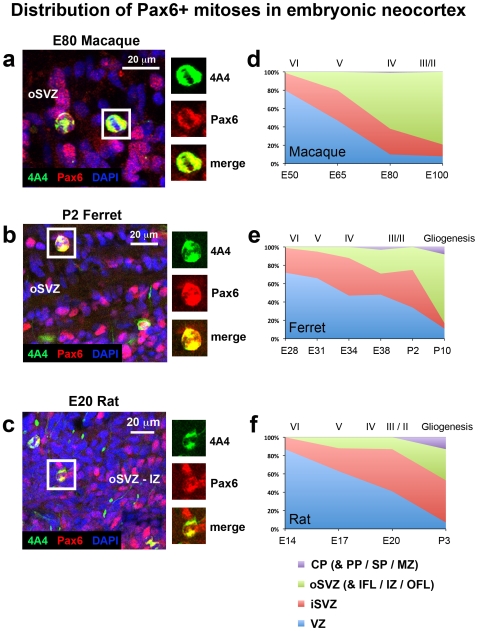
![Figure 12 The distribution of Pax6+ mitoses shifts from the ventricular zone (VZ) to the subventricular zone (oSVZ) during early stages of cortical development in macaque, but not in ferret or rat. (a–c) Images taken from coronal sections of the E80 macaque, P2 ferret and E20 rat oSVZ. Tissue was immunostained for 4A4 (green), Pax6 (red) and counterstained with DAPI (blue). Boxes to the right highlight examples of 4A4+ mitotic cells that express Pax6. (d–f) Graphs showing changes in the distribution of Pax6+ mitoses during development of the somatosensory cortex. The stage of development is shown at the bottom of the graphs, and the approximate cortical layer generated at each stage of development is indicated along the top of each graph. (d) At E50 in the macaque nearly 90% of Pax6+ mitoses were located in the VZ. There was a steady shift in the distribution of mitoses to the outer SVZ (oSVZ). By E80 the majority of Pax6+ mitoses (61%) were located in the oSVZ. (e) In ferret the largest proportion of Pax6+ mitoses were located in the VZ until late stages of neurogenesis. At E38, during neurogenesis of layer 2 neurons [19], 48% of Pax6+ mitoses were still located in the VZ, 23% were located in the iSVZ and 26% in the oSVZ. (f) A similar pattern was observed in the rat. The majority of Pax6+ mitoses were located in the VZ until E20. At E20 40% of Pax6+ mitoses were located in the VZ, 45% were in the iSVZ and 13% in the oSVZ. Legend indicates histological zones: VZ: blue; iSVZ: red; oSVZ: green; CP: purple. PP, preplate; SP, subplate; MZ, marginal zone.](/paper-figures/e8abceb82fea4066982e3b6cc3fac2ed/comparative-analysis-of-the-subventricular-zone-in-rat-ferret-and-macaque-figure-12.jpg)


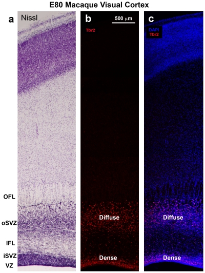
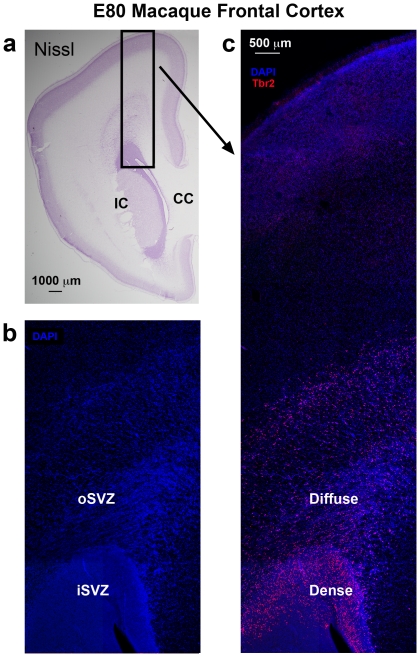
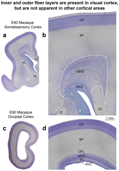
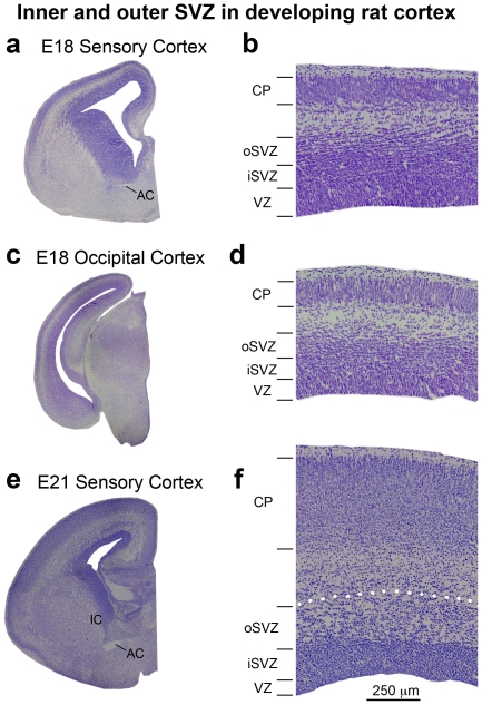
![Figure 20 The inner fiber layer (IFL) and outer fiber layer (OFL) are present in the E80 macaque visual cortex but are not apparent in other cortical areas. (a,c) Nissl-stained coronal sections of somatosensory and visual cortex taken from E80 macaque. (b,d) Higher magnification images taken from the sections shown in (a) and (c). Dotted line in (b) represents the outer boundary of the outer subventricular zone (oSVZ) in somatosensory cortex determined through Tbr2 immunostaining on adjacent sections (see Fig. 5). The outer fiber layer (OFL) and inner fiber layer (IFL) are visible in the visual cortex but are not apparent in cortical areas rostral to the occipital lobe. In somatosensory cortex the boundary between the inner SVZ (iSVZ) and oSVZ can be visualized in Nissl stained tissue as a sharp border created by differences in cell density. In the visual cortex the IFL divides the iSVZ from the oSVZ. In somatosensory cortex the oSVZ extends much farther from the ventricle than it does in visual cortex and is characterized by a striated appearance. In the E80 macaque the ventricular zone has become very thin and the cell dense proliferative zone that surrounds the lateral ventricle consists almost entirely of iSVZ. The subplate (SP) was identified according to Smart et al 2002 [9]. Scale bar in (b) applies to (d). CC, corpus callosum; IC, internal capsule; AC, anterior commissure; CP, cortical plate.](/paper-figures/7a7e0b4117ae43d596e4d1771803751a/comparative-analysis-of-the-subventricular-zone-in-rat-ferret-and-macaque-figure-20.jpg)


Verification
Claims the paper makes
The claims and statistics of this paper haven’t been checked yet.
Run the check — free, no account needed. It reads the open-access text and checks the sources it cites.
Claims other papers make about this one
Each is checked against this paper’s own text: does it say what the citing paper says it does?
In the citing paper
“oRGs are present in large numbers in gyrencephalic primates22,23,40–44 and are considered pivotal to the evolutionary increase in human cortex size and complexity45,46.”
From The use of brain organoids to investigate neural development and disease (2017) · cites this paper as [44]
Self-check
Percentages, sums and reported differences re-computed from the paper’s own numbers.
The self-check needs the paper’s full text.
Citations
Cites
Specification of Cerebral Cortical Areas
Rakic, P. (1988) · Science · doi:10.1126/science.3291116 · Cited by 3,127 · cited 1×
“: [43]), but remain to be fully understood.”
BackgroundNoctor, S. C., Martínez-Cerdeño, V., Ivic, L., & Kriegstein, A. R. (2004) · Nature Neuroscience · doi:10.1038/nn1172 · Cited by 2,229
Self-citationNeurons derived from radial glial cells establish radial units in neocortex
Noctor, S. C., Flint, A. C., Weissman, T. A., Dammerman, R. S., & Kriegstein, A. R. (2001) · Nature · doi:10.1038/35055553 · Cited by 2,016
Self-citationA tension-based theory of morphogenesis and compact wiring in the central nervous system
Essen, D. C. V. (1997) · Nature · doi:10.1038/385313a0 · Cited by 1,668
Neurogenic radial glia in the outer subventricular zone of human neocortex
Hansen, D. V., Lui, J. H., Parker, P. R. L., & Kriegstein, A. R. (2010) · Nature · doi:10.1038/nature08845 · Cited by 1,388
Englund, C., Fink, A., Lau, C., Pham, D., Daza, R. A. M., Bulfone, A., Kowalczyk, T., & Hevner, R. F. (2005) · The Journal of Neuroscience · doi:10.1523/jneurosci.2899-04.2005 · Cited by 1,261
Rakic, P. (1974) · Science · doi:10.1126/science.183.4123.425 · Cited by 1,161
Haubensak, W. E., Attardo, A., Denk, W. T., & Huttner, W. Β. (2004) · Proceedings of the National Academy of Sciences · doi:10.1073/pnas.0308600100 · Cited by 1,004
OSVZ progenitors of human and ferret neocortex are epithelial-like and expand by integrin signaling
Fietz, S. A., Kelava, I., Vogt, J., Wilsch-Bräuninger, M., Stenzel, D., Fish, J. L., Corbeil, D., Riehn, A., Distler, W., Nitsch, R., et al. (2010) · Nature Neuroscience · doi:10.1038/nn.2553 · Cited by 830
Asymmetric production of surface-dividing and non-surface-dividing cortical progenitor cells
Miyata, T., Kawaguchi, A., Saito, K., Kawano, M., Muto, T., & Ogawa, M. (2004) · Development · doi:10.1242/dev.01173 · Cited by 791
Cited by
Sorted by citations
Items 201–203 of 203
Sawada, K. (2026) · Frontiers in Neurology · doi:10.3389/fneur.2026.1796867 · Cited by 1
Cornelius, J. A., Shivakumar, M., Kim, T., Cervantes, O., Kapur, R. P., Huang, H., Zhao, H., Rosario, B. D., Li, A., Li, R., et al. (2026) · Frontiers in Neuroscience · doi:10.3389/fnins.2026.1785001
Coordinated neural progenitor adaptations contribute to primate neocortical downscaling
Tynianskaia, L., Betancourt, C. M. B., Grewe, E. M., Kniep, J., Liutikaite, N., Eşiyok, N., Göloğlu, İ. B., Heide, S., Rüger, N., Lindenwald, D. L., et al. (2026) · Science Advances · doi:10.1126/sciadv.aeg5563 · Cited by 1
Metadata
- Authors
- Martínez-Cerdeño, V., Cunningham, C. L., Camacho, J., Antczak, J. L., Prakash, A. N., Cziep, M. E., Walker, A. I., & Noctor, S. C.
- Affiliations
- University of California, Davis (US)
- Shriners Hospitals for Children - Northern California (US)
- Brigham Young University (US)
- University of California Davis Medical Center (US)
- Corresponding author
- Noctor, Stephen C.
- Venue
- PLoS ONE
- Publisher
- Public Library of Science (PLoS)
- Published
- 2012-01-17
- Volume
- 7
- Issue
- 1
- Pages
- e30178
- ISSN
- 1932-6203 (ISSN-L)
- PMC
- PMC3260244
- OpenAlex
- W2030135612
- Cited by
- 203
- References
- 49
- Citations by year
- 2026: 6 · 2025: 6 · 2024: 10 · 2023: 9 · 2022: 11 · 2021: 14 · 2020: 16 · 2019: 18 · 2018: 16 · 2017: 16 · 2016: 20 · 2015: 18 · 2014: 18 · 2013: 21 · 2012: 4
- FWCI
- 6.56
- Type
- journal article
- Language
- en
- Open access
- gold
- doi.org
- Locations
- PLoS ONE (publishedVersion, cc-by)
- Europe PMC (PubMed Central) (publishedVersion, cc-by)
- PubMed Central (publishedVersion, cc-by)
- PubMed (publishedVersion)
- eScholarship (California Digital Library) (submittedVersion, cc-by)
- DOAJ (DOAJ: Directory of Open Access Journals) (submittedVersion)
- eScholarship (California Digital Library) (submittedVersion)
- Funders
- National Science Foundation
- MIND Institute, University of California, Davis
- Children's Miracle Network Hospitals
- University of California, Davis
- National Institute of Mental Health (R01MH094681)
- Topics
- Life Sciences › Neuroscience › Developmental Neuroscience › Neurogenesis and neuroplasticity mechanisms
- Health Sciences › Medicine › Pediatrics, Perinatology and Child Health › Fetal and Pediatric Neurological Disorders
- Life Sciences › Biochemistry, Genetics and Molecular Biology › Molecular Biology › Developmental Biology and Gene Regulation
- Keywords
- subventricular zone, outer subventricular zone, cortical neurogenesis, radial glia, Pax6, Tbr2, Olig2, lissencephaly, gyrencephaly, neocortex evolution, cortical development, rats, ferrets, macaque
- MeSH terms
- PAX6 Transcription Factor, Anatomy, Comparative, Animals, Cell Count, Cerebral Cortex, Embryo, Mammalian, Eye Proteins, Female, Ferrets, Immunohistochemistry, Macaca mulatta, Male, Mitosis, Neurons, Pregnancy, Repressor Proteins, Somatosensory Cortex, Time Factors, Rats, Sprague-Dawley, Homeodomain Proteins, Neocortex, T-Box Domain Proteins, Mice, Rats, Paired Box Transcription Factors, Neurogenesis, Neural Stem Cells
About these checks
Registry, retraction, Cites and statistics results are computed, not judged, with one exception: a printed reference that no index lists is read once by a model before it counts as a citation error. Claims are extracted by a model and kept only when their quote appears verbatim in the text; each cited source is then checked, with a model’s help, for whether it says what the claim relies on. Those verdicts can be wrong — read the claim’s quote and the cited source before relying on one.
Statistics and arithmetic are recomputed, not judged.
Confidence bands are shown only for fields calibrated on our annotated test set. None is calibrated yet, so no band is shown.
What counts as a claim ›How we measure confidence ›How we check this ›
Are you an author, or do you see a problem with this page? Request a review