

The DOI matches its Crossref record; 47 of the 49 references it cites were found in a registry or an index.
Comparative Analysis of the Subventricular Zone in Rat, Ferret and Macaque: Evidence for an Outer Subventricular Zone in Rodents
Open accessAbstract
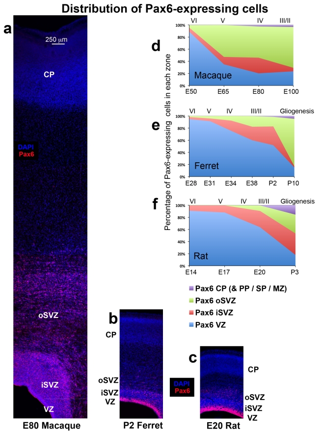
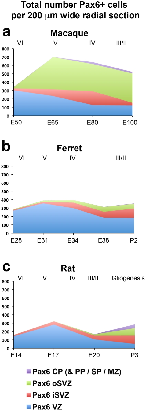
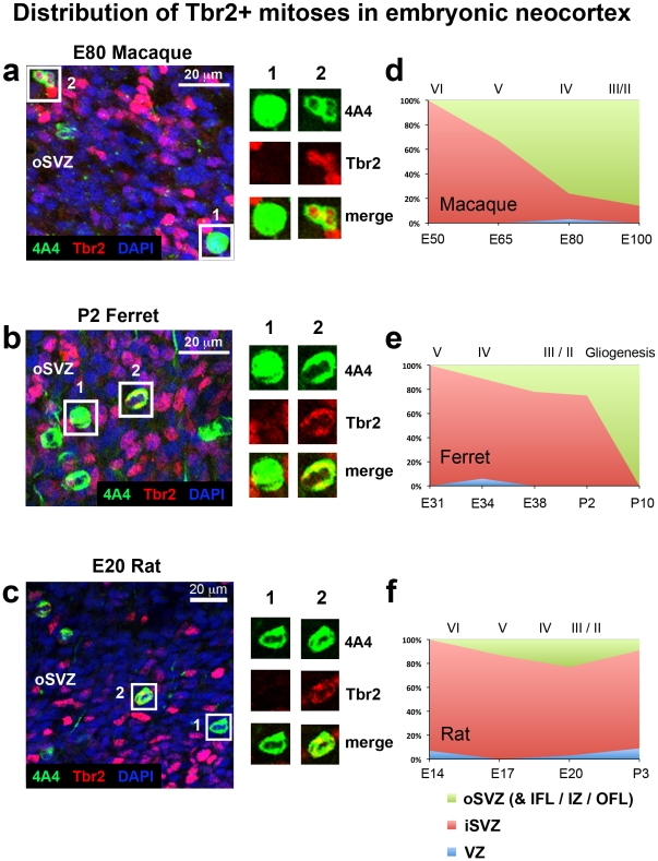
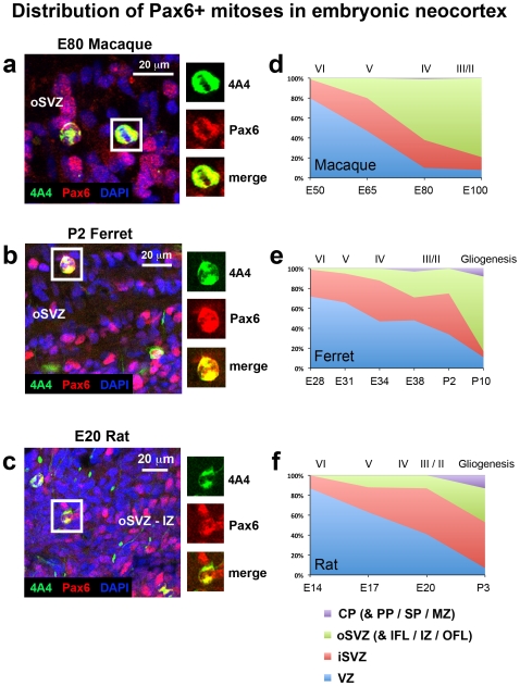
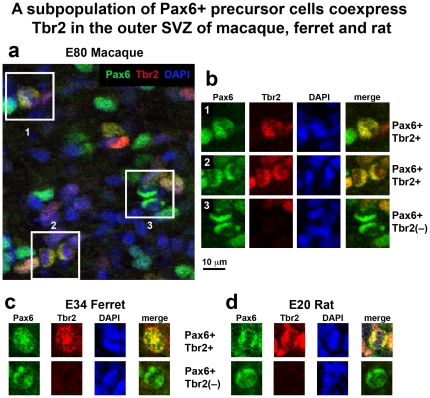
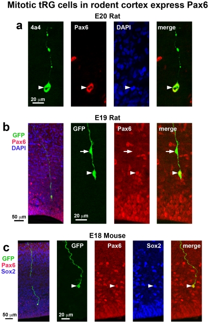
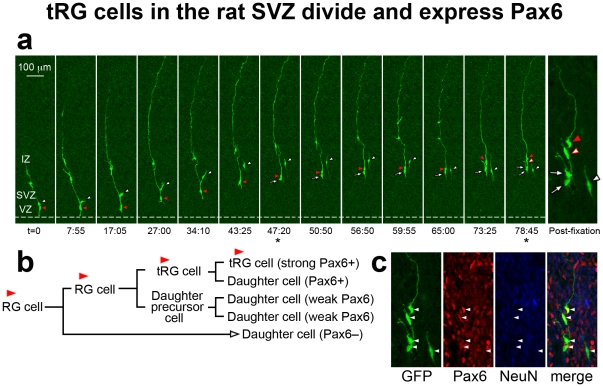
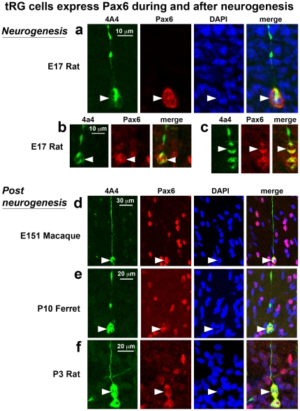
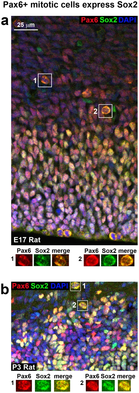
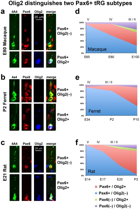
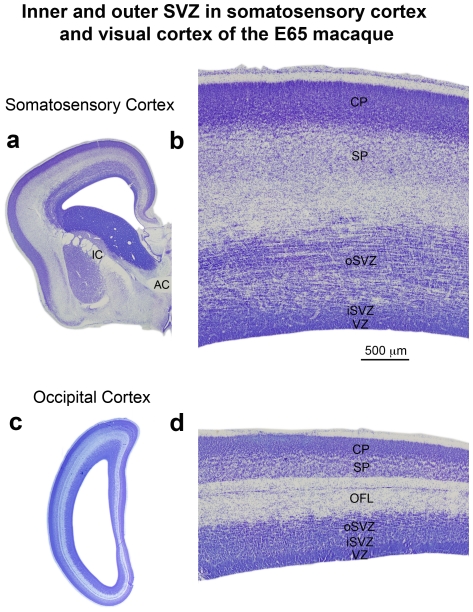
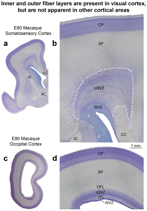
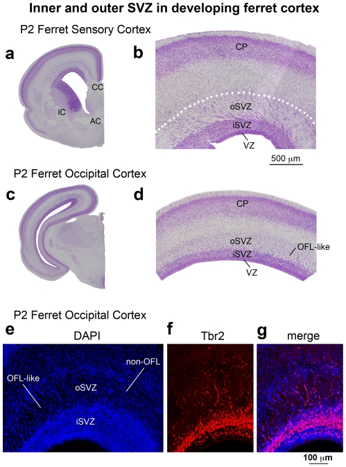
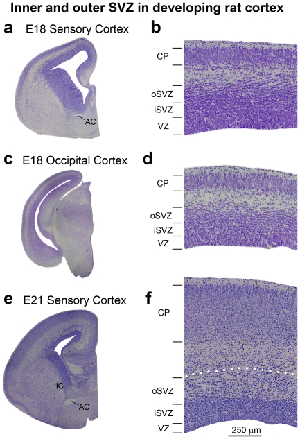
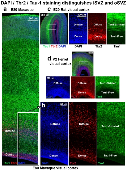
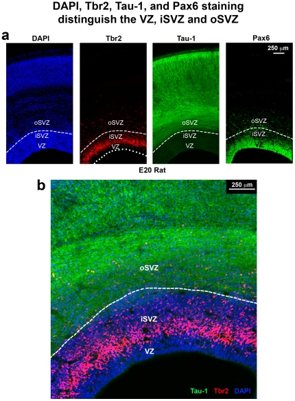
The mammalian cerebral cortex arises from precursor cells that reside in a proliferative region surrounding the lateral ventricles of the developing brain. Recent work has shown that precursor cells in the subventricular zone (SVZ) provide a major contribution to prenatal cortical neurogenesis, and that the SVZ is significantly thicker in gyrencephalic mammals such as primates than it is in lissencephalic mammals including rodents. Identifying characteristics that are shared by or that distinguish cortical precursor cells across mammalian species will shed light on factors that regulate cortical neurogenesis and may point toward mechanisms that underlie the evolutionary expansion of the neocortex in gyrencephalic mammals. We immunostained sections of the developing cerebral cortex from lissencephalic rats, and from gyrencephalic ferrets and macaques to compare the distribution of precursor cell types in each species. We also performed time-lapse imaging of precursor cells in the developing rat neocortex. We show that the distribution of Pax6+ and Tbr2+ precursor cells is similar in lissencephalic rat and gyrencephalic ferret, and different in the gyrencephalic cortex of macaque. We show that mitotic Pax6+ translocating radial glial cells (tRG) are present in the cerebral cortex of each species during and after neurogenesis, demonstrating that the function of Pax6+ tRG cells is not restricted to neurogenesis. Furthermore, we show that Olig2 expression distinguishes two distinct subtypes of Pax6+ tRG cells. Finally we present a novel method for discriminating the inner and outer SVZ across mammalian species and show that the key cytoarchitectural features and cell types that define the outer SVZ in developing primates are present in the developing rat neocortex. Our data demonstrate that the developing rat cerebral cortex possesses an outer subventricular zone during late stages of cortical neurogenesis and that the developing rodent cortex shares important features with that of primates.
Overview
The claims and statistics of this paper haven’t been checked yet.
Run the check — free, no account needed. It reads the open-access text and checks the sources it cites.
Integrity signals
Registry
Cites
Statistics
Figures
Verification
Claims the paper makes
The claims and statistics of this paper haven’t been checked yet.
Run the check — free, no account needed. It reads the open-access text and checks the sources it cites.
Claims other papers make about this one
Each is checked against this paper’s own text: does it say what the citing paper says it does?
In the citing paper
“oRGs are present in large numbers in gyrencephalic primates22,23,40–44 and are considered pivotal to the evolutionary increase in human cortex size and complexity45,46.”
From The use of brain organoids to investigate neural development and disease (2017) · cites this paper as [44]
Self-check
Percentages, sums and reported differences re-computed from the paper’s own numbers.
The self-check needs the paper’s full text.
Citations
Cites
Specification of Cerebral Cortical Areas
Rakic, P. (1988) · Science · doi:10.1126/science.3291116 · Cited by 3,127 · cited 1×
“: [43]), but remain to be fully understood.”
BackgroundNoctor, S. C., Martínez-Cerdeño, V., Ivic, L., & Kriegstein, A. R. (2004) · Nature Neuroscience · doi:10.1038/nn1172 · Cited by 2,229
Self-citationNeurons derived from radial glial cells establish radial units in neocortex
Noctor, S. C., Flint, A. C., Weissman, T. A., Dammerman, R. S., & Kriegstein, A. R. (2001) · Nature · doi:10.1038/35055553 · Cited by 2,016
Self-citationA tension-based theory of morphogenesis and compact wiring in the central nervous system
Essen, D. C. V. (1997) · Nature · doi:10.1038/385313a0 · Cited by 1,668
Neurogenic radial glia in the outer subventricular zone of human neocortex
Hansen, D. V., Lui, J. H., Parker, P. R. L., & Kriegstein, A. R. (2010) · Nature · doi:10.1038/nature08845 · Cited by 1,388
Englund, C., Fink, A., Lau, C., Pham, D., Daza, R. A. M., Bulfone, A., Kowalczyk, T., & Hevner, R. F. (2005) · The Journal of Neuroscience · doi:10.1523/jneurosci.2899-04.2005 · Cited by 1,261
Rakic, P. (1974) · Science · doi:10.1126/science.183.4123.425 · Cited by 1,161
Haubensak, W. E., Attardo, A., Denk, W. T., & Huttner, W. Β. (2004) · Proceedings of the National Academy of Sciences · doi:10.1073/pnas.0308600100 · Cited by 1,004
OSVZ progenitors of human and ferret neocortex are epithelial-like and expand by integrin signaling
Fietz, S. A., Kelava, I., Vogt, J., Wilsch-Bräuninger, M., Stenzel, D., Fish, J. L., Corbeil, D., Riehn, A., Distler, W., Nitsch, R., et al. (2010) · Nature Neuroscience · doi:10.1038/nn.2553 · Cited by 830
Asymmetric production of surface-dividing and non-surface-dividing cortical progenitor cells
Miyata, T., Kawaguchi, A., Saito, K., Kawano, M., Muto, T., & Ogawa, M. (2004) · Development · doi:10.1242/dev.01173 · Cited by 791
Cited by
Sorted by citations
Microglia Regulate the Number of Neural Precursor Cells in the Developing Cerebral Cortex
Cunningham, C. L., Martínez-Cerdeño, V., & Noctor, S. C. (2013) · The Journal of Neuroscience · doi:10.1523/jneurosci.3441-12.2013 · Cited by 976
Self-citationMolecular Identity of Human Outer Radial Glia during Cortical Development
Pollen, A. A., Nowakowski, T. J., Chen, J., Retallack, H., Sandoval-Espinosa, C., Nicholas, C. R., Shuga, J., Liu, S. J., Oldham, M. C., Diaz, A., et al. (2015) · Cell · doi:10.1016/j.cell.2015.09.004 · Cited by 908
Molecular logic of neocortical projection neuron specification, development and diversity
Greig, L. C., Woodworth, M. B., Galazo, M. J., Padmanabhan, H., & Macklis, J. D. (2013) · Nature Reviews Neuroscience · doi:10.1038/nrn3586 · Cited by 863
The use of brain organoids to investigate neural development and disease
Di Lullo, E., & Kriegstein, A. R. (2017) · Nature Reviews Neuroscience · doi:10.1038/nrn.2017.107 · Cited by 788
Neural progenitors, neurogenesis and the evolution of the neocortex
Florio, M., & Huttner, W. Β. (2014) · Development · doi:10.1242/dev.090571 · Cited by 654
Development of cortical folding during evolution and ontogeny
Zilles, K., Palomero‐Gallagher, N., & Amunts, K. (2013) · Trends in Neurosciences · doi:10.1016/j.tins.2013.01.006 · Cited by 567
Betizeau, M., Cortay, V., Patti, D., Pfister, S., Gautier, E., Bellemin-Ménard, A., Afanassieff, M., Huissoud, C., Douglas, R. J., Kennedy, H., et al. (2013) · Neuron · doi:10.1016/j.neuron.2013.09.032 · Cited by 462
Cerebral cortex expansion and folding: what have we learned?
Fernández, V., Llinares-Benadero, C., & Borrell, V. (2016) · The EMBO Journal · doi:10.15252/embj.201593701 · Cited by 382
Nowakowski, T. J., Pollen, A. A., Sandoval-Espinosa, C., & Kriegstein, A. R. (2016) · Neuron · doi:10.1016/j.neuron.2016.09.005 · Cited by 362
The outer subventricular zone and primate-specific cortical complexification
Dehay, C., Kennedy, H., & Kosik, K. S. (2015) · Neuron · doi:10.1016/j.neuron.2014.12.060 · Cited by 314
Single-cell analysis reveals transcriptional heterogeneity of neural progenitors in human cortex
Johnson, M. B., Wang, P. P., Atabay, K. D., Murphy, E. A., Doan, R. N., Hecht, J. L., & Walsh, C. A. (2015) · Nature Neuroscience · doi:10.1038/nn.3980 · Cited by 288
Stahl, R., Walcher, T., Romero, C. D. J., Pilz, G.-A., Cappello, S., Irmler, M., Sanz-Aquela, J. M., Beckers, J., Blum, R., Borrell, V., et al. (2013) · Cell · doi:10.1016/j.cell.2013.03.027 · Cited by 267
Deconstructing cortical folding: genetic, cellular and mechanical determinants
Llinares-Benadero, C., & Borrell, V. (2019) · Nature Reviews Neuroscience · doi:10.1038/s41583-018-0112-2 · Cited by 243
García‐Cabezas, M. Á., Zikopoulos, B., & Barbas, H. (2019) · Brain Structure and Function · doi:10.1007/s00429-019-01841-9 · Cited by 237
Mitotic spindle orientation predicts outer radial glial cell generation in human neocortex
Ostrem, B. E. L., Lui, J. H., Hansen, D. V., & Kriegstein, A. R. (2013) · Nature Communications · doi:10.1038/ncomms2647 · Cited by 244
Regulation of cerebral cortex size and folding by expansion of basal progenitors
Nonaka‐Kinoshita, M., Reillo, I., Artegiani, B., Ángeles Martínez‐Martínez, M., Nelson, M., Borrell, V., & Calegari, F. (2013) · The EMBO Journal · doi:10.1038/emboj.2013.96 · Cited by 221
Role of radial glial cells in cerebral cortex folding
Borrell, V., & Götz, M. (2014) · Current Opinion in Neurobiology · doi:10.1016/j.conb.2014.02.007 · Cited by 230
Cellular and molecular introduction to brain development
Jiang, X., & Nardelli, J. (2015) · Neurobiology of Disease · doi:10.1016/j.nbd.2015.07.007 · Cited by 222
Hedgehog signaling promotes basal progenitor expansion and the growth and folding of the neocortex
Wang, L., Hou, S., & Han, Y.-G. (2016) · Nature Neuroscience · doi:10.1038/nn.4307 · Cited by 196
Aspm knockout ferret reveals an evolutionary mechanism governing cerebral cortical size
Johnson, M. B., Sun, X., Kodani, A., Borges-Monroy, R., Girskis, K. M., Ryu, S. C., Wang, P. P., Patel, K., Gonzalez, D. M., Woo, Y. M., et al. (2018) · Nature · doi:10.1038/s41586-018-0035-0 · Cited by 172
Cortical neurogenesis in the absence of centrioles
Insolera, R., Bazzi, H., Shao, W., Anderson, K. V., & Shi, S. (2014) · Nature Neuroscience · doi:10.1038/nn.3831 · Cited by 189
Primary Cilia in Brain Development and Diseases
Youn, Y. H., & Han, Y.-G. (2017) · American Journal Of Pathology · doi:10.1016/j.ajpath.2017.08.031 · Cited by 182
Quantitative assessment of white matter injury in preterm neonates
Guo, T., Duerden, E. G., Adams, E., Chau, V., Branson, H. M., Chakravarty, M. M., Poskitt, K. J., Synnes, A. R., GRUNAU, R. E., & Miller, S. P. (2017) · Neurology · doi:10.1212/wnl.0000000000003606 · Cited by 166
Rash, B. G., Duque, A., Morozov, Y. M., Arellano, J. I., Micali, N., & Rakic, P. (2019) · Proceedings of the National Academy of Sciences · doi:10.1073/pnas.1822169116 · Cited by 156
Liu, J., Liu, W., Yang, L., Wu, Q., Zhang, H., Fang, A., Li, L., Xu, X., Sun, L., Zhang, J., et al. (2017) · Cell stem cell · doi:10.1016/j.stem.2017.08.013 · Cited by 143
Neural Progenitor Cell Terminology
Martínez‐Cerdeño, V., & Noctor, S. C. (2018) · Frontiers in Neuroanatomy · doi:10.3389/fnana.2018.00104 · Cited by 155
Self-citationHow Cells Fold the Cerebral Cortex
Borrell, V. (2018) · Journal of Neuroscience · doi:10.1523/jneurosci.1106-17.2017 · Cited by 143
Cortical Malformations: Lessons in Human Brain Development
Subramanian, L., Calcagnotto, M. E., & Paredes, M. F. (2020) · Frontiers in Cellular Neuroscience · doi:10.3389/fncel.2019.00576 · Cited by 138
OSVZ progenitors in the human cortex: an updated perspective on neurodevelopmental disease
Ostrem, B. E. L., Lui, J. H., Wang, X., & Kriegstein, A. R. (2012) · Current Opinion in Neurobiology · doi:10.1016/j.conb.2012.03.006 · Cited by 136
A restricted period for formation of outer subventricular zone defined by Cdh1 and Trnp1 levels
Martínez‐Martínez, M. Á., Romero, C. D. J., Fernández, V., Cárdenas, A., Götz, M., & Borrell, V. (2016) · Nature Communications · doi:10.1038/ncomms11812 · Cited by 133
Diverse Behaviors of Outer Radial Glia in Developing Ferret and Human Cortex
Gertz, C. C., Lui, J. H., Ostrem, B. E. L., Wang, X., & Kriegstein, A. R. (2014) · Journal of Neuroscience · doi:10.1523/jneurosci.2645-13.2014 · Cited by 122
Namba, T., & Huttner, W. Β. (2016) · Wiley Interdisciplinary Reviews Developmental Biology · doi:10.1002/wdev.256 · Cited by 120
Control of Outer Radial Glial Stem Cell Mitosis in the Human Brain
Ostrem, B. E. L., Lui, J. H., Gertz, C. C., & Kriegstein, A. R. (2014) · Cell Reports · doi:10.1016/j.celrep.2014.06.058 · Cited by 112
Probing human brain evolution and development in organoids
Giandomenico, S. L., & Lancaster, M. A. (2017) · Current Opinion in Cell Biology · doi:10.1016/j.ceb.2017.01.001 · Cited by 111
Role of ortho‐retronasal olfaction in mammalian cortical evolution
Rowe, T. B., & Shepherd, G. M. (2015) · The Journal of Comparative Neurology · doi:10.1002/cne.23802 · Cited by 111
Nelson, B. R., Hodge, R. D., Bedogni, F., & Hevner, R. F. (2013) · Journal of Neuroscience · doi:10.1523/jneurosci.0791-13.2013 · Cited by 108
Genes and Mechanisms Involved in the Generation and Amplification of Basal Radial Glial Cells
Penisson, M., Ladewig, J., Belvindrah, R., & Francis, F. (2019) · Frontiers in Cellular Neuroscience · doi:10.3389/fncel.2019.00381 · Cited by 97
Sustained Pax6 Expression Generates Primate-like Basal Radial Glia in Developing Mouse Neocortex
Wong, F. K., Fei, J., Mora‐Bermúdez, F., Taverna, E., Haffner, C., Fu, J., Anastassiadis, K., Stewart, A. F., & Huttner, W. Β. (2015) · PLoS Biology · doi:10.1371/journal.pbio.1002217 · Cited by 101
A novel population of Hopx-dependent basal radial glial cells in the developing mouse neocortex
Vaid, S., Camp, J. G., Hersemann, L., Oegema, C. E., Heninger, A., Winkler, S., Brandl, H., Sarov, M., Treutlein, B., Huttner, W. Β., et al. (2018) · Development · doi:10.1242/dev.169276 · Cited by 98
Coevolution of radial glial cells and the cerebral cortex
Romero, C. D. J., & Borrell, V. (2015) · Glia · doi:10.1002/glia.22827 · Cited by 102
Neuronal Migration Dynamics in the Developing Ferret Cortex
Gertz, C. C., & Kriegstein, A. R. (2015) · Journal of Neuroscience · doi:10.1523/jneurosci.2198-15.2015 · Cited by 84
The secondary loss of gyrencephaly as an example of evolutionary phenotypical reversal
Kelava, I., Lewitus, É., & Huttner, W. Β. (2013) · Frontiers in Neuroanatomy · doi:10.3389/fnana.2013.00016 · Cited by 93
Miyata, T., Okamoto, M., Shinoda, T., & Kawaguchi, A. (2015) · Frontiers in Cellular Neuroscience · doi:10.3389/fncel.2014.00473 · Cited by 103
Guo, T., Chau, V., Peyvandi, S., Latal, B., McQuillen, P. S., Knirsch, W., Synnes, A. R., Feldmann, M., Naef, N., Chakravarty, M. M., et al. (2018) · NeuroImage · doi:10.1016/j.neuroimage.2018.06.004 · Cited by 85
Molecular and cellular evolution of corticogenesis in amniotes
Cárdenas, A., & Borrell, V. (2019) · Cellular and Molecular Life Sciences · doi:10.1007/s00018-019-03315-x · Cited by 86
Arcila, M. L., Betizeau, M., Cambronne, X. A., Guzman, E., Doerflinger, N., Bouhallier, F., Zhou, H., Wu, B., Rani, N., Bassett, D. S., et al. (2014) · Neuron · doi:10.1016/j.neuron.2014.01.017 · Cited by 80
Lineage origins of GABAergic versus glutamatergic neurons in the neocortex
Marín, Ó., & Müller, U. (2014) · Current Opinion in Neurobiology · doi:10.1016/j.conb.2014.01.015 · Cited by 87
Folding brains: from development to disease modeling
Del-Valle-Anton, L., & Borrell, V. (2021) · Physiological Reviews · doi:10.1152/physrev.00016.2021 · Cited by 79
Evolution of the Chordate Telencephalon
Briscoe, S. D., & Ragsdale, C. W. (2019) · Current Biology · doi:10.1016/j.cub.2019.05.026 · Cited by 75
Neocortex expansion in development and evolution — from cell biology to single genes
Wilsch‐Bräuninger, M., Florio, M., & Huttner, W. Β. (2016) · Current Opinion in Neurobiology · doi:10.1016/j.conb.2016.05.004 · Cited by 76
Garcίa, M. T., Chang, Y., Arai, Y., & Huttner, W. Β. (2015) · The Journal of Comparative Neurology · doi:10.1002/cne.23801 · Cited by 77
Inside-Out Radial Migration Facilitates Lineage-Dependent Neocortical Microcircuit Assembly
He, S., Li, Z., Ge, S., Yu, Y., & Shi, S. (2015) · Neuron · doi:10.1016/j.neuron.2015.05.002 · Cited by 74
Kawasaki, H., Toda, T., & Tanno, K. (2012) · Biology Open · doi:10.1242/bio.20123160 · Cited by 66
Martínez-Cerdeño, V., Camacho, J., Fox, E., Miller, E., Ariza, J., Kienzle, D., Plank, K., Noctor, S. C., & Van de Water, J. (2016) · Cerebral Cortex · doi:10.1093/cercor/bhu291 · Cited by 63
Self-citationFine-Tuning of Neurogenesis is Essential for the Evolutionary Expansion of the Cerebral Cortex
Poluch, S., & Juliano, S. L. (2013) · Cerebral Cortex · doi:10.1093/cercor/bht232 · Cited by 67
Chen, C., Lee, G. A., Pourmorady, A., Sock, E., & Donoghue, M. J. (2015) · Journal of Neuroscience · doi:10.1523/jneurosci.1663-15.2015 · Cited by 59
Curzio, D. L. D., Buist, R. J., & Bigio, M. R. D. (2013) · Experimental Neurology · doi:10.1016/j.expneurol.2013.06.004 · Cited by 61
The Ferret as a Model System for Neocortex Development and Evolution
Gilardi, C., & Kalebic, N. (2021) · Frontiers in Cell and Developmental Biology · doi:10.3389/fcell.2021.661759 · Cited by 58
Béopoulos, A., Géa, M., Fasano, A., & Iris, F. (2022) · Frontiers in Neuroscience · doi:10.3389/fnins.2022.988735 · Cited by 60
The evolution of basal progenitors in the developing non-mammalian brain
Nomura, T., Ohtaka‐Maruyama, C., Yamashita, W., Wakamatsu, Y., Murakami, Y., Calegari, F., Suzuki, K., Gotoh, H., & Ono, K. (2016) · Development · doi:10.1242/dev.127100 · Cited by 52
Microglia, autonomic nervous system, immunity and hypertension: Is there a link?
Li, Y., Wei, B., Liu, X., Shen, X. Z., & Shi, P. D. (2019) · Pharmacological Research · doi:10.1016/j.phrs.2019.104451 · Cited by 54
Stepien, B. K., Vaid, S., & Huttner, W. Β. (2021) · Frontiers in Cell and Developmental Biology · doi:10.3389/fcell.2021.676911 · Cited by 59
The interaction between microglia and neural stem/precursor cells
Su, P., Zhang, J., Zhao, F., Aschner, M., Chen, J., & Luo, W. (2014) · Brain Research Bulletin · doi:10.1016/j.brainresbull.2014.09.005 · Cited by 50
Endogenous Proliferation after Spinal Cord Injury in Animal Models
McDonough, A., & Martínez‐Cerdeño, V. (2012) · Stem Cells International · doi:10.1155/2012/387513 · Cited by 51
Self-citationCellular Basis of Pineal Gland Development: Emerging Role of Microglia as Phenotype Regulator
Rodriguez, M. P. I., Noctor, S. C., & Muñoz, E. M. (2016) · PLoS ONE · doi:10.1371/journal.pone.0167063 · Cited by 50
Self-citationMartínez‐Cerdeño, V., Cunningham, C. L., Camacho, J. J., Keiter, J. A., Ariza, J., Lovern, M. B., & Noctor, S. C. (2015) · The Journal of Comparative Neurology · doi:10.1002/cne.23879 · Cited by 48
Self-citationOproescu, A.-M., Han, S., & Schuurmans, C. (2021) · Frontiers in Molecular Neuroscience · doi:10.3389/fnmol.2021.642016 · Cited by 54
Güven, A., Kalebic, N., Long, K. R., Florio, M., Vaid, S., Brandl, H., Stenzel, D., & Huttner, W. Β. (2020) · eLife · doi:10.7554/elife.49808 · Cited by 44
The Symmetry of Neural Stem Cell and Progenitor Divisions in the Vertebrate Brain
Gimeno, G. C., & Paridaen, J. T. M. L. (2022) · Frontiers in Cell and Developmental Biology · doi:10.3389/fcell.2022.885269 · Cited by 48
Chromatin Remodeling BAF155 Subunit Regulates the Genesis of Basal Progenitors in Developing Cortex
Narayanan, R., Pham, L., Kerimoglu, C., Watanabe, T., Castro-Hernández, R., Sokpor, G., Ulmke, P. A., Kiszka, K. A., Tonchev, A. B., Rosenbusch, J., et al. (2018) · iScience · doi:10.1016/j.isci.2018.05.014 · Cited by 44
Halley, A. C., & Krubitzer, L. A. (2019) · Current Opinion in Neurobiology · doi:10.1016/j.conb.2018.12.003 · Cited by 43
Krubitzer, L. A., & Dooley, J. C. (2013) · Frontiers in Human Neuroscience · doi:10.3389/fnhum.2013.00620 · Cited by 41
Neurogenesis in primates versus rodents and the value of non-human primate models
Zhang, R., Quan, H., Wang, Y., & Luo, F. (2023) · National Science Review · doi:10.1093/nsr/nwad248 · Cited by 47
Genetic maps and patterns of cerebral cortex folding
Romero, C. D. J., & Borrell, V. (2017) · Current Opinion in Cell Biology · doi:10.1016/j.ceb.2017.11.009 · Cited by 37
Barger, N., Keiter, J. A., Kreutz, A., Krishnamurthy, A., Weidenthaler, C., Martínez‐Cerdeño, V., Tarantal, A. F., & Noctor, S. C. (2018) · Cerebral Cortex · doi:10.1093/cercor/bhy145 · Cited by 38
Self-citationModeling genetic epilepsies in a dish
Niu, W., & Parent, J. M. (2019) · Developmental Dynamics · doi:10.1002/dvdy.79 · Cited by 38
Stick around: Cell–Cell Adhesion Molecules during Neocortical Development
de Agustín-Durán, D., Mateos-White, I., Fabra-Beser, J., & Gil-Sanz, C. (2021) · Cells · doi:10.3390/cells10010118 · Cited by 39
Multiple parallel cell lineages in the developing mammalian cerebral cortex
Del-Valle-Anton, L., Amin, S., Cimino, D., Neuhaus, F., Dvoretskova, E., Fernández, V., Babal, Y. K., Garcı́a-Frigola, C., Prieto‐Colomina, A., Murcia‐Ramón, R., et al. (2024) · Science Advances · doi:10.1126/sciadv.adn9998 · Cited by 35
Progenitor genealogy in the developing cerebral cortex
Laguesse, S., Peyre, E., & Nguyen, L. (2014) · Cell and Tissue Research · doi:10.1007/s00441-014-1979-5 · Cited by 40
GSK3β Inhibition Restores Impaired Neurogenesis in Preterm Neonates With Intraventricular Hemorrhage
Dohare, P., Kidwai, A., Kaur, J., Singla, P., Krishna, S., Klebe, D. W., Zhang, X., Hevner, R. F., & Ballabh, P. (2018) · Cerebral Cortex · doi:10.1093/cercor/bhy217 · Cited by 30
Role of intracortical neuropil growth in the gyrification of the primate cerebral cortex
Rash, B. G., Arellano, J. I., Duque, A., & RAKIC, P. T. (2022) · Proceedings of the National Academy of Sciences · doi:10.1073/pnas.2210967120 · Cited by 31
Clinton, B. K., Cunningham, C. L., Kriegstein, A. R., Noctor, S. C., & Martínez‐Cerdeño, V. (2014) · Neurogenesis · doi:10.4161/23262125.2014.970905 · Cited by 30
Self-citationCunningham, C. L., Martínez‐Cerdeño, V., & Noctor, S. C. (2013) · PLoS ONE · doi:10.1371/journal.pone.0063848 · Cited by 28
Self-citationOkamoto, M., Shinoda, T., Kawaue, T., Nagasaka, A., & Miyata, T. (2014) · Neuroscience Research · doi:10.1016/j.neures.2014.10.006 · Cited by 28
Development and evolution of the primate neocortex from a progenitor cell perspective
Dehay, C., & Huttner, W. Β. (2024) · Development · doi:10.1242/dev.199797 · Cited by 30
Wang, X., Pettersson, D. R., Studholme, C., & Kroenke, C. D. (2015) · Frontiers in Neuroanatomy · doi:10.3389/fnana.2015.00147 · Cited by 27
Human brain development and its in vitro recapitulation
Seto, Y., & Eiraku, M. (2018) · Neuroscience Research · doi:10.1016/j.neures.2018.09.011 · Cited by 27
Progenitor-Based Cell Biological Aspects of Neocortex Development and Evolution
Vaid, S., & Huttner, W. B. (2022) · Frontiers in Cell and Developmental Biology · doi:10.3389/fcell.2022.892922 · Cited by 27
Dantas, T. J., Carabalona, A., Hu, D. J. K., & Vallee, R. B. (2016) · Cytoskeleton · doi:10.1002/cm.21293 · Cited by 28
Sauerland, C., Menzies, B. R., Glatzle, M., Seeger, J., Renfree, M. B., & Fietz, S. A. (2016) · Cerebral Cortex · doi:10.1093/cercor/bhw360 · Cited by 28
Moreno, N., & González, A. (2017) · Frontiers in Neuroanatomy · doi:10.3389/fnana.2017.00024 · Cited by 26
Saito, K., Mizuguchi, K., Hamabe‐Horiike, T., Duong, T. A. D., Shinmyo, Y., & Kawasaki, H. (2018) · Cerebral Cortex · doi:10.1093/cercor/bhy312 · Cited by 26
Sawada, K., Hikishima, K., Murayama, A. Y., Okano, H. J., Sasaki, E., & Okano, H. (2013) · Neuroscience · doi:10.1016/j.neuroscience.2013.10.067 · Cited by 26
Genetic Mechanisms Underlying Cortical Evolution in Mammals
Franchini, L. F. (2021) · Frontiers in Cell and Developmental Biology · doi:10.3389/fcell.2021.591017 · Cited by 32
Gatto, R. G. (2020) · Journal of Integrative Neuroscience · doi:10.31083/j.jin.2020.03.165 · Cited by 27
Camacho, J. J., Moon, R., Smith, S. K., Lin, J. D., Randolph, C., Rasweiler, J. J., Behringer, R. R., & Abzhanov, A. (2020) · EvoDevo · doi:10.1186/s13227-020-00156-9 · Cited by 24
Self-citationIs the ferret a suitable species for studying perinatal brain injury?
Empie, K., Rangarajan, V., & Juul, S. E. (2015) · International Journal of Developmental Neuroscience · doi:10.1016/j.ijdevneu.2015.06.005 · Cited by 24
Sawada, K., Horiuchi‐Hirose, M., Saito, S., & Aoki, I. (2013) · NeuroImage · doi:10.1016/j.neuroimage.2013.06.024 · Cited by 23
González‐Arnay, E., González‐Gómez, M., & Meyer, G. (2017) · Frontiers in Neuroanatomy · doi:10.3389/fnana.2017.00111 · Cited by 25
Homman‐Ludiye, J. (2014) · Frontiers in Neural Circuits · doi:10.3389/fncir.2014.00079 · Cited by 24
Metadata
- Authors
- Martínez-Cerdeño, V., Cunningham, C. L., Camacho, J., Antczak, J. L., Prakash, A. N., Cziep, M. E., Walker, A. I., & Noctor, S. C.
- Affiliations
- University of California, Davis (US)
- Shriners Hospitals for Children - Northern California (US)
- Brigham Young University (US)
- University of California Davis Medical Center (US)
- Corresponding author
- Noctor, Stephen C.
- Venue
- PLoS ONE
- Publisher
- Public Library of Science (PLoS)
- Published
- 2012-01-17
- Volume
- 7
- Issue
- 1
- Pages
- e30178
- ISSN
- 1932-6203 (ISSN-L)
- PMC
- PMC3260244
- OpenAlex
- W2030135612
- Cited by
- 203
- References
- 49
- Citations by year
- 2026: 6 · 2025: 6 · 2024: 10 · 2023: 9 · 2022: 11 · 2021: 14 · 2020: 16 · 2019: 18 · 2018: 16 · 2017: 16 · 2016: 20 · 2015: 18 · 2014: 18 · 2013: 21 · 2012: 4
- FWCI
- 6.56
- Type
- journal article
- Language
- en
- Open access
- gold
- doi.org
- Locations
- PLoS ONE (publishedVersion, cc-by)
- Europe PMC (PubMed Central) (publishedVersion, cc-by)
- PubMed Central (publishedVersion, cc-by)
- PubMed (publishedVersion)
- eScholarship (California Digital Library) (submittedVersion, cc-by)
- DOAJ (DOAJ: Directory of Open Access Journals) (submittedVersion)
- eScholarship (California Digital Library) (submittedVersion)
- Funders
- National Science Foundation
- MIND Institute, University of California, Davis
- Children's Miracle Network Hospitals
- University of California, Davis
- National Institute of Mental Health (R01MH094681)
- Topics
- Life Sciences › Neuroscience › Developmental Neuroscience › Neurogenesis and neuroplasticity mechanisms
- Health Sciences › Medicine › Pediatrics, Perinatology and Child Health › Fetal and Pediatric Neurological Disorders
- Life Sciences › Biochemistry, Genetics and Molecular Biology › Molecular Biology › Developmental Biology and Gene Regulation
- Keywords
- subventricular zone, outer subventricular zone, cortical neurogenesis, radial glia, Pax6, Tbr2, Olig2, lissencephaly, gyrencephaly, neocortex evolution, cortical development, rats, ferrets, macaque
- MeSH terms
- PAX6 Transcription Factor, Anatomy, Comparative, Animals, Cell Count, Cerebral Cortex, Embryo, Mammalian, Eye Proteins, Female, Ferrets, Immunohistochemistry, Macaca mulatta, Male, Mitosis, Neurons, Pregnancy, Repressor Proteins, Somatosensory Cortex, Time Factors, Rats, Sprague-Dawley, Homeodomain Proteins, Neocortex, T-Box Domain Proteins, Mice, Rats, Paired Box Transcription Factors, Neurogenesis, Neural Stem Cells
About these checks
Registry, retraction, Cites and statistics results are computed, not judged, with one exception: a printed reference that no index lists is read once by a model before it counts as a citation error. Claims are extracted by a model and kept only when their quote appears verbatim in the text; each cited source is then checked, with a model’s help, for whether it says what the claim relies on. Those verdicts can be wrong — read the claim’s quote and the cited source before relying on one.
Statistics and arithmetic are recomputed, not judged.
Confidence bands are shown only for fields calibrated on our annotated test set. None is calibrated yet, so no band is shown.
What counts as a claim ›How we measure confidence ›How we check this ›
Are you an author, or do you see a problem with this page? Request a review
