

The DOI matches its Crossref record; 55 of the 55 references it cites were found in a registry or an index.
Osteocyte Network; a Negative Regulatory System for Bone Mass Augmented by the Induction of Rankl in Osteoblasts and Sost in Osteocytes at Unloading
Open accessAbstract
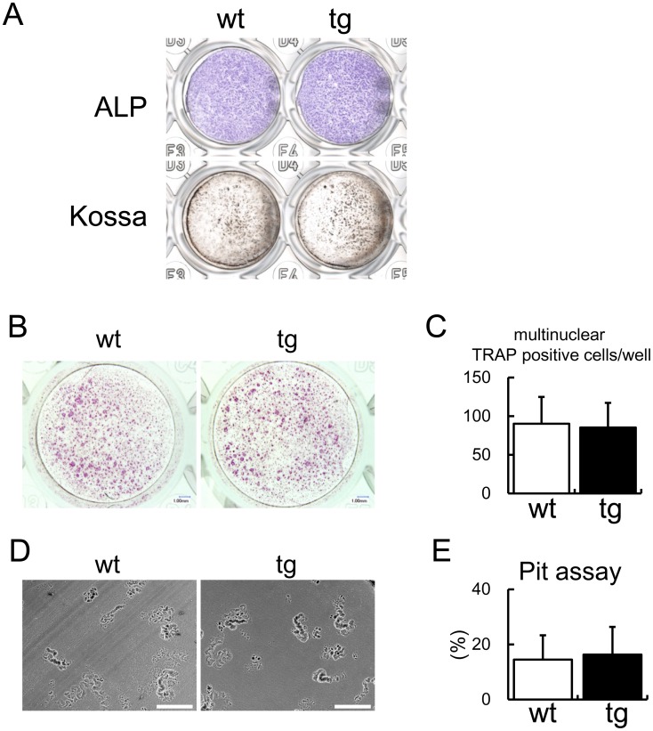
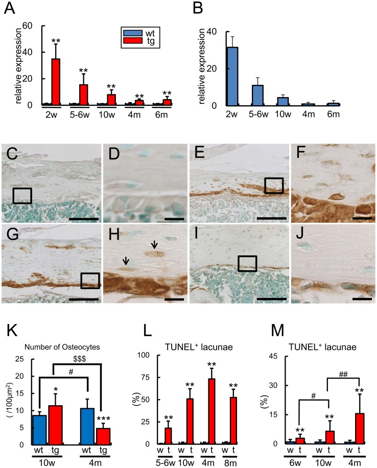
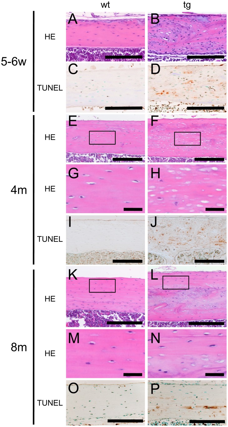
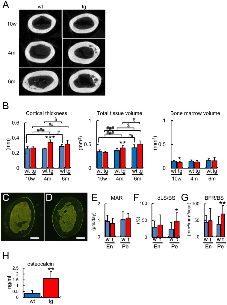
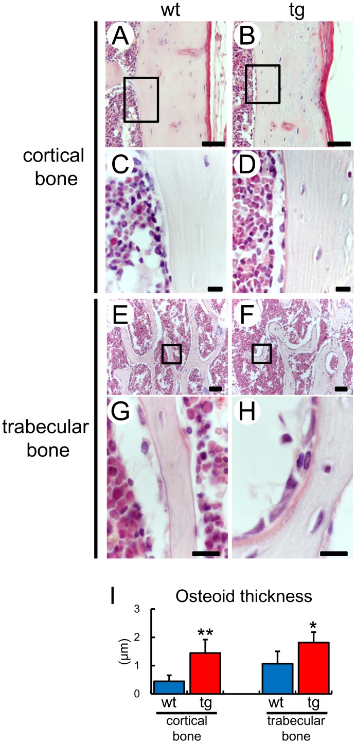
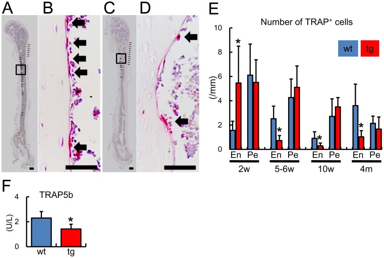
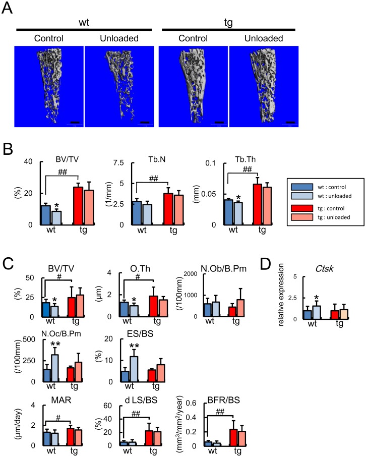
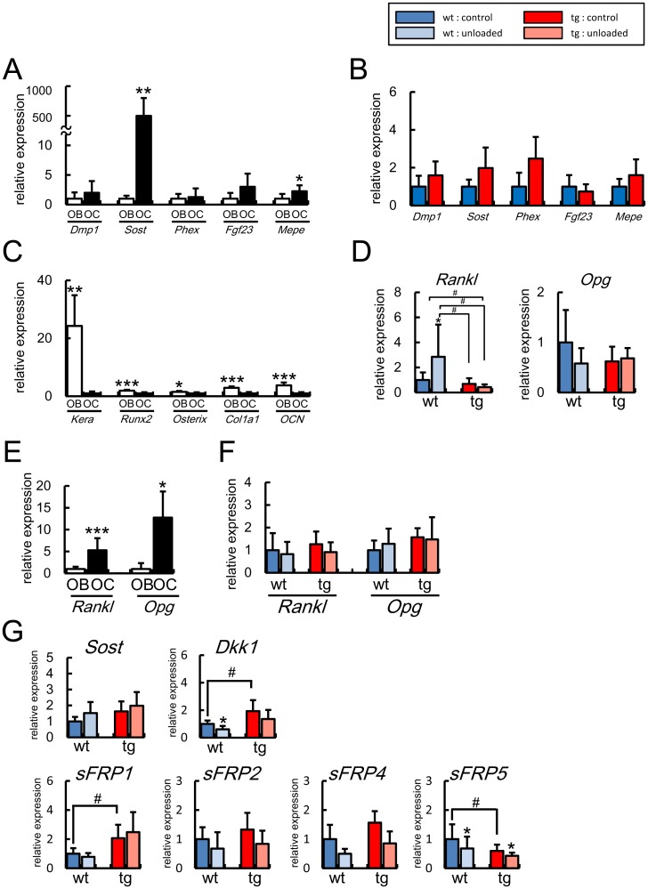
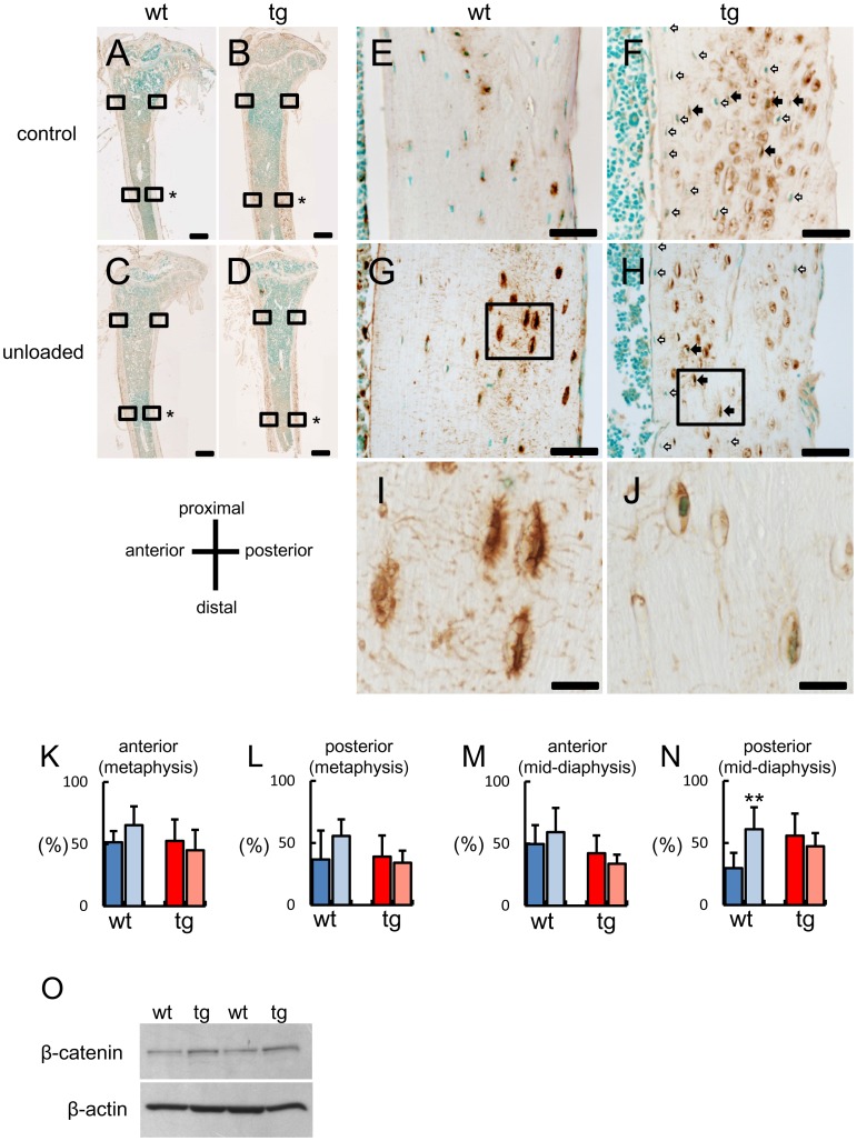
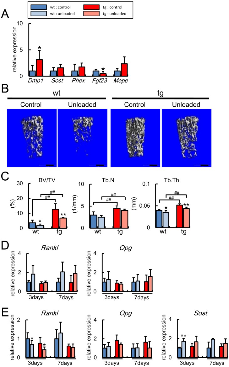
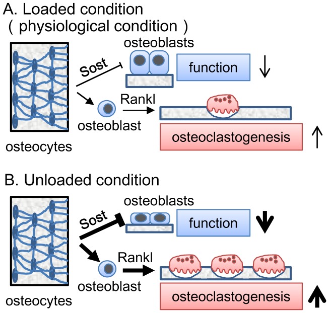
Reduced mechanical stress is a major cause of osteoporosis in the elderly, and the osteocyte network, which comprises a communication system through processes and canaliculi throughout bone, is thought to be a mechanosensor and mechanotransduction system; however, the functions of osteocytes are still controversial and remain to be clarified. Unexpectedly, we found that overexpression of BCL2 in osteoblasts eventually caused osteocyte apoptosis. Osteoblast and osteoclast differentiation were unaffected by BCL2 transgene in vitro. However, the cortical bone mass increased due to enhanced osteoblast function and suppressed osteoclastogenesis at 4 months of age, when the frequency of TUNEL-positive lacunae reached 75%. In the unloaded condition, the trabecular bone mass decreased in both wild-type and BCL2 transgenic mice at 6 weeks of age, while it decreased due to impaired osteoblast function and enhanced osteoclastogenesis in wild-type mice but not in BCL2 transgenic mice at 4 months of age. Rankl and Opg were highly expressed in osteocytes, but Rankl expression in osteoblasts but not in osteocytes was increased at unloading in wild-type mice but not in BCL2 transgenic mice at 4 months of age. Sost was locally induced at unloading in wild-type mice but not in BCL2 transgenic mice, and the dissemination of Sost was severely interrupted in BCL2 transgenic mice, showing the severely impaired osteocyte network. These findings indicate that the osteocyte network is required for the upregulation of Rankl in osteoblasts and Sost in osteocytes in the unloaded condition. These findings suggest that the osteocyte network negatively regulate bone mass by inhibiting osteoblast function and activating osteoclastogenesis, and these functions are augmented in the unloaded condition at least partly through the upregulation of Rankl expression in osteoblasts and that of Sost in osteocytes, although it cannot be excluded that low BCL2 transgene expression in osteoblasts contributed to the enhanced osteoblast function.
Overview
The claims and statistics of this paper haven’t been checked yet.
Run the check — free, no account needed. It reads the open-access text and checks the sources it cites.
Integrity signals
Registry
Cites
Statistics
Figures
Verification
Claims the paper makes
The claims and statistics of this paper haven’t been checked yet.
Run the check — free, no account needed. It reads the open-access text and checks the sources it cites.
Claims other papers make about this one
Each is checked against this paper’s own text: does it say what the citing paper says it does?
In the citing paper
“On the other hand, mechanical unloading downregulates anabolic factors and stimulates osteocytes to produce sclerostin and DKK-1, which are inhibitors of osteoblast activity [185–188], as well as specific factors that stimulate local osteoclastogenesis [189].”
From Biology of Bone Tissue: Structure, Function, and Factors That Influence Bone Cells (2015) · cites this paper as [188]
Self-check
Percentages, sums and reported differences re-computed from the paper’s own numbers.
The self-check needs the paper’s full text.
Citations
Cites
Bonewald, L. F. (2011) · Journal of Bone and Mineral Research · doi:10.1002/jbmr.320 · Cited by 2,287
Das Gesetz der Transformation der Knochen
Wolff, J. (1893) · DMW - Deutsche Medizinische Wochenschrift · doi:10.1055/s-0028-1144106 · Cited by 2,037
Evidence for osteocyte regulation of bone homeostasis through RANKL expression
Nakashima, T., Hayashi, M., Fukunaga, T., Kurata, K., Oh-hora, M., Feng, J. Q., Bonewald, L. F., Kodama, T., Wutz, A., Wagner, E. F., et al. (2011) · Nature Medicine · doi:10.1038/nm.2452 · Cited by 1,778
Sclerostin Binds to LRP5/6 and Antagonizes Canonical Wnt Signaling
Li, X., Zhang, Y., Kang, H., Liu, W., Liu, P., Zhang, J., Harris, S. E., & Wu, D. (2005) · Journal of Biological Chemistry · doi:10.1074/jbc.m413274200 · Cited by 1,366 · cited 1×
“encoding a collagenase-resistant form of type I collagen, showed osteocyte apoptosis and increased bone formation [53], and it has been shown that osteocytes secrete Sost, which is a potent antagonist of Wnt, and inhibits bone formation [54], [55], [56], [57]; therefore, there were contrasting observations that osteoc…”
Mechanical Stimulation of Bone in Vivo Reduces Osteocyte Expression of Sost/Sclerostin
Robling, A. G., Niziolek, P. J., Baldridge, L. A., Condon, K. W., Allen, M. R., Alam, I., Mantila, S. M., Gluhak-Heinrich, J., Bellido, T. M., Harris, S. E., et al. (2008) · Journal of Biological Chemistry · doi:10.1074/jbc.m705092200 · Cited by 1,350
Matrix-embedded cells control osteoclast formation
Xiong, J., Onal, M., Jilka, R. L., Weinstein, R. S., Manolagas, S. C., & O'Brien, C. A. (2011) · Nature Medicine · doi:10.1038/nm.2448 · Cited by 1,304
Martin, R. B., Burr, D. B., Sharkey, N. A., & Fyhrie, D. P. (2015) · doi:10.1007/978-1-4939-3002-9 · Cited by 1,159
Osteocyte control of bone formation via sclerostin, a novel BMP antagonist
Winkler, D. G., Sutherland, M. K., Geoghegan, J. C., Yu, C., Hayes, T., Skonier, J. E., Shpektor, D., Jonas, M., Kovacevich, B. R., Staehling‐Hampton, K., et al. (2003) · The EMBO Journal · doi:10.1093/emboj/cdg599 · Cited by 1,114
Osteocytes, mechanosensing and Wnt signaling
Bonewald, L. F., & Johnson, M. L. (2008) · Bone · doi:10.1016/j.bone.2007.12.224 · Cited by 1,076
Sclerostin is a delayed secreted product of osteocytes that inhibits bone formation
Poole, K. E. S., Van Bezooijen, R. L., Loveridge, N., Hamersma, H., Papapoulos, S. E., Löwik, C. W., & Reeve, J. (2005) · The FASEB Journal · doi:10.1096/fj.05-4221fje · Cited by 965
Necrotic death as a cell fatePreprint
Zong, W., & Thompson, C. B. (2006) · Genes & Development · doi:10.1101/gad.1376506 · Cited by 952
Li, X., Ominsky, M. S., Niu, Q., Sun, N., Daugherty, B., D’Agostin, D. M., Kurahara, C., Gao, Y., Cao, J., Gong, J., et al. (2008) · Journal of Bone and Mineral Research · doi:10.1359/jbmr.080216 · Cited by 941
Cell Death Independent of Caspases: A Review
Bröker, L. E., Kruyt, F. A. E., & Giaccone, G. (2005) · Clinical Cancer Research · doi:10.1158/1078-0432.ccr-04-2223 · Cited by 914
Mechanotransduction in bone—role of the lacunocanalicular network
Burger, E. H., & Klein‐Nulend, J. (1999) · The FASEB Journal · doi:10.1096/fasebj.13.9001.s101 · Cited by 897
van Bezooijen, R. L., Roelen, B. A. J., Visser, A., van der Wee-Pals, L. J. A., de Wilt, E., Karperien, M., Hamersma, H. A., Papapoulos, S. E., ten Dijke, P., & Löwik, C. W. G. M. (2004) · The Journal of Experimental Medicine · doi:10.1084/jem.20031454 · Cited by 893
Targeted ablation of osteocytes induces osteoporosis with defective mechanotransduction.
Tatsumi, S., Ishii, K., Amizuka, N., Li, M., Kobayashi, T., Kohno, K., Ito, M., Takeshita, S., & Ikeda, K. (2007) · Cell Metabolism · doi:10.1016/j.cmet.2007.05.001 · Cited by 848
Self-citationSOST Is a Ligand for LRP5/LRP6 and a Wnt Signaling Inhibitor
Semënov, M., Tamai, K., & He, X. (2005) · Journal of Biological Chemistry · doi:10.1074/jbc.m504308200 · Cited by 771
Verborgt, O., Gibson, G. J., & Schaffler, M. B. (2000) · Journal of Bone and Mineral Research · doi:10.1359/jbmr.2000.15.1.60 · Cited by 731
Lin, C., Jiang, X., Dai, Z., Guo, X., Weng, T., Wang, J., Li, Y., Feng, G., Gao, X., & He, L. (2009) · Journal of Bone and Mineral Research · doi:10.1359/jbmr.090411 · Cited by 648
Bellido, T., Ali, A., Gubrij, I. B., Plotkin, L. I., Fu, Q., O’Brien, C. A., Manolagas, S. C., & Jilka, R. L. (2005) · Endocrinology · doi:10.1210/en.2005-0239 · Cited by 640
Yoshida, C. A., Yamamoto, H., Fujita, T., Furuichi, T., Ito, K., Inoue, K., Yamana, K., Zanma, A., Takada, K., Ito, Y., et al. (2004) · Genes & Development · doi:10.1101/gad.1174704 · Cited by 588
Self-citationOsteocyte Wnt/β-Catenin Signaling Is Required for Normal Bone Homeostasis
Krämer, I., Halleux, C. M., Keller, H., Pegurri, M., Gooi, J. H., Weber, P. B., Feng, J. Q., Bonewald, L. F., & Kneissel, M. (2010) · Molecular and Cellular Biology · doi:10.1128/mcb.01428-09 · Cited by 575
Deletion of a Single Allele of the Dkk1 Gene Leads to an Increase in Bone Formation and Bone Mass
Morvan, F., Boulukos, K. E., Clément-Lacroix, P., Roman‐Roman, S., Suc-Royer, I., Vayssière, B., Ammann, P., Martin, P., Pinho, S., Pognonec, P., et al. (2006) · Journal of Bone and Mineral Research · doi:10.1359/jbmr.060311 · Cited by 533
Mechanical Strain and Bone Cell Function: A Review
Ehrlich, P. J., & Lanyon, L. E. (2002) · Osteoporosis International · doi:10.1007/s001980200095 · Cited by 509
Bodine, P. V. N., Zhao, W., Kharode, Y. P., Bex, F. J., Lambert, A.-J., Goad, M. B., Gaur, T., Stein, G. S., Lian, J. B., & Komm, B. S. (2004) · Molecular Endocrinology · doi:10.1210/me.2003-0498 · Cited by 456
Liu, W., Toyosawa, S., Furuichi, T., Kanatani, N., Yoshida, C. A., Liu, Y., Himeno, M., Narai, S., Yamaguchi, A., & Komori, T. (2001) · The Journal of Cell Biology · doi:10.1083/jcb.200105052 · Cited by 454
Self-citationApoptosis of Osteocytes in Glucocorticoid-Induced Osteonecrosis of the Hip
Weinstein, R. S., Nicholas, R. W., & Manolagas, S. C. (2000) · The Journal of Clinical Endocrinology & Metabolism · doi:10.1210/jcem.85.8.6714 · Cited by 446
Osteocytes as mechanosensors in the inhibition of bone resorption due to mechanical loading
You, L., Temiyasathit, S., Lee, P., Kim, C. H., Tummala, P., Yao, W., Kingery, W. S., Malone, A. M. D., Kwon, R. Y., & Jacobs, C. R. (2007) · Bone · doi:10.1016/j.bone.2007.09.047 · Cited by 361
Apoptosis of Osteocytes in Glucocorticoid-Induced Osteonecrosis of the Hip
Weinstein, R. S. (2000) · The Journal of Clinical Endocrinology & Metabolism · doi:10.1210/jc.85.8.2907 · Cited by 315
“Whither flows the fluid in bone?” An osteocyte's perspective
Knothe Tate, M. L. (2003) · Journal of Biomechanics · doi:10.1016/s0021-9290(03)00123-4 · Cited by 309
Toyosawa, S., Shintani, S., Fujiwara, T., Ooshima, T., Sato, A., Ijuhin, N., & Komori, T. (2001) · Journal of Bone and Mineral Research · doi:10.1359/jbmr.2001.16.11.2017 · Cited by 288
Self-citationvan Bezooijen, R. L., Svensson, J. P., Eefting, D., Visser, A., van der Horst, G., Karperien, M., Quax, P. H. A., Vrieling, H., Papapoulos, S. E., ten Dijke, P., et al. (2006) · Journal of Bone and Mineral Research · doi:10.1359/jbmr.061002 · Cited by 286
Identification of differentially expressed genes between osteoblasts and osteocytes
Paić, F., Igwe, J. C., Nori, R., Kronenberg, M. S., Franceschetti, T., Harrington, P., Kuo, L., Shin, D., Rowe, D. W., Harris, S. E., et al. (2009) · Bone · doi:10.1016/j.bone.2009.06.010 · Cited by 271
Moustafa, A., Sugiyama, T., Prasad, J., Zaman, G., Gross, T. S., Lanyon, L. E., & Price, J. S. (2011) · Osteoporosis International · doi:10.1007/s00198-011-1656-4 · Cited by 243
Secondary necrosis in multicellular animals: an outcome of apoptosis with pathogenic implications
Silva, M. T., do Vale, A., & dos Santos, N. M. S. (2008) · APOPTOSIS · doi:10.1007/s10495-008-0187-8 · Cited by 235
Perspective: Quantifying Osteoblast and Osteocyte Apoptosis: Challenges and Rewards
Jilka, R. L., Weinstein, R. S., PARFITT, A. M., & Manolagas, S. C. (2007) · Journal of Bone and Mineral Research · doi:10.1359/jbmr.070518 · Cited by 211
Heino, T. J., Hentunen, T. A., & Väänänen, H. K. (2002) · Journal of Cellular Biochemistry · doi:10.1002/jcb.10109 · Cited by 191
Noble, B. (2008) · Archives of Biochemistry and Biophysics · doi:10.1016/j.abb.2008.04.009 · Cited by 185
Structural adaptations to bone loss in aging men and women
Russo, C. R., Lauretani, F., Seeman, E., Bartali, B., Bandinelli, S., Iorio, A. D., Guralnik, J. M., & Ferrucci, L. (2005) · Bone · doi:10.1016/j.bone.2005.07.025 · Cited by 160
Zhao, W., Byrne, M. H., Wang, Y., & Krane, S. M. (2000) · Journal of Clinical Investigation · doi:10.1172/jci10158 · Cited by 151
Relationships between osteocyte density and bone formation rate in human cancellous bone
Qiu, S., Rao, D. S., Palnitkar, S., & PARFITT, A. M. (2002) · Bone · doi:10.1016/s8756-3282(02)00907-9 · Cited by 104
Clearance deficiency—A potential link between infections and autoimmunity
Schulze, C., Munoz, L. E., Franz, S., Sarter, K., Chaurio, R. A., Gaipl, U. S., & i, M. H. (2008) · Autoimmunity Reviews · doi:10.1016/j.autrev.2008.07.049 · Cited by 89
Seeman, E. (2001) · Calcified Tissue International · doi:10.1007/s00223-001-1040-z · Cited by 84
Quantitative Evaluation on Osteocyte Canalicular Density in Human Secondary Osteons
Marotti, G., Ferretti, M., Remaggi, F., & Palumbo, C. (1995) · Bone · doi:10.1016/8756-3282(95)80022-i · Cited by 84
Histomorphometric analysis of the effects of osteocyte density on osteonal morphology and remodeling
Metz, L. N., Martin, R. B., & Turner, A. S. (2003) · Bone · doi:10.1016/s8756-3282(03)00245-x · Cited by 81
Osteoclasts—the innate immune cells of the bone
Wu, Y., Humphrey, M. B., & Nakamura, M. C. (2008) · Autoimmunity · doi:10.1080/08916930701693180 · Cited by 69
Moriishi, T., Maruyama, Z., Fukuyama, R., Ito, M., Miyazaki, T., Kitaura, H., Ohnishi, H., Furuichi, T., Kawai, Y., Masuyama, R., et al. (2011) · PLoS ONE · doi:10.1371/journal.pone.0027487 · Cited by 68
Self-citationDoes osteocyte formation cause the nonlinear refilling of osteons?
Martin, R. B. (2000) · Bone · doi:10.1016/s8756-3282(99)00242-2 · Cited by 65
Lazenby, R. A. (1990) · American Journal of Physical Anthropology · doi:10.1002/ajpa.1330820408 · Cited by 63
Hedgecock, N. L., Hadi, T. M., Chen, A. A., Curtiss, S. B., Martin, R. B., & Hazelwood, S. J. (2006) · Bone · doi:10.1016/j.bone.2006.10.006 · Cited by 62
Nonpharmacological approaches to improve bone health and reduce osteoporosis
Schwab, P., & Klein, R. F. (2008) · Current Opinion in Rheumatology · doi:10.1097/bor.0b013e3282f3cbd3 · Cited by 41
Pyruvate dehydrogenase kinase 4 induces bone loss at unloading by promoting osteoclastogenesis
Wang, Y., Liu, W., Masuyama, R., Fukuyama, R., Ito, M., Zhang, Q., Komori, H., Murakami, T., Moriishi, T., Miyazaki, T., et al. (2011) · Bone · doi:10.1016/j.bone.2011.07.012 · Cited by 40
Self-citationChanges in trabecular bone turnover and bone marrow cell development in tail-suspended mice.
Sakai, A., & Nakamura, T. (2001) · PubMed · Cited by 31
Mueller, R., & Richards, R. G. (2004) · Journal of Materials Science Materials in Medicine · doi:10.1023/b:jmsm.0000021103.39114.cd · Cited by 13
Pyruvate dehydrogenase kinase 4 induces bone loss at unloading by promoting osteoclastogenesis
王, 宇. (2011) · Medical Entomology and Zoology · Cited by 2
Research works printed but not linked: 5
- J Xiong, 2011, Matrix-embedded cells control osteoclast formation., Nat Med, 17, 1235, 10.1038/nm.2448 · DOI · Paper page
- KE Poole, 2005, Sclerostin is a delayed secreted product of osteocytes that inhibits bone formation., FASEB J, 19, 1842, 10.1096/fj.05-4221fje · DOI · Paper page
- X Li, 2005, Sclerostin Binds to LRP5/6 and Antagonizes Canonical Wnt Signaling., J Biol Chem, 280, 19883, 10.1074/jbc.M413274200 · DOI · Paper page
- M Semenov, 2005, SOST Is a Ligand for LRP5/LRP6 and a Wnt Signaling Inhibitor., J Biol Chem, 280, 26770, 10.1074/jbc.M504308200 · DOI · Paper page
- X Li, 2008, Targeted Deletion of the Sclerostin Gene in Mice Results in Increased Bone Formation and Bone Strength., J Bone Miner Res, 23, 860, 10.1359/jbmr.080216 · DOI
Cited by
Sorted by citations
Biology of Bone Tissue: Structure, Function, and Factors That Influence Bone Cells
Florencio-Silva, R., Sasso, G. R. D. S., Sasso-Cerri, E., Simões, M. J., & Cerri, P. S. (2015) · BioMed Research International · doi:10.1155/2015/421746 · Cited by 2,044
Osteocytes: master orchestrators of bone
Schaffler, M. B., Cheung, W.-Y., Majeska, R., & Kennedy, O. (2014) · Calcified Tissue International · doi:10.1007/s00223-013-9790-y · Cited by 514
Runx2, an inducer of osteoblast and chondrocyte differentiation
Komori, T. (2018) · Histochemistry and Cell Biology · doi:10.1007/s00418-018-1640-6 · Cited by 468
Self-citationOsteoblast and osteocyte: Games without frontiers
Capulli, M., Paone, R., & Rucci, N. (2014) · Archives of Biochemistry and Biophysics · doi:10.1016/j.abb.2014.05.003 · Cited by 413
Xiong, J., Piemontese, M., Onal, M., Campbell, J., Goellner, J. J., Dusevich, V., Bonewald, L., Manolagas, S. C., & O’Brien, C. A. (2015) · PLOS ONE · doi:10.1371/journal.pone.0138189 · Cited by 323
Metadata
- Authors
- Moriishi, T., Fukuyama, R., Ito, M., Miyazaki, T., Maeno, T., Kawai, Y., Komori, H., & Komori, T.
- Affiliations
- Nagasaki University (JP)
- Hiroshima International University (JP)
- Osaka City University (JP)
- Corresponding author
- Komori, Toshihisa
- Venue
- PLoS ONE
- Publisher
- Public Library of Science (PLoS)
- Published
- 2012-06-29
- Volume
- 7
- Issue
- 6
- Pages
- e40143
- ISSN
- 1932-6203 (ISSN-L)
- PMC
- PMC3387151
- OpenAlex
- W1964279564
- Cited by
- 105
- References
- 55
- Citations by year
- 2026: 2 · 2025: 5 · 2024: 7 · 2023: 8 · 2022: 7 · 2021: 13 · 2020: 9 · 2019: 6 · 2018: 8 · 2017: 5 · 2016: 6 · 2015: 12 · 2014: 11 · 2013: 6
- FWCI
- 3.72
- Type
- journal article
- Language
- en
- Open access
- gold
- doi.org
- Locations
- PLoS ONE (publishedVersion, cc-by)
- Europe PMC (PubMed Central) (publishedVersion, cc-by)
- PubMed Central (publishedVersion, cc-by)
- PubMed (publishedVersion)
- Institutional Repositories DataBase (IRDB) (publishedVersion, other-oa)
- DOAJ (DOAJ: Directory of Open Access Journals) (submittedVersion, cc-by-sa)
- Figshare (submittedVersion, cc-by)
- Nagasaki University's Academic Output SITE (Nagasaki University) (submittedVersion, cc-by-nc-nd)
- Funders
- Nakatomi Foundation
- Ministry of Education, Culture, Sports, Science and Technology
- Japan Society for the Promotion of Science (24659829, 22249062)
- Topics
- Life Sciences › Biochemistry, Genetics and Molecular Biology › Molecular Biology › Bone Metabolism and Diseases
- Health Sciences › Medicine › Orthopedics and Sports Medicine › Bone health and osteoporosis research
- Health Sciences › Medicine › Physiology (medical) › Spaceflight effects on biology
- Keywords
- osteocyte network, osteocyte apoptosis, Bcl-2, osteoblast activity, osteoclastogenesis, RANKL, sclerostin, osteoporosis, mechanical unloading, mechanotransduction, bone mass regulation, trabecular bone, cortical bone
- MeSH terms
- Animals, Bone and Bones, Bone Resorption, Cell Count, Cell Differentiation, Glycoproteins, Humans, Immunohistochemistry, Mice, Transgenic, Models, Biological, Organ Size, Osteoblasts, Osteocytes, Osteogenesis, Up-Regulation, Apoptosis, Transgenes, Proto-Oncogene Proteins c-bcl-2, Hindlimb Suspension, Reverse Transcriptase Polymerase Chain Reaction, In Situ Nick-End Labeling, Intercellular Signaling Peptides and Proteins, Adaptor Proteins, Signal Transducing, Mice, RANK Ligand, X-Ray Microtomography, Real-Time Polymerase Chain Reaction
About these checks
Registry, retraction, Cites and statistics results are computed, not judged, with one exception: a printed reference that no index lists is read once by a model before it counts as a citation error. Claims are extracted by a model and kept only when their quote appears verbatim in the text; each cited source is then checked, with a model’s help, for whether it says what the claim relies on. Those verdicts can be wrong — read the claim’s quote and the cited source before relying on one.
Statistics and arithmetic are recomputed, not judged.
Confidence bands are shown only for fields calibrated on our annotated test set. None is calibrated yet, so no band is shown.
What counts as a claim ›How we measure confidence ›How we check this ›
Are you an author, or do you see a problem with this page? Request a review
