Student or educator? Sign up with your school email and get 6 months of Pro free.
CoChat

The DOI matches its Crossref record; 46 of the 46 references it cites were found in a registry or an index.

Cell Elasticity Determines Macrophage Function

Open access

Abstract

Macrophages serve to maintain organ homeostasis in response to challenges from injury, inflammation, malignancy, particulate exposure, or infection. Until now, receptor ligation has been understood as being the central mechanism that regulates macrophage function. Using macrophages of different origins and species, we report that macrophage elasticity is a major determinant of innate macrophage function. Macrophage elasticity is modulated not only by classical biologic activators such as LPS and IFN-γ, but to an equal extent by substrate rigidity and substrate stretch. Macrophage elasticity is dependent upon actin polymerization and small rhoGTPase activation, but functional effects of elasticity are not predicted by examination of gene expression profiles alone. Taken together, these data demonstrate an unanticipated role for cell elasticity as a common pathway by which mechanical and biologic factors determine macrophage function.

Overview

The claims and statistics of this paper haven’t been checked yet.

Free: it reads the open-access text and checks the first 5 claims that cite a source we can check. Create a free account to check every claim.

A few free checks a day · papers already verified open instantly · results are public

Integrity signals

Registry

Crossref · 10.1371/journal.pone.0041024

Cites

46 of 46 references verified0 citation errors · 0 not checkable

Statistics

Figures

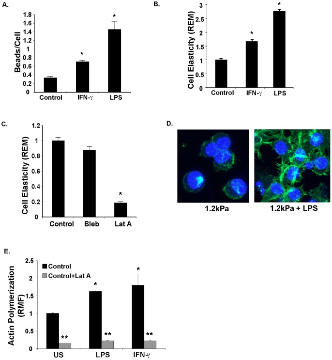
Figure 1 Bacterial PAMPs (LPS) and cytokines (IFNγ) increase macrophage phagocytosis and cell elasticity. A. LPS and IFN-γ increase phagocytosis by macrophages on a less rigid (1.2 kPa) substrate in proportion to changes in elasticity. Control, LPS-stimulated (1 µg.ml), or IFN-γ-stimulated (200 U/ml) macrophage were cultured on asubstrate rigidity corresponding to the lungs (1.2 kPa) for 24 hours, and then exposed to IgG-coated latex beads for 2 hours. Beads per cell were manually counted using confocal microscope images. *P = .008 LPS vs. control (n = 5), *P = 0.004, IFN-γ vs control (n = 5) Mann-Whitey U test. B. LPS and IFN-γ increase macrophage elasticity on a less rigid (1.2 kPa) substrate. Control unstimulated (US), LPS (1 µg/ml) stimulated, IFN-γ (200 U/ml) stimulated RAW macrophages were cultured on a less rigid substrate for 24 hours, and cell elasticity was measured via Optical Magnetic Twisting cytometry (OMTC). Data is shown as Relative Elastic Modulus (REM). Results depict data from 1 of 3 representative experiments with >100 magnetic beads assayed for each experiment. *P<0.0001 compared to control for both, Mann-Whitney U test. C. Macrophage elasticity is decreased by actin polymerization inhibitor. Optical Magnetic Twisting Cytometry (OMTC) measurement of RAW macrophages cultured on more rigid (150 kPa) surface and treated with the myosin inhibitor blebbistatin (Bleb, 50 µM) or the actin polymerization inhibitor latrunculin A (Lat A, 1 µM). Data is shown as Relative Elastic Modulus (REM). Results depict data from 1 of 3 representative experiments with >100 magnetic beads assayed for each experiment. *P<0.001, Mann Whitney U test D. LPS stimulation of macrophages on a less rigid (1.2 kPa) substrate increases cell spreading, filapodial projections, actin polymerization. RAW macrophages were cultured on less rigid substrate for 24 hours with or without LPS (1 µg/ml) stimulation for 24 hours. Cells were fixed, stained for polymerized actin (alexa-fluor phalloidin), and DNA (Hoechst nuclear stain), and visualized via confocal microscopy. Images represent collapsed stack of 7 confocal slices. E. LPS and IFN-γ increase polymerized actin in macrophages. RAW macrophages in suspension were treated with LPS (1 µg/ml), IFN-γ (200 U/ml), or latrunculin A (1 µM). After 24 hours, cells were fixed, stained for actin with Alexa-fluor phalloidin, and fluorescence was quantified via flow cytometry. Data is show as relative change in mean fluorescence (RMF) from control untreated cells (US). *P = 0.028, n = 4 compared to control, Mann-Whitney U test. **P = 0.028 compared to similar condition without Latrunculin A for each, n = 4, Mann-Whitney U test.
Figure 1 Bacterial PAMPs (LPS) and cytokines (IFNγ) increase macrophage phagocytosis and cell elasticity. A. LPS and IFN-γ increase phagocytosis by macrophages on a less rigid (1.2 kPa) substrate in proportion to changes in elasticity. Control, LPS-stimulated (1 µg.ml), or IFN-γ-stimulated (200 U/ml) macrophage were cultured on asubstrate rigidity corresponding to the lungs (1.2 kPa) for 24 hours, and then exposed to IgG-coated latex beads for 2 hours. Beads per cell were manually counted using confocal microscope images. *P = .008 LPS vs. control (n = 5), *P = 0.004, IFN-γ vs control (n = 5) Mann-Whitey U test. B. LPS and IFN-γ increase macrophage elasticity on a less rigid (1.2 kPa) substrate. Control unstimulated (US), LPS (1 µg/ml) stimulated, IFN-γ (200 U/ml) stimulated RAW macrophages were cultured on a less rigid substrate for 24 hours, and cell elasticity was measured via Optical Magnetic Twisting cytometry (OMTC). Data is shown as Relative Elastic Modulus (REM). Results depict data from 1 of 3 representative experiments with >100 magnetic beads assayed for each experiment. *P<0.0001 compared to control for both, Mann-Whitney U test. C. Macrophage elasticity is decreased by actin polymerization inhibitor. Optical Magnetic Twisting Cytometry (OMTC) measurement of RAW macrophages cultured on more rigid (150 kPa) surface and treated with the myosin inhibitor blebbistatin (Bleb, 50 µM) or the actin polymerization inhibitor latrunculin A (Lat A, 1 µM). Data is shown as Relative Elastic Modulus (REM). Results depict data from 1 of 3 representative experiments with >100 magnetic beads assayed for each experiment. *P<0.001, Mann Whitney U test D. LPS stimulation of macrophages on a less rigid (1.2 kPa) substrate increases cell spreading, filapodial projections, actin polymerization. RAW macrophages were cultured on less rigid substrate for 24 hours with or without LPS (1 µg/ml) stimulation for 24 hours. Cells were fixed, stained for polymerized actin (alexa-fluor phalloidin), and DNA (Hoechst nuclear stain), and visualized via confocal microscopy. Images represent collapsed stack of 7 confocal slices. E. LPS and IFN-γ increase polymerized actin in macrophages. RAW macrophages in suspension were treated with LPS (1 µg/ml), IFN-γ (200 U/ml), or latrunculin A (1 µM). After 24 hours, cells were fixed, stained for actin with Alexa-fluor phalloidin, and fluorescence was quantified via flow cytometry. Data is show as relative change in mean fluorescence (RMF) from control untreated cells (US). *P = 0.028, n = 4 compared to control, Mann-Whitney U test. **P = 0.028 compared to similar condition without Latrunculin A for each, n = 4, Mann-Whitney U test.CC BY · PubMed Central
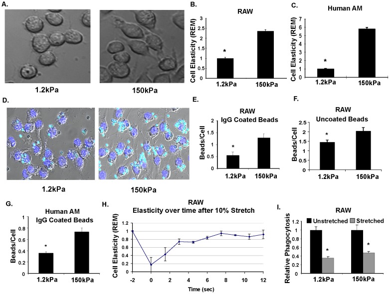
Figure 2: Changes in cell elasticity from physiologic substrate rigidity and stretch modulate macrophage phagocytosis. A. Increased cell spreading and filopodial projections of macrophages cultured on a rigid surface. Differential interference contrast (DIC) microscopy images of RAW 264.7 macrophages cultured on a less rigid (E = 1.2 kPa) versus rigid (E = 150 kPa) polyacrylamide gel coated with poly-L-lysine to facilitate attachment. B.&C. Increased elasticity of macrophages cultured on a rigid versus less rigid substrate. Optical Magnetic Twisting Cytometry (OMTC) measurement of mouse RAW macrophages (B.) and human alveolar macrophages (AM) (C.) cultured on less rigid (E = 1.2 kPa) versus rigid (E = 150 kPa) substrate. Data is displayed as relative elastic modulus (REM) as compared to less rigid condition. *P<0.001 (Mann Whitney U test) Results depict data from 1 of 3 representative experiments with >100 magnetic beads assayed for each experiment. Each experiment had similar results D. Increased phagocytosis of beads by macrophages cultured on a rigid versus less rigid surface. Merged DIC and fluorescent images of RAW macrophages cultured on a less rigid versus rigid surface, and exposed to blue-green fluorescent IgG coated latex beads for two hours at standard culture conditions. Unbound beads have been washed away, showing overall fewer beads per cell in soft versus stiff substrate. Nuclei have been stained blue with Hoechst stain. E&F. Quantification of phagocytosis in RAW macrophages, counting beads per cell in soft versus stiff macrophages. Results show significantly fewer beads per cell in macrophage cultured on less rigid substrate in both IgG-opsonized (*P = 0.03, n = 5; Mann Whitney U test), and unopsonized (*P = 0.03, n = 5; Mann Whitney U test) beads G. Phagocytosis of IgG-coated latex beads is also reduced in human alveolar macrophages (AM) comparing rigid versus less rigid surface. *P = 0.004, n = 5 (Mann Whitney U test) H. Substrate stretch decreases macrophage elasticity. Relative elasticity over time after a single 10% stretch of RAW macrophages shows initial 80% reduction in relative elasticity (REM) that returns back to steady state (prestretch) elasticity by about 8 seconds. P<0.0001 for stiffness over time using Spearman Rank Test. Results depict data from 1 of 3 representative experiments with >100 magnetic beads assayed for each time point. I. Periodic stretch reduces phagocytosis of uncoated latex beads on either less rigid (1.2 kPa) or more rigid substrate (150 kPa). Results show less phagocytosis (normalized to unstretched cells) in stretched cells on both more rigid and less rigid surface. *P<0.001 (Mann Whitney U test).
Figure 2: Changes in cell elasticity from physiologic substrate rigidity and stretch modulate macrophage phagocytosis. A. Increased cell spreading and filopodial projections of macrophages cultured on a rigid surface. Differential interference contrast (DIC) microscopy images of RAW 264.7 macrophages cultured on a less rigid (E = 1.2 kPa) versus rigid (E = 150 kPa) polyacrylamide gel coated with poly-L-lysine to facilitate attachment. B.&C. Increased elasticity of macrophages cultured on a rigid versus less rigid substrate. Optical Magnetic Twisting Cytometry (OMTC) measurement of mouse RAW macrophages (B.) and human alveolar macrophages (AM) (C.) cultured on less rigid (E = 1.2 kPa) versus rigid (E = 150 kPa) substrate. Data is displayed as relative elastic modulus (REM) as compared to less rigid condition. *P<0.001 (Mann Whitney U test) Results depict data from 1 of 3 representative experiments with >100 magnetic beads assayed for each experiment. Each experiment had similar results D. Increased phagocytosis of beads by macrophages cultured on a rigid versus less rigid surface. Merged DIC and fluorescent images of RAW macrophages cultured on a less rigid versus rigid surface, and exposed to blue-green fluorescent IgG coated latex beads for two hours at standard culture conditions. Unbound beads have been washed away, showing overall fewer beads per cell in soft versus stiff substrate. Nuclei have been stained blue with Hoechst stain. E&F. Quantification of phagocytosis in RAW macrophages, counting beads per cell in soft versus stiff macrophages. Results show significantly fewer beads per cell in macrophage cultured on less rigid substrate in both IgG-opsonized (*P = 0.03, n = 5; Mann Whitney U test), and unopsonized (*P = 0.03, n = 5; Mann Whitney U test) beads G. Phagocytosis of IgG-coated latex beads is also reduced in human alveolar macrophages (AM) comparing rigid versus less rigid surface. *P = 0.004, n = 5 (Mann Whitney U test) H. Substrate stretch decreases macrophage elasticity. Relative elasticity over time after a single 10% stretch of RAW macrophages shows initial 80% reduction in relative elasticity (REM) that returns back to steady state (prestretch) elasticity by about 8 seconds. P<0.0001 for stiffness over time using Spearman Rank Test. Results depict data from 1 of 3 representative experiments with >100 magnetic beads assayed for each time point. I. Periodic stretch reduces phagocytosis of uncoated latex beads on either less rigid (1.2 kPa) or more rigid substrate (150 kPa). Results show less phagocytosis (normalized to unstretched cells) in stretched cells on both more rigid and less rigid surface. *P<0.001 (Mann Whitney U test).CC BY · PubMed Central
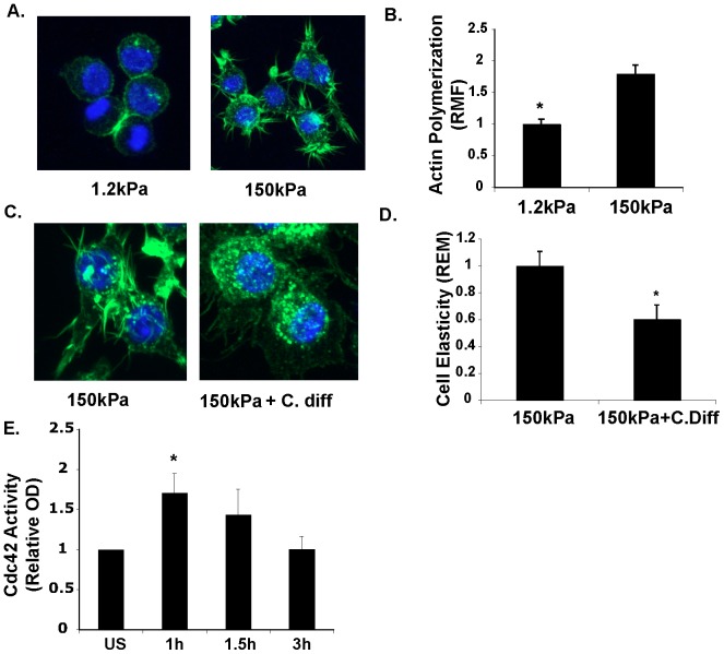
Figure 3 Macrophage elasticity is modulated by actin polymerization and rhoGTPase acitivity. A. Decreased polymerized actin staining and filapodia in macrophages cultured on a less rigid surface. Collapsed stack confocal (total of 7 slices) image of RAW macrophages cultured on less rigid (1.2 kPA) or rigid substrate (150 kPa). Green color represents actin staining (alexa-fluor 488 phalloidin) with blue stained nuclei (Hoechst). B. Decreased actin polymerization in macrophages cultured on less rigid substrate. RAW macrophages were cultured on less rigid versus more rigid substrate for 24 hours, and then lifted from surface using trypsin/EDTA solution, and immediately fixed, stained for polymerized actin (alexi-fluor-phalloidin), and staining was quantified using flow cytometry. Data displayed as changes in relative mean fluorescence. *P = 0.029, n = 4, Mann-Whitney U test. C. Decreased actin-rich filapodial projections and actin fibers in macrophages treated with rhoGTPase inhibitor. RAW macrophages were cultured on more rigid (150 kPa) substrate with or without C. difficile toxin (400 pM) for 24 hours. Cells were then fixed, stained for polymerized actin with alexi-fluor phalloidin and visualized via confocal microscopy. Image represent a collapsed stack of 7 confocal slices. D. RhoGTPase inhibitor decreases macrophage elasticity. OMTC measurement of RAW macrophages cultured on more rigid substrate (150 kPa) with or without C. diff toxin (400 pM). *P<0.0001, Mann Whitney U test. Representative experiment for >4 independent observations. E. Attachment to substrate leads to activation of cdc42. RAW macrophages in suspension were allowed to adhere to plastic tissue culture dish and protein lysates were assayed for cdc42 activation via ELISA after labeled incubation times. *P = 0.028, n = 4, Mann Whitney U test.
Figure 3 Macrophage elasticity is modulated by actin polymerization and rhoGTPase acitivity. A. Decreased polymerized actin staining and filapodia in macrophages cultured on a less rigid surface. Collapsed stack confocal (total of 7 slices) image of RAW macrophages cultured on less rigid (1.2 kPA) or rigid substrate (150 kPa). Green color represents actin staining (alexa-fluor 488 phalloidin) with blue stained nuclei (Hoechst). B. Decreased actin polymerization in macrophages cultured on less rigid substrate. RAW macrophages were cultured on less rigid versus more rigid substrate for 24 hours, and then lifted from surface using trypsin/EDTA solution, and immediately fixed, stained for polymerized actin (alexi-fluor-phalloidin), and staining was quantified using flow cytometry. Data displayed as changes in relative mean fluorescence. *P = 0.029, n = 4, Mann-Whitney U test. C. Decreased actin-rich filapodial projections and actin fibers in macrophages treated with rhoGTPase inhibitor. RAW macrophages were cultured on more rigid (150 kPa) substrate with or without C. difficile toxin (400 pM) for 24 hours. Cells were then fixed, stained for polymerized actin with alexi-fluor phalloidin and visualized via confocal microscopy. Image represent a collapsed stack of 7 confocal slices. D. RhoGTPase inhibitor decreases macrophage elasticity. OMTC measurement of RAW macrophages cultured on more rigid substrate (150 kPa) with or without C. diff toxin (400 pM). *P<0.0001, Mann Whitney U test. Representative experiment for >4 independent observations. E. Attachment to substrate leads to activation of cdc42. RAW macrophages in suspension were allowed to adhere to plastic tissue culture dish and protein lysates were assayed for cdc42 activation via ELISA after labeled incubation times. *P = 0.028, n = 4, Mann Whitney U test.CC BY · PubMed Central
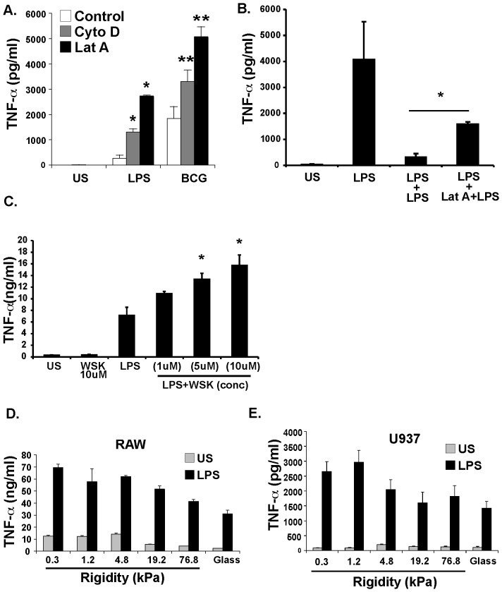
Figure 4 Macrophage elasticity affects response to LPS and LPS tolerance. A. Decreasing macrophage elasticity using actin inhibitors increases response to LPS. Human, PMA-differentiated U937 macrophage like cells were cultured on plastic tissue culture plate. Control cells, cells treated with Cytochalasin D (10 µM), or Latrunculin A (1 µM), were cultured for 24 hours with or without LPS (1 µg/ml). Culture supernatants were assayed for TNF-α via ELISA. *P = 0.015 versus control, (n = 6) Mann-Whitney U test. **P = 0.015 versus control, (n = 6), Mann-Whitney U test B. LPS tolerance is reduced by actin inhibition on U937 macrophages. Differentiated human U937 macrophages were stimulated with LPS, and had culture supernatant examined for TNFα via ELISA. Levels were compared to cells that were rechallenged with LPS for another 24 hours after 24 hours of initial LPS challenge, or treated Latrunculin A (1 µM) for 30 minutes before rechallenge with LPS. Results show that latrunculin enchanced LPS response with rechallenge. (n = 6) *P = 0.015 C. The WASP inhibitor, wiskostatin, increases LPS-mediated TNF-α release. Differentiated human U937 macrophages were stimulated with LPS with or without pretreatment with wiskostatin. Culture supernatants were assessed for TNFα release via ELISA. Results show a dose dependent increase in TNFα release in cells stimulated with LPS with wiskostatin, versus LPS alone. *P<0.01 (n = 6) D. Increasing substrate rigidity decreases TNF-α release in response to LPS in RAW macrophages. RAW macrophages were cultured in a 96 well plate in wells containing poly-L-lysine coated gels of increasing elastic modulus from 0.3 kPa-76.8 kPa, or on poly-L-lysine coated glass for 24 hours. Cells were then stimulated with LPS (1 µg/ml) for 24 hours and culture supernatants were assayed for TNF-α via ELISA. P<0.001 for correlation of TNF level with substrate rigidity. Spearman Rank Test. E. Increasing substrate rigidity decreases TNF-α release in response to LPS in human U937 macrophage-like cells. Protocol similar to D, except PMA-differentiated U937 cells are used. P<0.01, Spearman Rank Test.
Figure 4 Macrophage elasticity affects response to LPS and LPS tolerance. A. Decreasing macrophage elasticity using actin inhibitors increases response to LPS. Human, PMA-differentiated U937 macrophage like cells were cultured on plastic tissue culture plate. Control cells, cells treated with Cytochalasin D (10 µM), or Latrunculin A (1 µM), were cultured for 24 hours with or without LPS (1 µg/ml). Culture supernatants were assayed for TNF-α via ELISA. *P = 0.015 versus control, (n = 6) Mann-Whitney U test. **P = 0.015 versus control, (n = 6), Mann-Whitney U test B. LPS tolerance is reduced by actin inhibition on U937 macrophages. Differentiated human U937 macrophages were stimulated with LPS, and had culture supernatant examined for TNFα via ELISA. Levels were compared to cells that were rechallenged with LPS for another 24 hours after 24 hours of initial LPS challenge, or treated Latrunculin A (1 µM) for 30 minutes before rechallenge with LPS. Results show that latrunculin enchanced LPS response with rechallenge. (n = 6) *P = 0.015 C. The WASP inhibitor, wiskostatin, increases LPS-mediated TNF-α release. Differentiated human U937 macrophages were stimulated with LPS with or without pretreatment with wiskostatin. Culture supernatants were assessed for TNFα release via ELISA. Results show a dose dependent increase in TNFα release in cells stimulated with LPS with wiskostatin, versus LPS alone. *P<0.01 (n = 6) D. Increasing substrate rigidity decreases TNF-α release in response to LPS in RAW macrophages. RAW macrophages were cultured in a 96 well plate in wells containing poly-L-lysine coated gels of increasing elastic modulus from 0.3 kPa-76.8 kPa, or on poly-L-lysine coated glass for 24 hours. Cells were then stimulated with LPS (1 µg/ml) for 24 hours and culture supernatants were assayed for TNF-α via ELISA. P<0.001 for correlation of TNF level with substrate rigidity. Spearman Rank Test. E. Increasing substrate rigidity decreases TNF-α release in response to LPS in human U937 macrophage-like cells. Protocol similar to D, except PMA-differentiated U937 cells are used. P<0.01, Spearman Rank Test.CC BY · PubMed Central
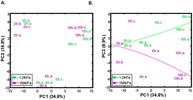
Figure 5 Macrophage gene expression is determined by duration of culture on substrate and substrate rigidity. Gene expression profile over time in RAW macrophages increasingly correlates with substrate rigidity. RAW macrophages, after removing from a plastic tissue culture dish, were plated on less rigid (E = 1.2 kPa) or more rigid (E = 150 kPa) substrate for 2 hours, 6 hours, and 18 hours, and RNA was extracted for microarray analysis at each time point. A. Principle component analysis (PCA) reveals a PC1 that well correlates with duration of culture on substrate and describes the largest variance (34.8%), while a PC2 distinguished 6 hours culture time from 2 hours and 18 hours culture times. B. The third larges source of variance was PC3 (9.9% of variance) that distinguishes by substrate rigidity increasingly over time.
Figure 5 Macrophage gene expression is determined by duration of culture on substrate and substrate rigidity. Gene expression profile over time in RAW macrophages increasingly correlates with substrate rigidity. RAW macrophages, after removing from a plastic tissue culture dish, were plated on less rigid (E = 1.2 kPa) or more rigid (E = 150 kPa) substrate for 2 hours, 6 hours, and 18 hours, and RNA was extracted for microarray analysis at each time point. A. Principle component analysis (PCA) reveals a PC1 that well correlates with duration of culture on substrate and describes the largest variance (34.8%), while a PC2 distinguished 6 hours culture time from 2 hours and 18 hours culture times. B. The third larges source of variance was PC3 (9.9% of variance) that distinguishes by substrate rigidity increasingly over time.CC BY · PubMed Central

Verification

Claims the paper makes

The claims and statistics of this paper haven’t been checked yet.

Free: it reads the open-access text and checks the first 5 claims that cite a source we can check. Create a free account to check every claim.

A few free checks a day · papers already verified open instantly · results are public

Claims other papers make about this one

Each is checked against this paper’s own text: does it say what the citing paper says it does?

Self-check

Percentages, sums and reported differences re-computed from the paper’s own numbers.

The self-check needs the paper’s full text.

Citations

Cites

Show all 46 references

Cited by

See all 275

Metadata

Authors
Patel, N. R., Bole, M., Chen, C., Hardin, C. C., Kho, A. T., Mih, J., Deng, L., Butler, J., Tschumperlin, D., Fredberg, J. J., Krishnan, R., & Koziel, H.
Affiliations
  • Beth Israel Deaconess Medical Center (US)
  • Harvard University (US)
  • Boston Children's Hospital (US)
  • Chongqing University (CN)
Corresponding author
Patel, Naimish R.
Venue
PLoS ONE
Published
2012-09-18
Volume
7
Issue
9
Pages
e41024
ISSN
1932-6203 (ISSN-L)
OpenAlex
W2114845386
Cited by
275
References
46
Citations by year
2026: 8 · 2025: 23 · 2024: 21 · 2023: 16 · 2022: 21 · 2021: 44 · 2020: 24 · 2019: 32 · 2018: 24 · 2017: 19 · 2016: 9 · 2015: 18 · 2014: 12 · 2013: 4
FWCI
5.15
Type
journal article
Language
en
Open access
Locations
  • PLoS ONE (publishedVersion, cc-by)
  • PubMed Central (publishedVersion, cc-by)
  • PubMed (publishedVersion)
  • Digital Access to Scholarship at Harvard (DASH) (Harvard University) (submittedVersion, cc-by)
  • DOAJ (DOAJ: Directory of Open Access Journals) (submittedVersion, cc-by-sa)
  • DSpace@MIT (Massachusetts Institute of Technology) (submittedVersion, cc-by-nc)
  • Figshare (submittedVersion, cc-by)
Funders
  • National Heart, Lung, and Blood Institute (K25HL111212, K25HL091124, R01HL102373)
  • National Institute of Allergy and Infectious Diseases
Topics
  • Life Sciences › Biochemistry, Genetics and Molecular Biology › Cell Biology › Cellular Mechanics and Interactions
  • Life Sciences › Immunology and Microbiology › Immunology › Immune cells in cancer
  • Physical Sciences › Engineering › Biomedical Engineering › Elasticity and Material Modeling
Keywords
macrophage function, substrate stiffness, actin polymerization, Rho GTPase activation, innate immunity, lipopolysaccharide, interferon-gamma, mechanobiology
MeSH terms
Actins, Animals, Cell Line, Elasticity, Humans, Inflammation, Interferon-gamma, Lipopolysaccharides, Macrophages, Phagocytosis, rho GTP-Binding Proteins, Gene Expression Profiling, Mice, Protein Multimerization, Transcriptome

About these checks

Registry, retraction, Cites and statistics results are computed, not judged, with one exception: a printed reference that no index lists is read once by a model before it counts as a citation error. Claims are extracted by a model and kept only when their quote appears verbatim in the text; each cited source is then checked, with a model’s help, for whether it says what the claim relies on. Those verdicts can be wrong — read the claim’s quote and the cited source before relying on one.

Statistics and arithmetic are recomputed, not judged.

Confidence bands are shown only for fields calibrated on our annotated test set. None is calibrated yet, so no band is shown.

What counts as a claim ›How we measure confidence ›How we check this ›

Are you an author, or do you see a problem with this page? Request a review

How to cite

Cite CoChat for the checks on this page, and the paper for its own findings.

This page
CoChat. (2026). Citation check: Cell Elasticity Determines Macrophage Function. https://app.cochat.ai/papers/10.1371/journal.pone.0041024
The paper
Patel, N. R., Bole, M., Chen, C., Hardin, C. C., Kho, A. T., Mih, J., Deng, L., Butler, J., Tschumperlin, D., Fredberg, J. J., Krishnan, R., & Koziel, H. (2012). Cell Elasticity Determines Macrophage Function. PLoS ONE. https://doi.org/10.1371/journal.pone.0041024

Data sources

Bibliographic data: Semantic Scholar, OpenAlex, Crossref.

Contains information from the Semantic Scholar Academic Graph Datasets, made available under the ODC Attribution License (ODC-BY 1.0).

Semantic Scholar’s data is described in: Kinney, R., et al. (2023). The Semantic Scholar Open Data Platform. arXiv:2301.10140.