

The DOI matches its Crossref record; 61 of the 61 references it cites were found in a registry or an index.
Brain Microbial Populations in HIV/AIDS: α-Proteobacteria Predominate Independent of Host Immune Status
Open accessAbstract
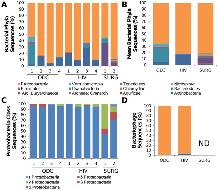
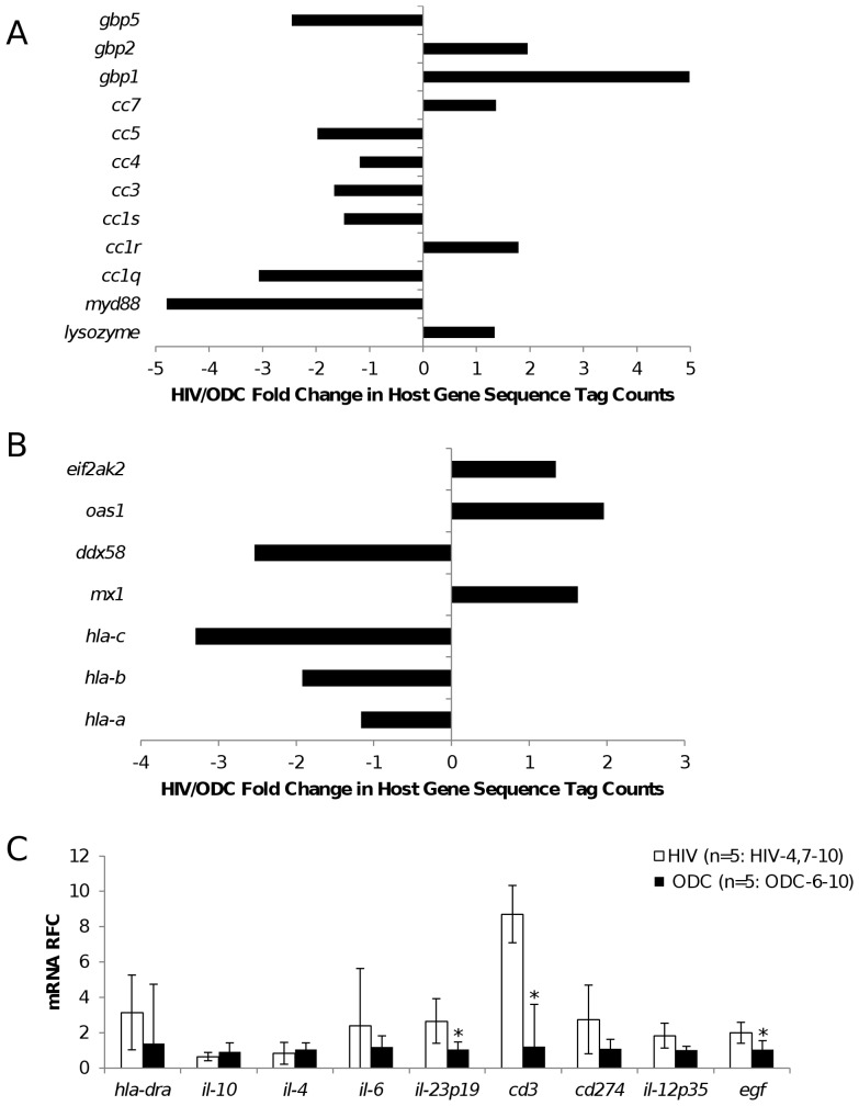
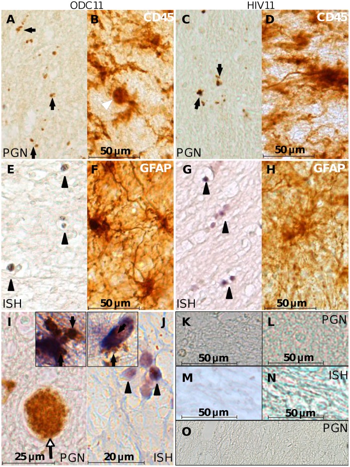
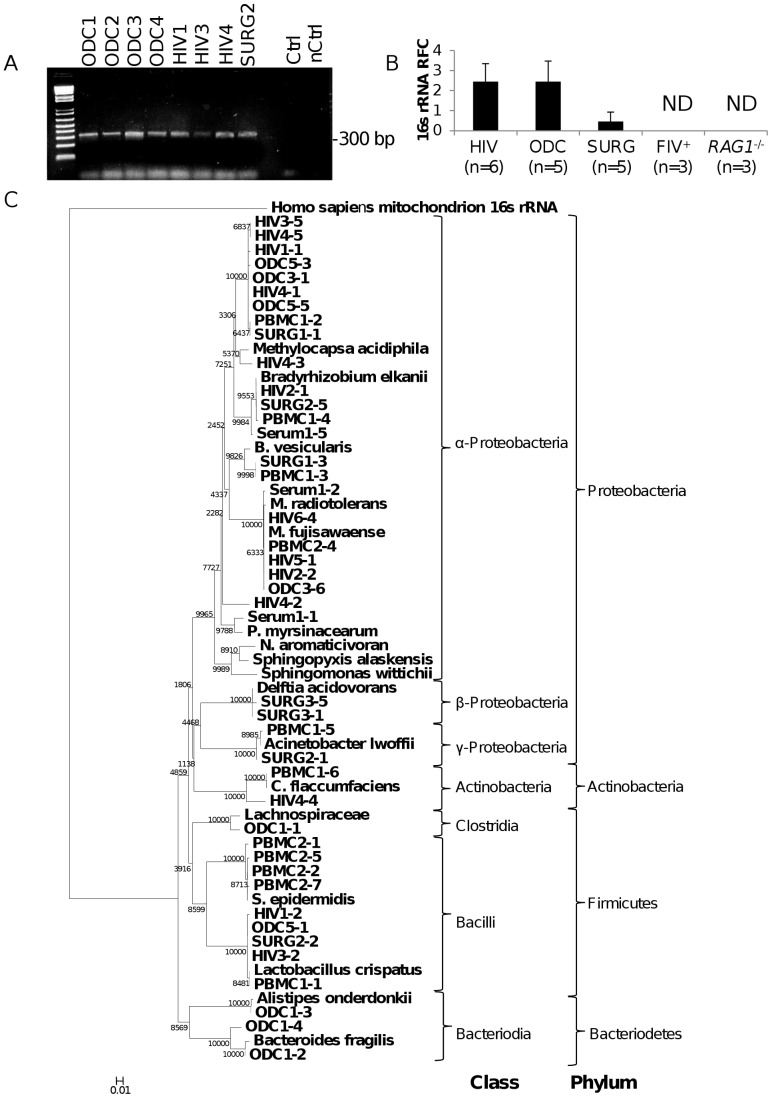
The brain is assumed to be a sterile organ in the absence of disease although the impact of immune disruption is uncertain in terms of brain microbial diversity or quantity. To investigate microbial diversity and quantity in the brain, the profile of infectious agents was examined in pathologically normal and abnormal brains from persons with HIV/AIDS [HIV] (n = 12), other disease controls [ODC] (n = 14) and in cerebral surgical resections for epilepsy [SURG] (n = 6). Deep sequencing of cerebral white matter-derived RNA from the HIV (n = 4) and ODC (n = 4) patients and SURG (n = 2) groups revealed bacterially-encoded 16 s RNA sequences in all brain specimens with α-proteobacteria representing over 70% of bacterial sequences while the other 30% of bacterial classes varied widely. Bacterial rRNA was detected in white matter glial cells by in situ hybridization and peptidoglycan immunoreactivity was also localized principally in glia in human brains. Analyses of amplified bacterial 16 s rRNA sequences disclosed that Proteobacteria was the principal bacterial phylum in all human brain samples with similar bacterial rRNA quantities in HIV and ODC groups despite increased host neuroimmune responses in the HIV group. Exogenous viruses including bacteriophage and human herpes viruses-4, -5 and -6 were detected variably in autopsied brains from both clinical groups. Brains from SIV- and SHIV-infected macaques displayed a profile of bacterial phyla also dominated by Proteobacteria but bacterial sequences were not detected in experimentally FIV-infected cat or RAG1⁻/⁻ mouse brains. Intracerebral implantation of human brain homogenates into RAG1⁻/⁻ mice revealed a preponderance of α-proteobacteria 16 s RNA sequences in the brains of recipient mice at 7 weeks post-implantation, which was abrogated by prior heat-treatment of the brain homogenate. Thus, α-proteobacteria represented the major bacterial component of the primate brain's microbiome regardless of underlying immune status, which could be transferred into naïve hosts leading to microbial persistence in the brain.
Overview
The claims and statistics of this paper haven’t been checked yet.
Run the check — free, no account needed. It reads the open-access text and checks the sources it cites.
Integrity signals
Registry
Cites
Statistics
Figures
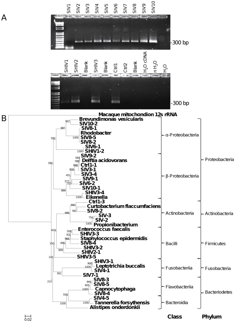
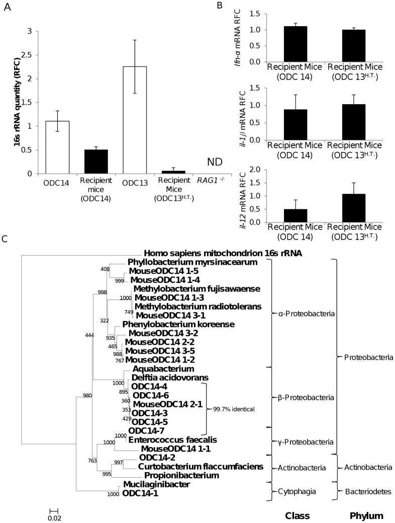
Verification
Claims the paper makes
The claims and statistics of this paper haven’t been checked yet.
Run the check — free, no account needed. It reads the open-access text and checks the sources it cites.
Claims other papers make about this one
Each is checked against this paper’s own text: does it say what the citing paper says it does?
The source backs part of the claim, not all of it — or backs it for a different population, condition or comparison.
In the citing paper
“The same bacterial sequences were detected in the brains of immunocompromised mice, weeks after human brain tissue transplantation but not in mice given heat-treated tissue, suggesting the bacterial sequences in the human brain samples were viable [133].”
From What would it take to prove that a chronic infection is a causal agent in Alzheimer’s disease? (2025) · cites this paper as [133]
Experimental — accuracy not yet measuredThe source backs part of the claim, not all of it — or backs it for a different population, condition or comparison.
In the citing paper
“A pioneering study investigating microbial infiltration into the brains of people living with HIV, found bacterial sequences present in both “healthy” and HIV brains at a similar distribution [133].”
From What would it take to prove that a chronic infection is a causal agent in Alzheimer’s disease? (2025) · cites this paper as [133]
Experimental — accuracy not yet measured
Self-check
Percentages, sums and reported differences re-computed from the paper’s own numbers.
The self-check needs the paper’s full text.
Citations
Cites
Enterotypes of the human gut microbiome
MetaHIT Consortium (additional members), Arumugam, M., Raes, J., Pelletier, E., Le Paslier, D., Yamada, T., Mende, D. R., Fernandes, G. R., Tap, J., Bruls, T., et al. (2011) · Nature · doi:10.1038/nature09944 · Cited by 7,548 · cited 1×
“The composition of the human microbiome at several body sites including skin, oral cavity, gastrointestinal tract, nasopharyngeal passage and urogenital tract are subjects of intensive study [2, 5, 6, 7] , revealing both local effects at these sites but also systemic effects, including influencing inflammatory or meta…”
BackgroundGrice, E. A., & Segre, J. A. (2011) · Nature Reviews Microbiology · doi:10.1038/nrmicro2537 · Cited by 3,143
Human oral, gut, and plaque microbiota in patients with atherosclerosis
Koren, O., Spor, A., Felin, J., Fåk, F., Stombaugh, J., Tremaroli, V., Behre, C. J., Knight, R., Fagerberg, B., Ley, R. E., et al. (2011) · Proceedings of the National Academy of Sciences · doi:10.1073/pnas.1011383107 · Cited by 1,196 · cited 1×
“In contrast, diseases such as biliary cirrhosis [13,14], atherosclerosis [15] and aortic”
BackgroundMicrobial community profiling for human microbiome projects: Tools, techniques, and challengesPreprint
Hamady, M., & Knight, R. D. (2009) · Genome Research · doi:10.1101/gr.085464.108 · Cited by 1,031 · cited 1×
“intensive study [2,5,6,7], revealing both local effects at these sites”
BackgroundGreisen, K. S., Loeffelholz, M. J., Purohit, A. P., & Leong, D. (1994) · Journal of Clinical Microbiology · doi:10.1128/jcm.32.2.335-351.1994 · Cited by 626 · cited 1×
“An amplicon from the V8 region of the 16 s gene was generated from brain-derived cDNA using the universal bacterial primers RW01 or RDR080 with DG74 [54].”
MethodNeuroinvasion by Human Respiratory Coronaviruses
Arbour, N., Day, R. W., Newcombe, J., & Talbot, P. J. (2000) · Journal of Virology · doi:10.1128/jvi.74.19.8913-8921.2000 · Cited by 526 · cited 1×
“Saffold [58] and Corona [59] viruses were detected in brain-derived cDNA by nested RT-PCR using the primers listed in Table S4.”
MethodAncuta, P., Kamat, A., Kunstman, K. J., Kim, E.-Y., Autissier, P., Wurcel, A., Zaman, T., Stone, D., Mefford, M., Morgello, S., et al. (2008) · PLoS ONE · doi:10.1371/journal.pone.0002516 · Cited by 499 · cited 1×
“These events occur because of reduced immune surveillance and damaged barrier function at epithelial and endothelial sites, including damage to the blood-brain barrier [19].”
BackgroundPower, C., Kong, P., Crawford, T. O., Wesselingh, S. L., Glass, J. D., McArthur, J. C., & Trapp, B. D. (1993) · Annals of Neurology · doi:10.1002/ana.410340307 · Cited by 332 · cited 1×
“Multiple studies have shown the blood-brain barrier to be more permeable in HIV/ AIDS, implying this circumstance might contribute to HIVassociated neurological disease [20,21].”
BackgroundSelf-citationWatzinger, F., Suda, M., Preuner, S., Baumgartinger, R., Ebner, K., Bašková, L., Niesters, H. G. M., Lawitschka, Á., & Lion, T. (2004) · Journal of Clinical Microbiology · doi:10.1128/jcm.42.11.5189-5198.2004 · Cited by 326
Comparative analyses of the bacterial microbiota of the human nostril and oropharynx
Lemon, K. P., Klepac-Ceraj, V., Schiffer, H. K., Brodie, E. L., Lynch, S. V., & Kolter, R. (2010) · mBio · doi:10.1128/mbio.00129-10 · Cited by 322 · cited 1×
“intensive study [2,5,6,7], revealing both local effects at these sites”
Background
Cited by
Sorted by citations
Reagent and laboratory contamination can critically impact sequence-based microbiome analyses
Salter, S. J., Cox, M. J., Turek, E. M., Calus, S. T., Cookson, W. O., Moffatt, M. F., Turner, P., Parkhill, J., Loman, N. J., & Walker, A. W. (2014) · BMC Biology · doi:10.1186/s12915-014-0087-z · Cited by 3,463
“Many recent publications [19-37] describe important or core microbiota members, often members that are biologically unexpected, which overlap with previouslydescribed contaminant genera.”
BackgroundContamination in Low Microbial Biomass Microbiome Studies: Issues and Recommendations
Eisenhofer, R., Minich, J. J., Marotz, C., Cooper, A., Knight, R., & Weyrich, L. S. (2019) · Trends in Microbiology · doi:10.1016/j.tim.2018.11.003 · Cited by 1,146
The role of microbiome in central nervous system disorders
Wang, Y., & Kasper, L. H. (2014) · Brain Behavior and Immunity · doi:10.1016/j.bbi.2013.12.015 · Cited by 803
“Interestingly, microbiota have been identified within immune-privileged sites such as the CNS. a-proteobacteria class is reported to be the major commensals persistent http:// in the human brain regardless of immune status (Branton et al., 2013).”
The microbiome of the urinary tract—a role beyond infection
Whiteside, S. A., RAZVI, H. A., Davé, S., Reid, G., & Burton, J. P. (2015) · Nature Reviews Urology · doi:10.1038/nrurol.2014.361 · Cited by 657
“(2013)27 Healthy men (6) Healthy women (10) Jonquetella, Parvimonas, Proteiniphilum, Saccharofermentans”
BackgroundGram-negative bacterial molecules associate with Alzheimer disease pathology
Zhan, X., Stamova, B., Jin, L.-W., DeCarli, C., Phinney, B., & Sharp, F. R. (2016) · Neurology · doi:10.1212/wnl.0000000000003391 · Cited by 521
“Our findings complement a recent RNAseq study that showed bacterially encoded 16s RNA sequences in all human brains with Gram-negativea-Proteobacteria representing over 70% of bacterial sequences.(16) The other 30% of bacterial classes varied widely, with g-Proteobacteria, including E coli, being present at ,5% of tot…”
BackgroundResultInfluential
Metadata
- Authors
- Branton, W. G., Ellestad, K. K., Maingat, F., Wheatley, B. M., Rud, E., Warren, R. L., Holt, R. A., Surette, M. G., & Power, C.
- Affiliations
- University of Alberta (CA)
- Health Canada (CA)
- BC Cancer Agency (CA)
- McMaster University (CA)
- Corresponding author
- Power, Christopher
- ORCID
- Venue
- PLoS ONE
- Publisher
- Public Library of Science (PLoS)
- Published
- 2013-01-23
- Volume
- 8
- Issue
- 1
- Pages
- e54673
- ISSN
- 1932-6203 (ISSN-L)
- PMC
- PMC3552853
- OpenAlex
- W2057381259
- Cited by
- 149
- References
- 60
- Citations by year
- 2026: 5 · 2025: 6 · 2024: 14 · 2023: 17 · 2022: 15 · 2021: 10 · 2020: 12 · 2019: 12 · 2018: 16 · 2017: 11 · 2016: 12 · 2015: 13 · 2014: 2 · 2013: 4
- FWCI
- 4.75
- Type
- journal article
- Language
- en
- Open access
- gold
- doi.org
- Locations
- PLoS ONE (publishedVersion, cc-by)
- PubMed Central (publishedVersion, cc-by)
- PubMed (publishedVersion)
- DOAJ (DOAJ: Directory of Open Access Journals) (submittedVersion, cc-by-sa)
- Figshare (submittedVersion, cc-by)
- Funders
- Alberta Innovates
- Health Canada
- Canadian Institutes of Health Research (G118160525)
- Topics
- Life Sciences › Immunology and Microbiology › Virology › HIV Research and Treatment
- Life Sciences › Biochemistry, Genetics and Molecular Biology › Molecular Biology › Gut microbiota and health
- Health Sciences › Medicine › Infectious Diseases › Infectious Diseases and Tuberculosis
- Keywords
- HIV/AIDS, Alphaproteobacteria, Proteobacteria, bacterial 16S rRNA, peptidoglycan, herpesvirus, bacteriophages, simian immunodeficiency virus, SHIV, FIV, Rag1-deficient mice, microbial persistence, neuroimmune response, epilepsy surgery
- MeSH terms
- Acquired Immunodeficiency Syndrome, Animals, Autopsy, Cats, Female, Humans, Male, Neuroglia, RNA, Bacterial, RNA, Ribosomal, Feline Acquired Immunodeficiency Syndrome, Mice, Knockout, Alphaproteobacteria, Central Nervous System Bacterial Infections, Mice, Cerebrum
About these checks
Registry, retraction, Cites and statistics results are computed, not judged, with one exception: a printed reference that no index lists is read once by a model before it counts as a citation error. Claims are extracted by a model and kept only when their quote appears verbatim in the text; each cited source is then checked, with a model’s help, for whether it says what the claim relies on. Those verdicts can be wrong — read the claim’s quote and the cited source before relying on one.
Statistics and arithmetic are recomputed, not judged.
Confidence bands are shown only for fields calibrated on our annotated test set. None is calibrated yet, so no band is shown.
What counts as a claim ›How we measure confidence ›How we check this ›
Are you an author, or do you see a problem with this page? Request a review
