

The DOI matches its Crossref record; 32 of the 32 references it cites were found in a registry or an index.
Impact of the 3D Microenvironment on Phenotype, Gene Expression, and EGFR Inhibition of Colorectal Cancer Cell Lines
Open accessAbstract
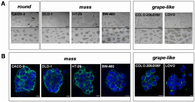
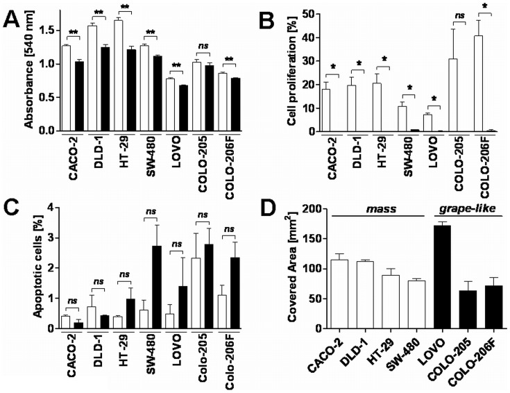
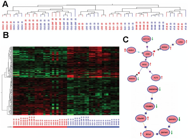
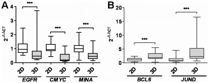
Three-dimensional (3D) tumor cell cultures grown in laminin-rich-extracellular matrix (lrECM) are considered to reflect human tumors more realistic as compared to cells grown as monolayer on plastic. Here, we systematically investigated the impact of ECM on phenotype, gene expression, EGFR signaling pathway, and on EGFR inhibition in commonly used colorectal cancer (CRC) cell lines. LrECM on-top (3D) culture assays were performed with the CRC cell lines SW-480, HT-29, DLD-1, LOVO, CACO-2, COLO-205 and COLO-206F. Morphology of lrECM cultivated CRC cell lines was determined by phase contrast and confocal laser scanning fluorescence microscopy. Proliferation of cells was examined by MTT assay, invasive capacity of the cell lines was assayed using Matrigel-coated Boyden chambers, and migratory activity was determined employing the Fence assay. Differential gene expression was analyzed at the transcriptional level by the Agilent array platform. EGFR was inhibited by using the specific small molecule inhibitor AG1478. A specific spheroid growth pattern was observed for all investigated CRC cell lines. DLD-1, HT-29 and SW-480 and CACO-2 exhibited a clear solid tumor cell formation, while LOVO, COLO-205 and COLO-206F were characterized by forming grape-like structures. Although the occurrence of a spheroid morphology did not correlate with an altered migratory, invasive, or proliferative capacity of CRC cell lines, gene expression was clearly altered in cells grown on lrECM as compared to 2D cultures. Interestingly, in KRAS wild-type cell lines, inhibition of EGFR was less effective in lrECM (3D) cultures as compared to 2D cell cultures. Thus, comparing both 2D and 3D cell culture models, our data support the influence of the ECM on cancer growth. Compared to conventional 2D cell culture, the lrECM (3D) cell culture model offers the opportunity to investigate permanent CRC cell lines under more physiological conditions, i.e. in the context of molecular therapeutic targets and their pharmacological inhibition.
Overview
The claims and statistics of this paper haven’t been checked yet.
Run the check — free, no account needed. It reads the open-access text and checks the sources it cites.
Integrity signals
Registry
Cites
Statistics
Figures
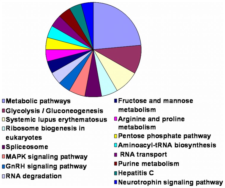
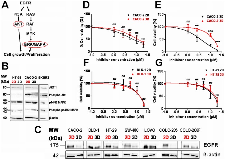
Verification
Claims the paper makes
The claims and statistics of this paper haven’t been checked yet.
Run the check — free, no account needed. It reads the open-access text and checks the sources it cites.
Claims other papers make about this one
Each is checked against this paper’s own text: does it say what the citing paper says it does?
The source backs part of the claim, not all of it — or backs it for a different population, condition or comparison.
In the citing paper
“The proliferation rates of cells cultured in 3D and 2D are usually different, and are cell line and matrix dependent.”
From Three-dimensional cell culture systems and their applications in drug discovery and cell-based biosensors (2014) · cites this paper as [29]
Experimental — accuracy not yet measuredThe cited source says what the claim attributes to it.
In the citing paper
“Overall, the morphological appearance of 3D spheroids is primarily cell line dependent. 29,35,37”
From Three-dimensional cell culture systems and their applications in drug discovery and cell-based biosensors (2014) · cites this paper as [29]
The cited source says what the claim attributes to it.
In the citing paper
“A variety of cell lines showed reduced proliferation rate in 3D cultures compared to those cultured in 2D. 17,27-31”
From Three-dimensional cell culture systems and their applications in drug discovery and cell-based biosensors (2014) · cites this paper as [29]
The check wasn’t confident either way, so it gives no verdict — never disputed.
In the citing paper
“A similar trend was observed in hepatocellular carcinoma models, where monolayers showed enrichment in structural gene expression, whereas spheroids upregulated genes related to metabolism and biosynthesis [196,197,198].”
From Modeling Tumor Microenvironment Complexity In Vitro: Spheroids as Physiologically Relevant Tumor Models and Strategies for Their Analysis (2025) · cites this paper as [197]
Experimental — accuracy not yet measured
Self-check
Percentages, sums and reported differences re-computed from the paper’s own numbers.
The self-check needs the paper’s full text.
Citations
Cites
Analysis of Relative Gene Expression Data Using Real-Time Quantitative PCR and the 2−ΔΔCT Method
Livak, K. J., & Schmittgen, T. D. (2001) · Methods · doi:10.1006/meth.2001.1262 · Cited by 180,860
Integrins: Versatility, modulation, and signaling in cell adhesion
Hynes, R. O. (1992) · Cell · doi:10.1016/0092-8674(92)90115-s · Cited by 9,857
Wild-Type BRAF Is Required for Response to Panitumumab or Cetuximab in Metastatic Colorectal Cancer
Di Nicolantonio, F., Martini, M., Molinari, F., Sartore-Bianchi, A., Arena, S., Saletti, P., De Dosso, S., Mazzucchelli, L., Frattini, M., Siena, S., et al. (2008) · Journal of Clinical Oncology · doi:10.1200/jco.2008.18.0786 · Cited by 1,591 · cited 1×
“KRAS and BRAF mutational status of patients is usually assessed before EGFR treatment on the basis that only patients exhibiting BRAF and KRAS wild type profit from treatments with anti-EGFR therapies [29,30].”
BackgroundBasement membrane complexes with biological activity
Kleinman, H. K., McGarvey, M. L., Hassell, J. R., Star, V. L., Cannon, F. B., Laurie, G. W., & Martin, G. R. (1986) · Biochemistry · doi:10.1021/bi00350a005 · Cited by 1,502
Three-dimensional culture models of normal and malignant breast epithelial cells
Lee, G. Y., Kenny, P. A., Lee, E. H., & Bissell, M. J. (2007) · Nature Methods · doi:10.1038/nmeth1015 · Cited by 1,336
Kleinman, H. K., McGarvey, M. L., Liotta, L. A., Robey, P. G., Tryggvason, K., & Martin, G. R. (1982) · Biochemistry · doi:10.1021/bi00267a025 · Cited by 1,231
PETERSEN, O. W., Rønnov‐Jessen, L., Howlett, A. R., & Bissell, M. J. (1992) · Proceedings of the National Academy of Sciences · doi:10.1073/pnas.89.19.9064 · Cited by 1,096
Kenny, P. A., Lee, G. Y., Myers, C. A., Neve, R. M., Semeiks, J. R., Spellman, P. T., Lorenz, K., Lee, E. H., Barcellos‐Hoff, M. H., PETERSEN, O. W., et al. (2008) · eScholarship (California Digital Library) · Cited by 1,039
Benvenuti, S., Sartore-Bianchi, A., Di Nicolantonio, F., Zanon, C., Moroni, M., Veronese, S., Siena, S., & Bardelli, A. (2007) · Cancer Research · doi:10.1158/0008-5472.can-06-4158 · Cited by 830
Weaver, V. M., Lelièvre, S. A., Lakins, J. N., Chrenek, M. A., Jones, J., Giancotti, F. G., Werb, Z., & Bissell, M. J. (2002) · Cancer Cell · doi:10.1016/s1535-6108(02)00125-3 · Cited by 794
Research works printed but not linked: 1
- PD Yurchenco, 1992, Laminin forms an independent network in basement membranes, J Cell Biol, 117, 1119
Cited by
Sorted by citations
Items 101–200 of 339
Phenotypic Analysis of Organoids by Proteomics
Gonneaud, A., Asselin, C., Boudreau, F., & Boisvert, F. (2017) · PROTEOMICS · doi:10.1002/pmic.201700023 · Cited by 43
Gomez-Roman, N., Chong, M. Y., Chahal, S. K., Caragher, S. P., Jackson, M. R., Stevenson, K. H., Dongre, S. A., & Chalmers, A. J. (2019) · Molecular Cancer Therapeutics · doi:10.1158/1535-7163.mct-18-1320 · Cited by 42
Freckmann, E. C., Sandilands, E., Cumming, E. M., Neilson, M., Román‐Fernández, Á., Nikolatou, K., Nacke, M., Lannagan, T. R. M., Hedley, A., Strachan, D., et al. (2022) · Nature Communications · doi:10.1038/s41467-022-32958-x · Cited by 40
Modeling glioblastoma complexity with organoids for personalized treatments
Pawlowski, K. D., Duffy, J. T., Babak, M. V., & Balyasnikova, I. V. (2023) · Trends in Molecular Medicine · doi:10.1016/j.molmed.2023.01.002 · Cited by 42
Scaffold-free generation of heterotypic cell spheroids using acoustofluidics
Wu, Z., Chen, B., Wu, Y., Xia, Y., Chen, H., Gong, Z., Hu, H., Ding, Z., & Guo, S. (2021) · Lab on a Chip · doi:10.1039/d1lc00496d · Cited by 40
Aouad, P., Saikali, M., Abdel‐Samad, R., Fostok, S. F., El–Houjeiri, L., Pisano, C., Talhouk, R., & Darwiche, N. (2017) · Anti-Cancer Drugs · doi:10.1097/cad.0000000000000511 · Cited by 39
Katiyar, A., Tocco, V. J., Li, Y., Aggarwal, V., Tamashunas, A. C., Dickinson, R. B., & Lele, T. P. (2019) · Soft Matter · doi:10.1039/c9sm01666j · Cited by 41
Zaszczyńska, A., Niemczyk-Soczynska, B., & Sajkiewicz, P. (2022) · Polymers · doi:10.3390/polym14235278 · Cited by 37
Transcriptional Landscape of 3D vs. 2D Ovarian Cancer Cell Models
Kerslake, R., Belay, B., Panfilov, S., Hall, M., Kyrou, I., Randeva, H. S., Hyttinen, J., Karteris, E., & Sisu, C. (2023) · Cancers · doi:10.3390/cancers15133350 · Cited by 36
Yapa, K. T. D. S., Deuis, J. R., Peters, A. A., Kenny, P. A., Roberts‐Thomson, S. J., Vetter, I., & Monteith, G. R. (2018) · Life Sciences · doi:10.1016/j.lfs.2018.02.030 · Cited by 36
Pallegar, N. K., Garland, C., Mahendralingam, M. J., Viloria‐Petit, A., & Christian, S. L. (2018) · Journal of Mammary Gland Biology and Neoplasia · doi:10.1007/s10911-018-9420-4 · Cited by 36
Park, H. J., & Helfman, D. M. (2019) · Scientific Reports · doi:10.1038/s41598-019-56276-3 · Cited by 36
3D Scaffolds to Model the Hematopoietic Stem Cell Niche: Applications and Perspectives
Congrains, A., Bianco, J., Rosa, R. G., Mancuso, R. I., & Saad, S. T. O. (2021) · Materials · doi:10.3390/ma14030569 · Cited by 40
Collins, T., Pyne, E. S., Christensen, M., Iles, A., Pamme, N., & Pires, I. M. (2021) · Biomicrofluidics · doi:10.1063/5.0061373 · Cited by 36
ErbB Family Signalling: A Paradigm for Oncogene Addiction and Personalized Oncology
Jacobi, N., Seeboeck, R., Hofmann, E., & Eger, A. (2017) · Cancers · doi:10.3390/cancers9040033 · Cited by 36
Anderson, M. Z., Gerstein, A. C., Wigen, L., Baller, J. A., & Berman, J. G. (2014) · PLoS Genetics · doi:10.1371/journal.pgen.1004436 · Cited by 34
Ganjibakhsh, M., Mehraein, F., Koruji, M., Aflatoonian, R., & Farzaneh, P. (2019) · Materials Science and Engineering C · doi:10.1016/j.msec.2019.02.090 · Cited by 34
In Vitro 3D Modeling of Neurodegenerative Diseases
Louit, A., Galbraith, T., & Berthod, F. (2023) · Bioengineering · doi:10.3390/bioengineering10010093 · Cited by 37
PAR2-dependent activation of GSK3β regulates the survival of colon stem/progenitor cells
Nasri, I., Bonnet, D., Zwarycz, B., d'Aldebert, E., Khou, S., Jarraya, R. M., Quaranta, M., Rolland, C., Bonnart, C., Mas, E., et al. (2016) · American Journal of Physiology-Gastrointestinal and Liver Physiology · doi:10.1152/ajpgi.00328.2015 · Cited by 34
Assessing chemotherapeutic effectiveness using a paper-based tumor model
Boyce, M. W., LaBonia, G. J., Hummon, A. B., & Lockett, M. R. (2017) · The Analyst · doi:10.1039/c7an00806f · Cited by 34
LRP-1 Promotes Colon Cancer Cell Proliferation in 3D Collagen Matrices by Mediating DDR1 Endocytosis
Le, C., Bennasroune, A., Collin, G., Hachet, C., Lehrter, V., Rioult, D., Dedieu, S., Morjani, H., & Appert-Collin, A. (2020) · Frontiers in Cell and Developmental Biology · doi:10.3389/fcell.2020.00412 · Cited by 33
The third dimension: new developments in cell culture models for colorectal research
Pereira, J., Awatade, N. T., Loureiro, C. A., Matos, P., Amaral, M. D., & Jordan, P. (2016) · Cellular and Molecular Life Sciences · doi:10.1007/s00018-016-2258-2 · Cited by 32
Advances in Modeling the Immune Microenvironment of Colorectal Cancer
Yoon, P., Piccolo, N. D., Shirure, V. S., Peng, Y., Kirane, A., Canter, R. J., Fields, R. C., George, S. C., & Gholami, S. (2021) · Frontiers in Immunology · doi:10.3389/fimmu.2020.614300 · Cited by 33
Buhrmann, C., Brockmueller, A., Harsha, C., Kunnumakkara, A. B., Kubatka, P., Aggarwal, B. B., & Shakibaei, M. (2021) · Frontiers in Pharmacology · doi:10.3389/fphar.2021.699842 · Cited by 32
Wu, Y., Zhou, Y., Qin, X., & Liu, Y. (2021) · Biomicrofluidics · doi:10.1063/5.0062697 · Cited by 33
Wu, Y., Li, K., Li, Y., Sun, T., Liu, C., Dong, C., Zhao, T., Tang, D., Chen, X., Chen, X., et al. (2021) · Nucleic Acids Research · doi:10.1093/nar/gkab1201 · Cited by 31
Koban, R., Neumann, M., Daugs, A., Bloch, O., Nitsche, A., Langhammer, S., & Ellerbrok, H. (2017) · Antiviral Research · doi:10.1016/j.antiviral.2017.12.005 · Cited by 30
Dissecting cellular mechanics: Implications for aging, cancer, and immunity
Harris, M. J., Wirtz, D., & Wu, P.-H. (2018) · Seminars in Cell and Developmental Biology · doi:10.1016/j.semcdb.2018.10.008 · Cited by 29
Uniformity of spheroids-on-a-chip by surface treatment of PDMS microfluidic platforms
Azizipour, N., Avazpour, R., Sawan, M., Rosenzweig, D. H., & Ajji, A. (2022) · Sensors & Diagnostics · doi:10.1039/d2sd00004k · Cited by 29
Ulusoy, M., Lavrentieva, A. V., Walter, J., Sambale, F., Green, M., Stahl, F., & Scheper, T. H. (2015) · Toxicology Research · doi:10.1039/c5tx00236b · Cited by 30
Ong, L. J. Y., Chia, S., Wong, S. Q. R., Zhang, X., Chua, H., Loo, J. M., Chua, W. Y., Chua, C., Tan, E., Hentze, H., et al. (2022) · Frontiers in Bioengineering and Biotechnology · doi:10.3389/fbioe.2022.952726 · Cited by 28
Biomaterial-based in vitro 3D modeling of glioblastoma multiforme
Ahmed, T. (2023) · Cancer Pathogenesis and Therapy · doi:10.1016/j.cpt.2023.01.002 · Cited by 31
Lingard, E., Dong, S., Hoyle, A., Appleton, E., Hales, A., Skaria, E., Lawless, C., Taylor-Hearn, I., Saadati, S., Chu, Q., et al. (2024) · Biomaterials Advances · doi:10.1016/j.bioadv.2024.213847 · Cited by 30
Kwakwa, K. A., Vanderburgh, J. P., Guelcher, S. A., & Sterling, J. A. R. (2017) · Current Osteoporosis Reports · doi:10.1007/s11914-017-0385-9 · Cited by 28
Astrocytes Decreased the Sensitivity of Glioblastoma Cells to Temozolomide and Bay 11-7082
Pustchi, S. E., Avci, N. G., Akay, Y. M., & Akay, M. (2020) · International Journal of Molecular Sciences · doi:10.3390/ijms21197154 · Cited by 28
Sreepadmanabh, M., Ganesh, M., Bhat, R., & Bhattacharjee, T. (2023) · Biomedical Materials · doi:10.1088/1748-605x/acd315 · Cited by 28
Krieg, A. H., Mersch, S., Boeck, I., Dizdar, L., Weihe, E., Hilal, Z., Krausch, M., Möhlendick, B., Topp, S. A., Piekorz, R. P., et al. (2014) · PLoS ONE · doi:10.1371/journal.pone.0088713 · Cited by 27
Self-citationKedaria, D., & Vasita, R. (2017) · Journal of Tissue Engineering · doi:10.1177/2041731417718391 · Cited by 27
Therapeutic response differences between 2D and 3D tumor models of magnetic hyperthermia
Gupta, R., & Sharma, D. (2021) · Nanoscale Advances · doi:10.1039/d1na00224d · Cited by 27
Seo, J., Li, X., Le, Y., Mei, N., Zhou, T., & Guo, X. (2023) · Archives of Toxicology · doi:10.1007/s00204-023-03461-z · Cited by 27
Park, M., Jeong, H., Son, S. H., Kim, Y., Han, D., Goughnour, P. C., Kang, T., Kwon, N. H., Moon, H. E., Paek, S. H., et al. (2015) · Cancer Research · doi:10.1158/0008-5472.can-14-2611 · Cited by 26
Circulating pEGFR Is a Candidate Response Biomarker of Cetuximab Therapy in Colorectal Cancer
Κάτσιλα, Θ., Juliachs, M., Gregori, J., Macarulla, T. M., Villarreal, L., Bardelli, A., Torrance, C. J., Élez, E., Tabernero, J. M., & Villanueva, J. (2014) · Clinical Cancer Research · doi:10.1158/1078-0432.ccr-14-0361 · Cited by 25
Anti-gastric cancer activity in three-dimensional tumor spheroids of bufadienolides
Wang, J., Zhang, X., Li, X., Zhang, Y., Hou, T., Wei, L., Qu, L., Shi, L., Liu, Y., Zou, L., et al. (2016) · Scientific Reports · doi:10.1038/srep24772 · Cited by 26
Padhye, A., Ungewiss, C., Fradette, J. J., Rodriguez, B. L., Albritton, J. L., Miller, J. S., & Gibbons, D. L. (2019) · Scientific Reports · doi:10.1038/s41598-019-41301-2 · Cited by 26
Coaxial Electrospun Nanofibers with Different Shell Contents to Control Cell Adhesion and Viability
Park, J.-K., Pham‐Nguyen, O., & Yoo, H. S. (2020) · ACS Omega · doi:10.1021/acsomega.0c03902 · Cited by 24
3D Organoids for Regenerative Endodontics
Li, F.-C., & Kishen, A. (2023) · Biomolecules · doi:10.3390/biom13060900 · Cited by 24
Evaporation-based microfluidic production of oil-free cell-containing hydrogel particles
Fan, R., Naqvi, K. F., Patel, K., Sun, J., & Wan, J. (2015) · Biomicrofluidics · doi:10.1063/1.4916508 · Cited by 24
Dynamic in vitro models for tumor tissue engineering
Karami, D., Richbourg, N. R., & Sikavitsas, V. I. (2019) · Cancer Letters · doi:10.1016/j.canlet.2019.01.043 · Cited by 27
Harnessing the Potential of Stem Cells for Disease Modeling: Progress and Promises
Argentati, C., Tortorella, I., Bazzucchi, M., Morena, F., & Martino, S. (2020) · Journal of Personalized Medicine · doi:10.3390/jpm10010008 · Cited by 24
ErbB3 upregulation by the HNSCC 3D microenvironment modulates cell survival and growth
Humtsoe, J. O., Pham, E., Louie, R. J., Chan, D. A., & Kramer, R. H. (2015) · Oncogene · doi:10.1038/onc.2015.220 · Cited by 24
Penarete-Acosta, D., Stading, R. E., Emerson, L., Horn, M., Chakraborty, S., Han, A., & Jayaraman, A. (2024) · Lab on a Chip · doi:10.1039/d4lc00013g · Cited by 23
Label-Free Single Cell Kinetics of the Invasion of Spheroidal Colon Cancer Cells through 3D Matrigel
Febles, N. K., Ferrie, A. M., & Ye, F. (2014) · Analytical Chemistry · doi:10.1021/ac502269v · Cited by 22
Role of LRP-1 in cancer cell migration in 3-dimensional collagen matrix
Appert-Collin, A., Bennasroune, A., Jeannesson, P., Terryn, C., Fuhrmann, G., Morjani, H., & Dedieu, S. (2016) · Cell Adhesion & Migration · doi:10.1080/19336918.2016.1215788 · Cited by 22
Chernyavska, M., Schmid, M., Freitag, P. C., Palacio-Castañeda, V., Piruska, A., Huck, W. T. S., Plückthun, A., & Verdurmen, W. P. R. (2019) · Scientific Reports · doi:10.1038/s41598-019-54939-9 · Cited by 22
Advancements in 3D In Vitro Models for Colorectal Cancer
Vitale, S., Calapà, F., Colonna, F., Luongo, F., Biffoni, M., Maria, R. D., & Fiori, M. E. (2024) · Advanced Science · doi:10.1002/advs.202405084 · Cited by 22
Pal, A., & Kleer, C. G. (2014) · Journal of Visualized Experiments · doi:10.3791/51311 · Cited by 24
Comparative proteome analysis of monolayer and spheroid culture of canine osteosarcoma cells
Gebhard, C., Miller, I., Hummel, K., Ondrovics, M., Schlosser, S., & Walter, I. (2018) · Journal of Proteomics · doi:10.1016/j.jprot.2018.01.006 · Cited by 21
In Vitro 3D Cultures to Reproduce the Bone Marrow Niche
Ham, J., Lever, L., Fox, M., & Reagan, M. R. (2019) · JBMR Plus · doi:10.1002/jbm4.10228 · Cited by 22
PTEN deletion potentiates invasion of colorectal cancer spheroidal cells through 3D Matrigel
Chandrasekaran, S., Deng, H., & Ye, F. (2015) · Integrative Biology · doi:10.1039/c4ib00298a · Cited by 21
Multiphoton Imaging of Melanoma 3D Models with Plasmonic Nanocapsules
Zamora-Pérez, P., Xiao, C., Sobrido, M. S., Rovira-Esteva, M., Conesa, J. J., Mulens‐Arias, V., Jaque, D., & Rivera-Gil, P. (2021) · Acta Biomaterialia · doi:10.1016/j.actbio.2022.01.052 · Cited by 20
Spatiotemporal T cell dynamics in a 3D bioprinted immunotherapy model
Morley, C. D., Flores, C., Drake, J., Moore, G. L., Mitchell, D. A., & Angelini, T. E. (2022) · Bioprinting · doi:10.1016/j.bprint.2022.e00231 · Cited by 21
Spheroids as a 3D Model of the Hypoxic Tumor Microenvironment
Kirsh, S. M., Pascetta, S. A., & Uniacke, J. (2023) · Methods in molecular biology · doi:10.1007/978-1-0716-2914-7_17 · Cited by 20
Nguyen, K., Nuß, B., Mühlberger, M., Unterweger, H., Friedrich, R. P., Alexiou, C., & Janko, C. (2020) · Nanomaterials · doi:10.3390/nano10081577 · Cited by 20
Duś‐Szachniewicz, K., Gdesz-Birula, K., & Rymkiewicz, G. (2022) · OncoTargets and Therapy · doi:10.2147/ott.s363994 · Cited by 21
Challenges and opportunities for cell line secretomes in cancer proteomics
Méndez, O., & Villanueva, J. (2014) · PROTEOMICS - CLINICAL APPLICATIONS · doi:10.1002/prca.201400131 · Cited by 20
Ono, K., Sato, K., Nakamura, T., Yoshida, Y., Murata, S., Yoshida, K., Kanemoto, H., Umemori, K., Kawai, H., Obata, K., et al. (2022) · International Journal of Medical Sciences · doi:10.7150/ijms.74109 · Cited by 20
Emerging Biomimetic Materials for Studying Tumor and Immune Cell Behavior
Northcutt, L. A., Suarez‐Arnedo, A., & Rafat, M. (2019) · Annals of Biomedical Engineering · doi:10.1007/s10439-019-02384-0 · Cited by 20
Towards an advanced cell-based in vitro glioma model system
Mikhailova, V., Gulaia, V. S., Tiasto, V., Александрович, Р. С., Yatsunskaya, M., & Kagansky, A. (2018) · Global Medical Genetics · doi:10.3934/genet.2018.2.91 · Cited by 18
Stankevičius, V., Vasauskas, G., Bulotienė, D., Butkytė, S., Jarmalaitė, S., Rotomskis, R., & Sužiedėlis, K. (2016) · BMC Cancer · doi:10.1186/s12885-016-2825-9 · Cited by 18
Plant Cellulose as a Substrate for 3D Neural Stem Cell Culture
Couvrette, L. J., Walker, K. L. A., Bui, T. V., & Pelling, A. E. (2023) · Bioengineering · doi:10.3390/bioengineering10111309 · Cited by 19
Impact of spheroid culture on molecular and functional characteristics of bladder cancer cell lines
Yoshida, T., Sopko, N. A., Kates, M. R., Liu, X., Joice, G. A., McConkey, D. J., & Bivalacqua, T. J. (2019) · Oncology Letters · doi:10.3892/ol.2019.10786 · Cited by 17
KRAS and BRAF mutations induce anoikis resistance and characteristic 3D phenotypes in Caco‑2 cells
Patankar, M., Eskelinen, S. M., Tuomisto, A., M�kinen, M. J., & Karttunen, T. J. (2019) · Molecular Medicine Reports · doi:10.3892/mmr.2019.10693 · Cited by 15
Boulifa, A., Raftery, M., Franzén, A. S., Radecke, C., Stintzing, S., Blohmer, J., & Pecher, G. (2024) · BMC Cancer · doi:10.1186/s12885-024-11979-3 · Cited by 16
Comprehensive analysis of signal transduction in three-dimensional ECM-based tumor cell cultures
Eke, I., Hehlgans, S., Zong, Y., & Cordes, N. (2015) · Journal of Biological Methods · doi:10.14440/jbm.2015.96 · Cited by 15
Sato, Y., Tateno, H., Adachi, J., Okuyama, H., Endo, H., Tomonaga, T., & Inoue, M. (2016) · Scientific Reports · doi:10.1038/srep24823 · Cited by 15
Light-sheet Microscopy for Three-dimensional Visualization of Human Immune Cells
Schoppmeyer, R., Zhao, R., Hoth, M., & Qu, B. (2018) · Journal of Visualized Experiments · doi:10.3791/57651 · Cited by 16
Three-dimensional models: a novel approach for lymphoma research
Rossi, M., Alviano, F., Righi, S., Sabattini, E., & Agostinelli, C. (2022) · Journal of Cancer Research and Clinical Oncology · doi:10.1007/s00432-021-03897-9 · Cited by 16
Stankevičius, V., Vasauskas, G., NOREIKIENE, R., Kuodyte, K., Valius, M., & Sužiedėlis, K. (2016) · Anticancer Research · doi:10.21873/anticanres.11004 · Cited by 15
CNC-Milled Superhydrophobic Macroporous Monoliths for 3D Cell Culture
Hayase, G., & Yoshino, D. (2020) · ACS Applied Bio Materials · doi:10.1021/acsabm.0c00719 · Cited by 15
Zhang, G., Liang, Q., Wu, Y., & Wang, Y. (2025) · International Journal of Molecular Medicine · doi:10.3892/ijmm.2025.5626 · Cited by 17
Borah, A., & Kumar, D. S. (2022) · Elsevier eBooks · doi:10.1016/b978-0-12-823966-7.00003-7 · Cited by 16
Human reconstructed kidney models
Kishi, S., Matsumoto, T., Ichimura, T., & Brooks, C. R. (2021) · In Vitro Cellular & Developmental Biology - Animal · doi:10.1007/s11626-021-00548-8 · Cited by 14
Roudsari, L. C., Jeffs, S. E., & West, J. L. (2017) · ACS Biomaterials Science & Engineering · doi:10.1021/acsbiomaterials.7b00011 · Cited by 14
Clavero, A. L., Boqvist, P. L., Ingelshed, K., Bosdotter, C., Sedimbi, S. K., Jiang, L., Wermeling, F., Vojtěšek, B., Lane, D. P., & Kannan, P. (2023) · Scientific Reports · doi:10.1038/s41598-023-31484-0 · Cited by 13
A High Throughput Apoptosis Assay using 3D Cultured Cells
Lee, S., Doh, I., & Lee, D. W. (2019) · Molecules · doi:10.3390/molecules24183362 · Cited by 13
N’deh, K. P. U., Kim, G.-J., Chung, K., Shin, J., Lee, K.-S., Choi, J., Lee, K.-J., & An, J. H. (2020) · Nanomaterials · doi:10.3390/nano10030529 · Cited by 13
Toussaint, M., Deuther‐Conrad, W., Kranz, M., Fischer, S., Ludwig, F., Juratli, T. A., Patt, M., Wünsch, B., Schackert, G., Sabri, O., et al. (2020) · Molecules · doi:10.3390/molecules25092170 · Cited by 13
da Mata, S., Franchi-Mendes, T., Abreu, S. C., Filipe, B., Morgado, S., Mesquita, M., Albuquerque, C. M., Fonseca, R., Santo, V. E., Boghaert, E. R., et al. (2021) · Cancers · doi:10.3390/cancers13184695 · Cited by 13
Payne, M. C., Ho, S. Y., Hashimoto, T., Imboden, S., Diaz, J. A., Lee, B. S., Rupert, M. J., Cai, N., Goldstein, A. S., & Lin, N. Y. C. (2023) · Biotechnology Journal · doi:10.1002/biot.202200434 · Cited by 11
A novel interleukin‐13 receptor alpha 2‐targeted hybrid peptide for effective glioblastoma therapy
Kurihara, R., Horibe, T., Shimizu, E., Torisawa, A., Gaowa, A., Kohno, M., & Kawakami, K. (2019) · Chemical Biology & Drug Design · doi:10.1111/cbdd.13517 · Cited by 13
In Vitro and In Vivo Tumor Models for the Evaluation of Anticancer Nanoparticles
Abreu, T. R., Biscaia-Caleiras, M., Gonçalves, N., Fonseca, N. A., & Moreira, J. N. (2021) · Advances in experimental medicine and biology · doi:10.1007/978-3-030-58174-9_12 · Cited by 14
Karimpoor, M., Yebra‐Fernandez, E., Parhizkar, M., Orlu, M., Craig, D. Q. M., Khorashad, J. S., & Edirisinghe, M. J. (2018) · Journal of The Royal Society Interface · doi:10.1098/rsif.2017.0928 · Cited by 13
A microfluidic electrochemical immunosensor for detection of CEA and Ki67 in 3D tumor spheroids
Kim, S., Kim, S., Ko, C., Lee, W., & Kim, H. D. (2025) · Materials Today Bio · doi:10.1016/j.mtbio.2025.101768 · Cited by 12
O’Donnell, N., & Dmitriev, R. I. (2017) · Advances in experimental medicine and biology · doi:10.1007/978-3-319-67358-5_4 · Cited by 12
Tissue transglutaminase 2 regulates tumor cell tensional homeostasis by increasing contractility
Bordeleau, F., Wang, W., Simmons, A., Antonyak, M. A., Cerione, R. A., & Reinhart‐King, C. A. (2019) · Journal of Cell Science · doi:10.1242/jcs.231134 · Cited by 12
Adams, J., Casali, A., & Campbell, K. (2021) · International Journal of Molecular Sciences · doi:10.3390/ijms22105101 · Cited by 12
Zhuang, Y., Grainger, J., Vedell, P. T., Yu, J., Moyer, A. M., Gao, H., Fan, X.-Y., Qin, S., Liu, D., Kalari, K. R., et al. (2021) · npj Breast Cancer · doi:10.1038/s41523-021-00285-x · Cited by 12
Meier-Hubberten, J. C., & Sanderson, M. P. (2019) · Methods in molecular biology · doi:10.1007/978-1-4939-9145-7_11 · Cited by 12
Kopyeva, I., Bretherton, R. C., Ayers, J., Yu, M., Grady, W. M., & DeForest, C. A. (2025) · ACS Biomaterials Science & Engineering · doi:10.1021/acsbiomaterials.4c01632 · Cited by 11
Cristini, N., Tavakoli, M., Sanati, M., & Yavari, S. A. (2025) · Journal of bone oncology · doi:10.1016/j.jbo.2025.100698 · Cited by 13
Metadata
- Authors
- Luca, A. C., Mersch, S., Deenen, R., Schmidt, S., Messner, I., Schäfer, K.-L., Baldus, S. E., Huckenbeck, W., Piekorz, R. P., Knoefel, W. T., Krieg, A., & Stoecklein, N. H.
- Affiliations
- Düsseldorf University Hospital (DE)
- Heinrich Heine University Düsseldorf (DE)
- Corresponding author
- Krieg, Andreas
- Stoecklein, Nikolas H.
- ORCID
- Venue
- PLoS ONE
- Publisher
- Public Library of Science (PLoS)
- Published
- 2013-03-26
- Volume
- 8
- Issue
- 3
- Pages
- e59689
- ISSN
- 1932-6203 (ISSN-L)
- PMC
- PMC3608563
- OpenAlex
- W1983051794
- Cited by
- 341
- References
- 31
- Citations by year
- 2026: 11 · 2025: 14 · 2024: 18 · 2023: 29 · 2022: 27 · 2021: 39 · 2020: 40 · 2019: 29 · 2018: 25 · 2017: 33 · 2016: 29 · 2015: 18 · 2014: 25 · 2013: 4
- FWCI
- 13.56
- Type
- journal article
- Language
- en
- Open access
- gold
- doi.org
- Locations
- PLoS ONE (publishedVersion, cc-by)
- Europe PMC (PubMed Central) (publishedVersion, other-oa)
- PubMed Central (publishedVersion, cc-by)
- PubMed (publishedVersion)
- DOAJ (DOAJ: Directory of Open Access Journals) (submittedVersion, cc-by-sa)
- Univ. Duesseldorf: Duesseldorfer Dokumenten- und Publikationsserver (submittedVersion)
- Figshare (submittedVersion, cc-by)
- Funders
- Deutsche Forschungsgemeinschaft (SFB728)
- Topics
- Health Sciences › Medicine › Oncology › Cancer Cells and Metastasis
- Life Sciences › Biochemistry, Genetics and Molecular Biology › Cell Biology › Cellular Mechanics and Interactions
- Physical Sciences › Engineering › Biomedical Engineering › 3D Printing in Biomedical Research
- Keywords
- colorectal cancer, 3D cell culture, tumor microenvironment, EGFR signaling, EGFR inhibition, AG1478, gene expression, cell morphology, spheroid formation, cell invasion, cell migration, KRAS wild-type, 2D vs 3D culture comparison, colorectal cancer cell lines
- MeSH terms
- Extracellular Matrix, Humans, Laminin, Phenotype, Colorectal Neoplasms, Gene Expression Regulation, Neoplastic, Spheroids, Cellular, Cell Culture Techniques, Cell Line, Tumor, Protein Kinase Inhibitors, Cell Proliferation, Tumor Microenvironment, ErbB Receptors
About these checks
Registry, retraction, Cites and statistics results are computed, not judged, with one exception: a printed reference that no index lists is read once by a model before it counts as a citation error. Claims are extracted by a model and kept only when their quote appears verbatim in the text; each cited source is then checked, with a model’s help, for whether it says what the claim relies on. Those verdicts can be wrong — read the claim’s quote and the cited source before relying on one.
Statistics and arithmetic are recomputed, not judged.
Confidence bands are shown only for fields calibrated on our annotated test set. None is calibrated yet, so no band is shown.
What counts as a claim ›How we measure confidence ›How we check this ›
Are you an author, or do you see a problem with this page? Request a review
