

The DOI matches its Crossref record; 58 of the 58 references it cites were found in a registry or an index.
Probiotic Microbes Sustain Youthful Serum Testosterone Levels and Testicular Size in Aging Mice
Open accessAbstract
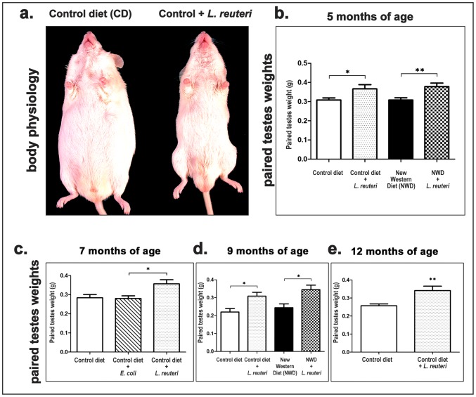
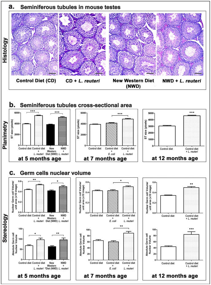
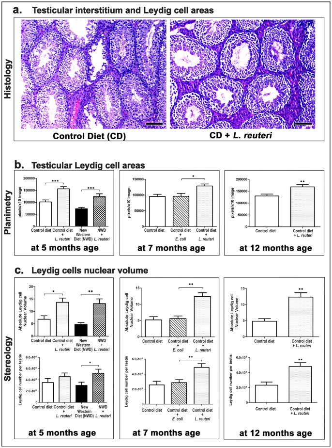
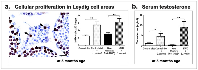
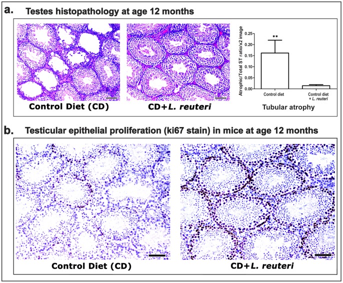
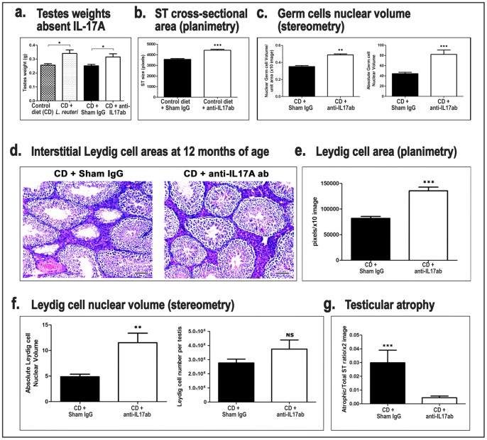
The decline of circulating testosterone levels in aging men is associated with adverse health effects. During studies of probiotic bacteria and obesity, we discovered that male mice routinely consuming purified lactic acid bacteria originally isolated from human milk had larger testicles and increased serum testosterone levels compared to their age-matched controls. Further investigation using microscopy-assisted histomorphometry of testicular tissue showed that mice consuming Lactobacillus reuteri in their drinking water had significantly increased seminiferous tubule cross-sectional profiles and increased spermatogenesis and Leydig cell numbers per testis when compared with matched diet counterparts This showed that criteria of gonadal aging were reduced after routinely consuming a purified microbe such as L. reuteri. We tested whether these features typical of sustained reproductive fitness may be due to anti-inflammatory properties of L. reuteri, and found that testicular mass and other indicators typical of old age were similarly restored to youthful levels using systemic administration of antibodies blocking pro-inflammatory cytokine interleukin-17A. This indicated that uncontrolled host inflammatory responses contributed to the testicular atrophy phenotype in aged mice. Reduced circulating testosterone levels have been implicated in many adverse effects; dietary L. reuteri or other probiotic supplementation may provide a viable natural approach to prevention of male hypogonadism, absent the controversy and side-effects of traditional therapies, and yield practical options for management of disorders typically associated with normal aging. These novel findings suggest a potential high impact for microbe therapy in public health by imparting hormonal and gonad features of reproductive fitness typical of much younger healthy individuals.
Overview
The claims and statistics of this paper haven’t been checked yet.
Run the check — free, no account needed. It reads the open-access text and checks the sources it cites.
Integrity signals
Registry
Cites
Statistics
Figures
Verification
Claims the paper makes
The claims and statistics of this paper haven’t been checked yet.
Run the check — free, no account needed. It reads the open-access text and checks the sources it cites.
Claims other papers make about this one
Each is checked against this paper’s own text: does it say what the citing paper says it does?
In the citing paper
“Further, aging mice supplemented with the probiotic bacterium L. reuteri had larger testicles and increased serum testosterone levels compared to their age-matched controls [237, 254].”
From International Society of Sports Nutrition Position Stand: Probiotics (2019) · cites this paper as [237]
In the citing paper
“In animals, probiotics have been associated with benefits including normalizing age-related drops in testosterone levels [237], increasing neurotransmitter synthesis [238], reducing stress-induced cortisol levels [239], reducing inflammation [100] and improving mood [240].”
From International Society of Sports Nutrition Position Stand: Probiotics (2019) · cites this paper as [237]
Self-check
Percentages, sums and reported differences re-computed from the paper’s own numbers.
The self-check needs the paper’s full text.
Citations
Cites
Bettelli, E., Carrier, Y., Gao, W., Korn, T., Strom, T. B., Oukka, M., Weiner, H. L., & Kuchroo, V. K. (2006) · Nature · doi:10.1038/nature04753 · Cited by 6,992
Bravo, J. A., Forsythe, P., Chew, M. V., Escaravage, E., Savignac, H. M., Dinan, T. G., Bienenstock, J., & Cryan, J. F. (2011) · Proceedings of the National Academy of Sciences · doi:10.1073/pnas.1102999108 · Cited by 3,888
Longitudinal Effects of Aging on Serum Total and Free Testosterone Levels in Healthy Men
Harman, S. M., Metter, E. J., Tobin, J. D., Pearson, J., & Blackman, M. R. (2001) · The Journal of Clinical Endocrinology & Metabolism · doi:10.1210/jcem.86.2.7219 · Cited by 2,760
Changes in diet and lifestyle and long-term weight gain in women and men
Mozaffarian, D., Hao, T., Rimm, E. B., Willett, W. C., & Hu, F. B. (2011) · New England Journal of Medicine · doi:10.1056/nejmoa1014296 · Cited by 2,527
Feldman, H. A., Longcope, C., Derby, C. A., Johannes, C. B., Araujo, A. B., Coviello, A. D., Bremner, W. J., & McKinlay, J. B. (2002) · The Journal of Clinical Endocrinology & Metabolism · doi:10.1210/jcem.87.2.8201 · Cited by 1,861
Park, E. J., Lee, J. H., Yu, G.-Y., He, G., Ali, S. R., Holzer, R. G., Österreicher, C. H., Takahashi, H., & Karin, M. (2010) · Cell · doi:10.1016/j.cell.2009.12.052 · Cited by 1,764
Diet, gut microbiota and immune responses
Maslowski, K. M., & Mackay, C. R. (2011) · Nature Immunology · doi:10.1038/ni0111-5 · Cited by 1,261
Has the Microbiota Played a Critical Role in the Evolution of the Adaptive Immune System?
Lee, Y. K., & Mazmanian, S. K. (2010) · Science · doi:10.1126/science.1195568 · Cited by 1,147
Salama, P. R., Phillips, M. R., Grieu, F., Morris, M. J., Zeps, N., Joseph, D. J., Platell, C. F. E., & Iacopetta, B. J. (2008) · Journal of Clinical Oncology · doi:10.1200/jco.2008.18.7229 · Cited by 1,025
Testosterone: a metabolic hormone in health and disease
Kelly, D. M., & Jones, T. H. (2013) · Journal of Endocrinology · doi:10.1530/joe-12-0455 · Cited by 612
Tajar, A., Forti, G., O'Neill, T. W., Lee, D. M., Silman, A. J., Finn, J. D., Bartfai, G., Boonen, S., Casanueva, F. F., Giwercman, A., et al. (2010) · The Journal of Clinical Endocrinology & Metabolism · doi:10.1210/jc.2009-1796 · Cited by 604
IL-23 plays a key role in Helicobacter hepaticus –induced T cell–dependent colitis
Kullberg, M. C., Jankovic, D. L., Feng, C. G., Hüe, S., Gorelick, P. L., McKenzie, B. S., Cua, D. J., Powrie, F. M., Cheever, A. W., Maloy, K. J., et al. (2006) · The Journal of Experimental Medicine · doi:10.1084/jem.20061082 · Cited by 592
Andropause: clinical implications of the decline in serum testosterone levels with aging in men
Matsumoto, A. M. (2002) · The Journals of Gerontology Series A: Biological Sciences and Medical Sciences · doi:10.1093/gerona/57.2.m76 · Cited by 490
Is vitamin D deficiency to blame for the asthma epidemic?
Litonjua, A. A., & Weiss, S. T. (2007) · Journal of Allergy and Clinical Immunology · doi:10.1016/j.jaci.2007.08.028 · Cited by 477
Testosterone Acts Directly on CD4+ T Lymphocytes to Increase IL-10 Production
Liva, S. M., & Voskuhl, R. R. (2001) · The Journal of Immunology · doi:10.4049/jimmunol.167.4.2060 · Cited by 395
The benefits and risks of testosterone replacement therapy: a review
Morley, J. E. (2009) · Therapeutics and Clinical Risk Management · doi:10.2147/tcrm.s3025 · Cited by 381
Cell Biology of Leydig Cells in the Testis
Haider, S. G. (2004) · International review of cytology · doi:10.1016/s0074-7696(04)33005-6 · Cited by 373
Walter, J., Britton, R. A., & Roos, S. (2010) · Proceedings of the National Academy of Sciences · doi:10.1073/pnas.1000099107 · Cited by 341
Morphometric analysis of Leydig cells in the normal rat testis.
Mori, H., & Christensen, A. K. (1980) · The Journal of Cell Biology · doi:10.1083/jcb.84.2.340 · Cited by 335
Microbial symbionts accelerate wound healing via the neuropeptide hormone oxytocin
Poutahidis, T., Kearney, S. M., Levkovich, T., Qi, P., Varian, B. J., Lakritz, J. R., Ibrahim, Y. M., Chatzigiagkos, A., Alm, E. J., & Erdman, S. E. (2013) · PLoS ONE · doi:10.1371/journal.pone.0078898 · Cited by 310
Self-citationCD4+ CD25+ Regulatory T Lymphocytes Inhibit Microbially Induced Colon Cancer in Rag2-Deficient Mice
Erdman, S. E., Poutahidis, T., Tomczak, M. F., Rogers, A. B., Cormier, K. S., Plank, B., Horwitz, B. H., & Fox, J. G. (2003) · American Journal Of Pathology · doi:10.1016/s0002-9440(10)63863-1 · Cited by 303
Self-citationRook, G. A. W. (2009) · Immunology · doi:10.1111/j.1365-2567.2008.03007.x · Cited by 295
Leydig cells: From stem cells to aging
Chen, H., Ge, R., & Zirkin, B. R. (2009) · Molecular and Cellular Endocrinology · doi:10.1016/j.mce.2009.01.023 · Cited by 278
Atanassova, N. N., McKinnell, C., Walker, M., Turner, K. J., Fisher, J. S., Morley, M., Millar, M. R., Groome, N. P., & Sharpe, R. M. (1999) · Endocrinology · doi:10.1210/endo.140.11.7108 · Cited by 251
Axling, U., Olsson, C., Xu, J., Fernandez, C., Larsson, S. M., Ström, K., Ahrné, S., Holm, C., Molin, G., & Berger, K. (2012) · Nutrition & Metabolism · doi:10.1186/1743-7075-9-105 · Cited by 234
Leydig cell aging and the mechanisms of reduced testosterone synthesis
Midzak, A. S., Chen, H., Papadopoulos, V., & Zirkin, B. R. (2008) · Molecular and Cellular Endocrinology · doi:10.1016/j.mce.2008.07.016 · Cited by 225
Vitamin D Suppresses Th17 Cytokine Production by Inducing C/EBP Homologous Protein (CHOP) Expression
Chang, S. H., Chung, Y., & Dong, C. (2010) · Journal of Biological Chemistry · doi:10.1074/jbc.c110.185777 · Cited by 216
Kim, S.-W., Park, K.-Y., Kim, B., Kim, E., & Hyun, C. (2013) · Biochemical and Biophysical Research Communications · doi:10.1016/j.bbrc.2012.12.121 · Cited by 215
Naito, E., Yoshida, Y., Makino, K., KOUNOSHI, Y., Kunihiro, S., Takahashi, R., Matsuzaki, T., Miyazaki, K., & Ishikawa, F. (2010) · Journal of Applied Microbiology · doi:10.1111/j.1365-2672.2010.04922.x · Cited by 213
Obesity, body fat distribution and sex hormones in men.
Haffner, S. M., Valdez, R. A., D, M. P. S. M., & Katz, M. S. (1993) · PubMed · Cited by 213
CD4+CD25+ Regulatory Lymphocytes Induce Regression of Intestinal Tumors in ApcMin/+ Mice
Erdman, S. E., Sohn, J. J., Rao, V. P., Nambiar, P. R., Ge, Z., Fox, J. G., & Schauer, D. B. (2005) · Cancer Research · doi:10.1158/0008-5472.can-04-3104 · Cited by 211
Self-citationGhanayem, B. I., Bai, R., Kissling, G. E., Travlos, G. S., & Hoffler, U. (2009) · Biology of Reproduction · doi:10.1095/biolreprod.109.078915 · Cited by 207
Testosterone for the aging male; current evidence and recommended practice
Jones, H. (2008) · Clinical Interventions in Aging · doi:10.2147/cia.s190 · Cited by 204
Ak, C., & Kc, P. (1980) · Biology of Reproduction · doi:10.1093/biolreprod/22.2.383 · Cited by 202
Probiotic Bacteria Induce a ‘Glow of Health’
Levkovich, T., Poutahidis, T., Smillie, C. S., Varian, B. J., Ibrahim, Y. M., Lakritz, J. R., Alm, E. J., & Erdman, S. E. (2013) · PLoS ONE · doi:10.1371/journal.pone.0053867 · Cited by 201
Self-citationGounaris, E. G., Blatner, N. R., Dennis, K. L., Magnusson, F. C., Gurish, M. F., Strom, T. B., Beckhove, P., Gounari, F., & Khazaie, K. (2009) · Cancer Research · doi:10.1158/0008-5472.can-09-0304 · Cited by 194
Obesity and testicular function
Mah, P., & Wittert, G. (2009) · Molecular and Cellular Endocrinology · doi:10.1016/j.mce.2009.06.007 · Cited by 183
The Leydig Cell in Health and Disease
Payne, A. H., & Hardy, M. P. (2007) · Humana Press eBooks · doi:10.1007/978-1-59745-453-7 · Cited by 181
Saulnier, D. M. A., dos Santos, F. B., Roos, S., Mistretta, T., Spinler, J. K., Molenaar, D., Teusink, B., & Versalovic, J. A. (2011) · PLoS ONE · doi:10.1371/journal.pone.0018783 · Cited by 181
Baker, P., & O'Shaughnessy, P. J. (2001) · Reproduction · doi:10.1530/rep.0.1220227 · Cited by 179
Fåk, F., & Bäckhed, F. (2012) · PLoS ONE · doi:10.1371/journal.pone.0046837 · Cited by 166
Powrie, F. M., & Maloy, K. J. (2003) · Science · doi:10.1126/science.1082031 · Cited by 138
Kang, J.-H., Yun, S.-I., & Park, H.-O. (2010) · The Journal of Microbiology · doi:10.1007/s12275-010-0363-8 · Cited by 135
Vitamin D and Colon Carcinogenesis
Harris, D. M., & Go, V. L. W. (2004) · Journal of Nutrition · doi:10.1093/jn/134.12.3463s · Cited by 116
Andropause: is androgen replacement therapy indicated for the aging male?
Hijazi, R. A., & Cunningham, G. R. (2005) · Annual Review of Medicine · doi:10.1146/annurev.med.56.082103.104518 · Cited by 105
Thornton, C. E., Brown, S. D. M., & Glenister, P. H. (1999) · Mammalian Genome · doi:10.1007/s003359901145 · Cited by 104
High fertilizing ability of mouse spermatozoa diluted slowly after cryopreservation
Nakagata, N., & Takeshima, T. (1992) · Theriogenology · doi:10.1016/0093-691x(92)90183-r · Cited by 88
Poutahidis, T., Haigis, K. M., Rao, V. P., Nambiar, P. R., Taylor, C. L., Ge, Z., Watanabe, K., Davidson, A., Horwitz, B. H., Fox, J. G., et al. (2007) · Carcinogenesis · doi:10.1093/carcin/bgm180 · Cited by 64
Self-citationSelenium-Enriched Probiotics Improves Murine Male Fertility Compromised by High Fat Diet
Ibrahim, H. A. M., Zhu, Y., Wu, C., Lu, C., Ezekwe, M. O., Liao, S. F., & Haung, K. (2012) · Biological Trace Element Research · doi:10.1007/s12011-011-9308-2 · Cited by 62
Late-Life Onset Hypogonadism: A Review
Bassil, N., & Morley, J. E. (2010) · Clinics in Geriatric Medicine · doi:10.1016/j.cger.2010.02.003 · Cited by 59
Age-related histologic changes in the adult mouse testis.
Takano, H., & Abe, K. (1987) · Archivum histologicum japonicum · doi:10.1679/aohc.50.533 · Cited by 51
Stimulatory effect of follicle-stimulating hormone on rat Leydig cells
KERR, J. B., & Sharpe, R. M. (1985) · Cell and Tissue Research · doi:10.1007/bf00218021 · Cited by 49
Oksaharju, A., Kooistra, T., Kleemann, R., van Duyvenvoorde, W., Miettinen, M., Lappalainen, J., Lindstedt, K. A., Kovanen, P. T., Korpela, R., & Kekkonen, R. A. (2012) · British Journal Of Nutrition · doi:10.1017/s0007114512004801 · Cited by 31
DUCKETT, R. J., Hedger, M. P., MCLACHLAN, R. I., & Wreford, N. G. (1997) · Journal of Andrology · doi:10.1002/j.1939-4640.1997.tb01947.x · Cited by 30
Male menopause: is it a real clinical syndrome?
Pines, A. (2010) · Climacteric · doi:10.3109/13697137.2010.507442 · Cited by 26
Dombrowicz, D., Sente, B., Reiter, É., Closset, J., & Hennen, G. P. (1996) · Journal of Andrology · doi:10.1002/j.1939-4640.1996.tb01848.x · Cited by 21
Cryopreservation of Embryos and Gametes in Mice
Nakagata, N. (1994) · EXPERIMENTAL ANIMALS · doi:10.1538/expanim1978.43.1_11 · Cited by 12
Testosterone replacement therapy to improve health in older males
Baer, J. T. (2012) · The Nurse Practitioner · doi:10.1097/01.npr.0000415874.61843.a6 · Cited by 4
Cited by
Sorted by citations
Flandroy, L., Poutahidis, T., Berg, G., Clarke, G. A., Dao, M. C., Decaestecker, E., Furman, E., Haahtela, T., Massart, S., Plovier, H., et al. (2018) · The Science of The Total Environment · doi:10.1016/j.scitotenv.2018.01.288 · Cited by 417
Self-citationSerum level of sex steroid hormone is associated with diversity and profiles of human gut microbiome
Shin, J.-H., Park, Y.-H., Sim, M., Kim, S.-A., Joung, H., & Shin, D.-M. (2019) · Research in Microbiology · doi:10.1016/j.resmic.2019.03.003 · Cited by 334
The neuroendocrinology of the microbiota-gut-brain axis: a behavioural perspective
Cussotto, S., Sandhu, K. V., Dinan, T. G., & Cryan, J. F. (2018) · Frontiers in Neuroendocrinology · doi:10.1016/j.yfrne.2018.04.002 · Cited by 326
“Interestingly, administration of the probiotic L. reuteri has been shown to increase testosterone levels in aging mice (Poutahidis et al., 2014).”
BackgroundThe relationship between circulating testosterone and inflammatory cytokines in men
Mohamad, N.-V., Wong, S. K., Hasan, W. N. W., Jolly, J. J., Farhana, M. F. N., Ima-Nirwana, S., & Chin, K. (2018) · The Aging Male · doi:10.1080/13685538.2018.1482487 · Cited by 319
Roles of Sex Hormones and Gender in the Gut Microbiota
Yoon, K., & Kim, N. (2021) · Journal of Neurogastroenterology and Motility · doi:10.5056/jnm20208 · Cited by 318
Metadata
- Authors
- Poutahidis, T., Springer, A., Levkovich, T., Qi, P., Varian, B. J., Lakritz, J. R., Ibrahim, Y. M., Chatzigiagkos, A., Alm, E. J., & Erdman, S. E.
- Affiliations
- Aristotle University of Thessaloniki (GR)
- Massachusetts Institute of Technology (US)
- Broad Institute (US)
- Corresponding author
- Alm, Eric J.
- Erdman, Susan E.
- ORCID
- Venue
- PLoS ONE
- Publisher
- Public Library of Science (PLoS)
- Published
- 2014-01-02
- Volume
- 9
- Issue
- 1
- Pages
- e84877
- ISSN
- 1932-6203 (ISSN-L)
- PMC
- PMC3879365
- OpenAlex
- W2045577016
- Cited by
- 177
- References
- 58
- Citations by year
- 2026: 16 · 2025: 17 · 2024: 24 · 2023: 13 · 2022: 22 · 2021: 20 · 2020: 14 · 2019: 10 · 2018: 8 · 2017: 10 · 2016: 11 · 2015: 7 · 2014: 5
- FWCI
- 15.74
- Type
- journal article
- Language
- en
- Open access
- gold
- doi.org
- Licence
- Locations
- PLoS ONE (publishedVersion, cc-by)
- PubMed Central (publishedVersion, cc-by)
- PubMed (publishedVersion)
- DOAJ (DOAJ: Directory of Open Access Journals) (submittedVersion, cc-by-sa)
- DSpace@MIT (Massachusetts Institute of Technology) (submittedVersion, cc-by-nc)
- Figshare (submittedVersion, cc-by)
- Funders
- National Institutes of Health (ES002109)
- National Cancer Institute (U01CA164337, R01CA108854)
- National Institute of Environmental Health Sciences (P30ES002109)
- Topics
- Life Sciences › Agricultural and Biological Sciences › Food Science › Probiotics and Fermented Foods
- Health Sciences › Medicine › Reproductive Medicine › Sperm and Testicular Function
- Health Sciences › Medicine › Endocrinology, Diabetes and Metabolism › Hormonal and reproductive studies
- Keywords
- probiotics, Lactobacillus reuteri, testosterone, male hypogonadism, testicular aging, seminiferous tubules, spermatogenesis, Leydig cells, IL-17A, inflammation, lactic acid bacteria, reproductive fitness, older men
- MeSH terms
- Age Factors, Animals, Atrophy, Diet, Leydig Cells, Male, Organ Size, Seminiferous Tubules, Spermatogenesis, Testis, Testosterone, Probiotics, Interleukin-17, Mice, Limosilactobacillus reuteri
About these checks
Registry, retraction, Cites and statistics results are computed, not judged, with one exception: a printed reference that no index lists is read once by a model before it counts as a citation error. Claims are extracted by a model and kept only when their quote appears verbatim in the text; each cited source is then checked, with a model’s help, for whether it says what the claim relies on. Those verdicts can be wrong — read the claim’s quote and the cited source before relying on one.
Statistics and arithmetic are recomputed, not judged.
Confidence bands are shown only for fields calibrated on our annotated test set. None is calibrated yet, so no band is shown.
What counts as a claim ›How we measure confidence ›How we check this ›
Are you an author, or do you see a problem with this page? Request a review
