

Emotion-Motion Interactions in Conversion Disorder: An fMRI Study
Open accessThe DOI matches its Crossref record; 42 of the 44 references it cites were found in a registry or an index.
Abstract
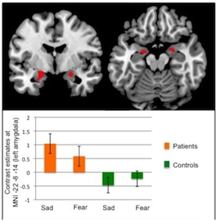
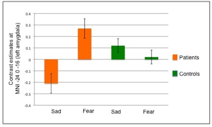
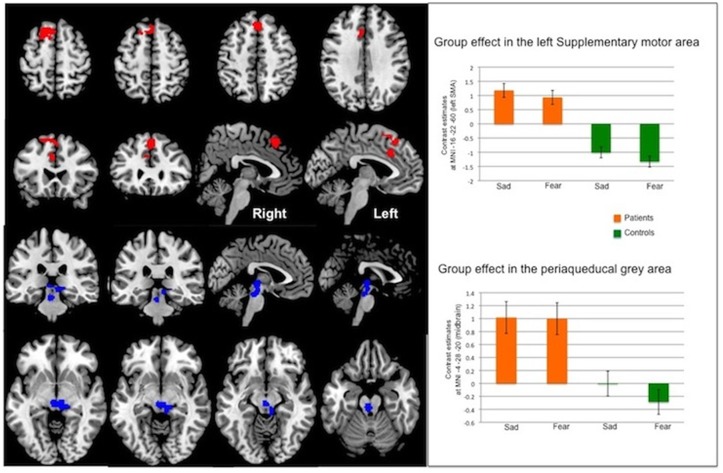
OBJECTIVES: To evaluate the neural correlates of implicit processing of negative emotions in motor conversion disorder (CD) patients. METHODS: An event related fMRI task was completed by 12 motor CD patients and 14 matched healthy controls using standardised stimuli of faces with fearful and sad emotional expressions in comparison to faces with neutral expressions. Temporal changes in the sensitivity to stimuli were also modelled and tested in the two groups. RESULTS: We found increased amygdala activation to negative emotions in CD compared to healthy controls in region of interest analyses, which persisted over time consistent with previous findings using emotional paradigms. Furthermore during whole brain analyses we found significantly increased activation in CD patients in areas involved in the 'freeze response' to fear (periaqueductal grey matter), and areas involved in self-awareness and motor control (cingulate gyrus and supplementary motor area). CONCLUSIONS: In contrast to healthy controls, CD patients exhibited increased response amplitude to fearful stimuli over time, suggesting abnormal emotional regulation (failure of habituation / sensitization). Patients with CD also activated midbrain and frontal structures that could reflect an abnormal behavioral-motor response to negative including threatening stimuli. This suggests a mechanism linking emotions to motor dysfunction in CD.
Overview
The claims and statistics of this paper haven’t been checked yet.
Free: it reads the open-access text and checks the first 5 claims that cite a source we can check. Create a free account to check every claim.
Integrity signals
Registry
Cites
Statistics
Figures
Verification
Claims the paper makes
The claims and statistics of this paper haven’t been checked yet.
Free: it reads the open-access text and checks the first 5 claims that cite a source we can check. Create a free account to check every claim.
Claims other papers make about this one
Each is checked against this paper’s own text: does it say what the citing paper says it does?
The source backs part of the claim, not all of it — or backs it for a different population, condition or comparison.
In the citing paper
“While findings have not been entirely consistent across studies and FND sub-populations, between-group differences in default mode, salience and sensorimotor network areas have been identified in a range FND populations compared to controls.45,207,[209][210][211][212]”
From A new science of emotion: implications for functional neurological disorder (2022) · cites this paper as [45]
Experimental — accuracy not yet measuredThe source backs part of the claim, not all of it — or backs it for a different population, condition or comparison.
In the citing paper
“Additionally, heightened arousal is linked to defensive behaviours driven by the periaqueductal grey, such as tonic immobility and fight or flight responses-behavioural programmes that share overlapping semiological features with functional seizures.45,[171][172][173][174]”
From A new science of emotion: implications for functional neurological disorder (2022) · cites this paper as [45]
Experimental — accuracy not yet measured
Self-check
Percentages, sums and reported differences re-computed from the paper’s own numbers.
The self-check needs the paper’s full text.
Citations
Cites
The Hospital Anxiety and Depression Scale
Zigmond, A. S., & Snaith, R. P. (1983) · Acta Psychiatrica Scandinavica · doi:10.1111/j.1600-0447.1983.tb09716.x · Cited by 47,139
Depression and quality of life in older persons: A review
Sivertsen, H., Bjørkløf, G. H., Engedal, K., Selbæk, G., & Helvik, A.-S. (2015) · Dementia and Geriatric Cognitive Disorders · doi:10.1159/000437299 · Cited by 14,152
Diagnostic and Statistical Manual of Mental Disorders(DSM)
(2008) · Encyclopedia of Counseling · doi:10.4135/9781412963978.n182 · Cited by 10,830
Meeting of minds: the medial frontal cortex and social cognition
Amodio, D. M., & Frith, C. D. (2006) · Nature Reviews Neuroscience · doi:10.1038/nrn1884 · Cited by 3,782
Social Origins of Depression: A Study of Psychiatric Disorder in Women.
Schnitzer, P. K., Brown, G. W., & Harris, T. (1980) · Contemporary Sociology · doi:10.2307/2065631 · Cited by 3,053
Cognitive and emotional control of pain and its disruption in chronic pain
Bushnell, M. C., Čeko, M., & Low, L. A. (2013) · Nature Reviews Neuroscience · doi:10.1038/nrn3516 · Cited by 2,076
Social Origins of Depression: A Study of Psychiatric Disorder in Women
McDaniel, E. (1980) · The Journal of Nervous and Mental Disease · doi:10.1097/00005053-198009000-00013 · Cited by 1,896
LeDoux, J., Iwata, J., Cicchetti, P., & Reis, D. (1988) · The Journal of Neuroscience · doi:10.1523/jneurosci.08-07-02517.1988 · Cited by 1,580
Adolphs, R., Tranel, D., Damasio, H., & Damasio, A. (1995) · The Journal of Neuroscience · doi:10.1523/jneurosci.15-09-05879.1995 · Cited by 1,138
A Bayesian account of ‘hysteria’
Edwards, M. J., Adams, R. A., Brown, H., Parees, I., & Friston, K. J. (2012) · Brain · doi:10.1093/brain/aws129 · Cited by 825
Research works printed but not linked: 1
- V Voon, 2010, Emotional stimuli and motor conversion disorder, Brain, 133, 1526, 10.1093/brain/awq054 · DOI · Paper page
Cited by
Current Concepts in Diagnosis and Treatment of Functional Neurological Disorders
Espay, A. J., Aybek, S., Carson, A., Edwards, M. J., Goldstein, L. H., Hallett, M., LaFaver, K., LaFrance, W. C., Lang, A. E., Nicholson, T., et al. (2018) · JAMA Neurology · doi:10.1001/jamaneurol.2018.1264 · Cited by 786
Self-citationPick, S., Goldstein, L. H., Perez, D. L., & Nicholson, T. R. (2019) · Journal of Neurology, Neurosurgery & Psychiatry · doi:10.1136/jnnp-2018-319201 · Cited by 296
Self-citationNeuroimaging in functional neurological disorder: State of the field and research agenda
Perez, D. L., Nicholson, T. R., Asadi-Pooya, A. A., Bègue, I., Butler, M., Carson, A. J., David, A. S., Deeley, Q., Diez, I., Edwards, M. J., et al. (2021) · NeuroImage: Clinical · doi:10.1016/j.nicl.2021.102623 · Cited by 216
Self-citationA framework for understanding the pathophysiology of functional neurological disorder
Drane, D. L., Fani, N., Hallett, M., Khalsa, S. S., Perez, D. L., & Roberts, N. A. (2021) · CNS Spectrums · doi:10.1017/s1092852920001789 · Cited by 189
A new science of emotion: implications for functional neurological disorder
Jungilligens, J., Paredes-Echeverri, S., Popkirov, S., Barrett, L. F., & Perez, D. L. (2022) · Brain · doi:10.1093/brain/awac204 · Cited by 164
Metadata
- Authors
- Aybek, S., Nicholson, T. R., O’Daly, O., Zelaya, F., Kanaan, R. A., & David, A. S.
- Affiliations
- University of Geneva (CH)
- King's College London (GB)
- The University of Melbourne (AU)
- Corresponding author
- Aybek, Selma
- ORCID
- Venue
- PLOS ONE
- Publisher
- Public Library of Science (PLoS)
- Published
- 2015-04-10
- Volume
- 10
- Issue
- 4
- Pages
- e0123273
- ISSN
- 1932-6203 (ISSN-L)
- PMC
- PMC4393246
- OpenAlex
- W2056387840
- Cited by
- 183
- References
- 44
- Citations by year
- 2026: 20 · 2025: 19 · 2024: 17 · 2023: 13 · 2022: 17 · 2021: 16 · 2020: 20 · 2019: 13 · 2018: 18 · 2017: 18 · 2016: 9 · 2015: 3
- FWCI
- 12.91
- Type
- journal article
- Language
- en
- Open access
- gold
- doi.org
- Licence
- Locations
- PLoS ONE (publishedVersion, cc-by)
- PubMed Central (publishedVersion, cc-by)
- PubMed (publishedVersion)
- Minerva Access (University of Melbourne) (submittedVersion, cc-by)
- Research Portal (King's College London) (submittedVersion, cc-by)
- DOAJ (DOAJ: Directory of Open Access Journals) (submittedVersion, cc-by-sa)
- Funders
- South London and Maudsley NHS Foundation Trust
- National Institute for Health and Care Research (CL-2009-17-006, CS-2014-14-016)
- King's College London
- Université de Genève
- Medical Research Council (MR/J004162/1)
- Topics
- Health Sciences › Medicine › Psychiatry and Mental health › Psychosomatic Disorders and Their Treatments
- Health Sciences › Medicine › Clinical Neurology › Deep Brain Stimulation and Movement Disorders
- Life Sciences › Neuroscience › Cognitive Neuroscience › Neural and Behavioral Psychology Studies
- Keywords
- conversion disorder, amygdala, fear, emotional face processing, freezing response, periaqueductal gray, habituation, emotion regulation, supplementary motor area, cingulate gyrus, self-awareness, neural correlates
- MeSH terms
- Adult, Brain Mapping, Conversion Disorder, Emotions, Female, Humans, Magnetic Resonance Imaging, Male, Middle Aged, Motion, Psychomotor Performance, Models, Statistical, Case-Control Studies, Young Adult
About these checks
Registry, retraction, Cites and statistics results are computed, not judged, with one exception: a printed reference that no index lists is read once by a model before it counts as a citation error. Claims are extracted by a model and kept only when their quote appears verbatim in the text; each cited source is then checked, with a model’s help, for whether it says what the claim relies on. Those verdicts can be wrong — read the claim’s quote and the cited source before relying on one.
Statistics and arithmetic are recomputed, not judged.
Confidence bands are shown only for fields calibrated on our annotated test set. None is calibrated yet, so no band is shown.
What counts as a claim ›How we measure confidence ›How we check this ›
Are you an author, or do you see a problem with this page? Request a review
How to cite
Cite CoChat for the checks on this page, and the paper for its own findings.
- This page
- CoChat. (2026). Citation check: Emotion-Motion Interactions in Conversion Disorder: An fMRI Study. https://app.cochat.ai/papers/10.1371/journal.pone.0123273
- The paper
- Aybek, S., Nicholson, T. R., O’Daly, O., Zelaya, F., Kanaan, R. A., & David, A. S. (2015). Emotion-Motion Interactions in Conversion Disorder: An fMRI Study. PLOS ONE. https://doi.org/10.1371/journal.pone.0123273
Data sources
Bibliographic data: Semantic Scholar, OpenAlex, Crossref.
Contains information from the Semantic Scholar Academic Graph Datasets, made available under the ODC Attribution License (ODC-BY 1.0).
Semantic Scholar’s data is described in: Kinney, R., et al. (2023). The Semantic Scholar Open Data Platform. arXiv:2301.10140.
