

A machine learning algorithm predicts molecular subtypes in pancreatic ductal adenocarcinoma with differential response to gemcitabine-based versus FOLFIRINOX chemotherapy
Open accessPublished version of a preprint
The DOI matches its Crossref record; 40 of the 40 references it cites were found in a registry or an index.
Abstract
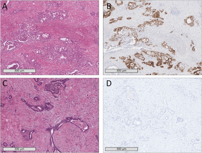
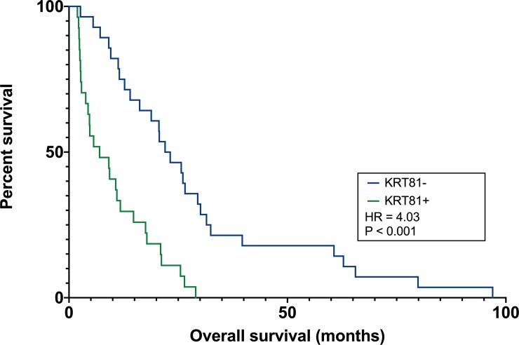
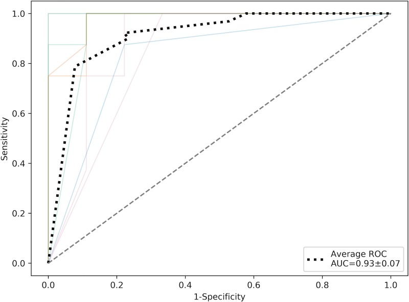
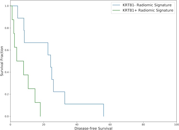
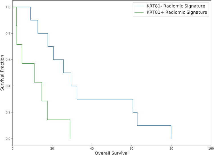
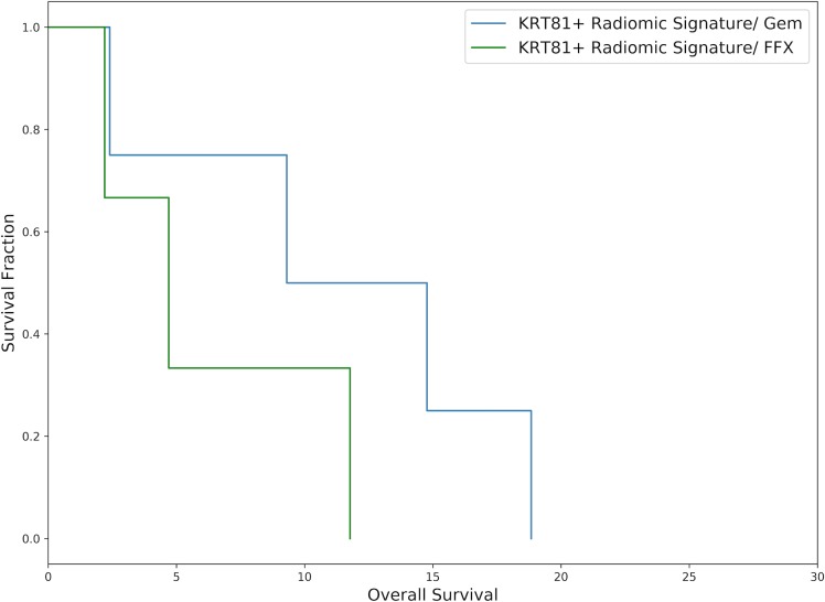
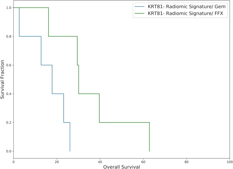
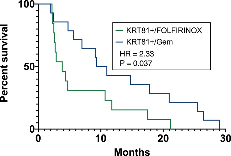
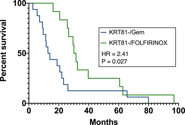
PURPOSE: Development of a supervised machine-learning model capable of predicting clinically relevant molecular subtypes of pancreatic ductal adenocarcinoma (PDAC) from diffusion-weighted-imaging-derived radiomic features. METHODS: The retrospective observational study assessed 55 surgical PDAC patients. Molecular subtypes were defined by immunohistochemical staining of KRT81. Tumors were manually segmented and 1606 radiomic features were extracted with PyRadiomics. A gradient-boosted-tree algorithm was trained on 70% of the patients (N = 28) and tested on 30% (N = 17) to predict KRT81+ vs. KRT81- tumor subtypes. A gradient-boosted survival regression model was fit to the disease-free and overall survival data. Chemotherapy response and survival were assessed stratified by subtype and radiomic signature. Radiomic feature importance was ranked. RESULTS: The mean±STDEV sensitivity, specificity and ROC-AUC were 0.90±0.07, 0.92±0.11, and 0.93±0.07, respectively. The mean±STDEV concordance indices between the disease-free and overall survival predicted by the model based on the radiomic parameters and actual patient survival were 0.76±0.05 and 0.71±0.06, respectively. Patients with a KRT81+ subtype experienced significantly diminished median overall survival compared to KRT81- patients (7.0 vs. 22.6 months, HR 4.03, log-rank-test P = <0.001) and a significantly improved response to gemcitabine-based chemotherapy over FOLFIRINOX (10.14 vs. 3.8 months median overall survival, HR 2.33, P = 0.037) compared to KRT81- patients, who responded significantly better to FOLFIRINOX over gemcitabine-based treatment (30.8 vs. 13.4 months median overall survival, HR 2.41, P = 0.027). Entropy was ranked as the most important radiomic feature. CONCLUSIONS: The machine-learning based analysis of radiomic features enables the prediction of subtypes of PDAC, which are highly relevant for disease-free and overall patient survival and response to chemotherapy.
Overview
The claims and statistics of this paper haven’t been checked yet.
Free: it reads the open-access text and checks the first 5 claims that cite a source we can check. Create a free account to check every claim.
Integrity signals
Registry
Cites
Statistics
Figures
Verification
Claims the paper makes
The claims and statistics of this paper haven’t been checked yet.
Free: it reads the open-access text and checks the first 5 claims that cite a source we can check. Create a free account to check every claim.
Claims other papers make about this one
Each is checked against this paper’s own text: does it say what the citing paper says it does?
The source backs part of the claim, not all of it — or backs it for a different population, condition or comparison.
In the citing paper
“AI has also been used to predict treatment responses, including chemotherapy, radiotherapy, immunotherapy, and surgery.”
From Artificial intelligence in pancreatic cancer (2022) · cites this paper as [81]
Experimental — accuracy not yet measuredThe cited source says what the claim attributes to it.
In the citing paper
“Particularly in oncology, success has been reported in machine-learning-based tumour detection3,37, subtyping38, and outcome prediction39”
From Swarm Learning for decentralized and confidential clinical machine learning (2021) · cites this paper as [38]
The cited source says what the claim attributes to it.
In the citing paper
“Both dependent and independent frameworks have applications in diagnosing 75-77 and prognosis 78-81 PC.”
From Artificial intelligence in pancreatic cancer (2022) · cites this paper as [81]
Self-check
Percentages, sums and reported differences re-computed from the paper’s own numbers.
The self-check needs the paper’s full text.
Citations
Cites
XGBoost: A Scalable Tree Boosting System
Chen, T., & Guestrin, C. (2016) · Proceedings of the 22nd ACM SIGKDD International Conference on Knowledge Discovery and Data Mining · doi:10.1145/2939672.2939785 · Cited by 54,569
Ror2 signaling regulates Golgi structure and transport through IFT20 for tumor invasiveness
Nishita, M., Park, S.-Y., Nishio, T., Kamizaki, K., Wang, Z., Tamada, K., Takumi, T., Hashimoto, R., Otani, H., Pazour, G. J., et al. (2017) · Scientific Reports · doi:10.1038/s41598-016-0028-x · Cited by 15,947
Ultrastructural Characterization of the Lower Motor System in a Mouse Model of Krabbe Disease
Cappello, V., Marchetti, L., Parlanti, P., Landi, S., Tonazzini, I., Cecchini, M., Piazza, V., & Gemmi, M. (2016) · Scientific Reports · doi:10.1038/s41598-016-0001-8 · Cited by 13,097
FOLFIRINOX versus Gemcitabine for Metastatic Pancreatic Cancer
Conroy, T., Desseigne, F., Ychou, M., Bouché, O., Guimbaud, R., Bécouarn, Y., Adenis, A., Raoul, J.-L., Gourgou-Bourgade, S., de la Fouchardière, C., et al. (2011) · New England Journal of Medicine · doi:10.1056/nejmoa1011923 · Cited by 7,810
Increased Survival in Pancreatic Cancer with nab-Paclitaxel plus Gemcitabine
Von Hoff, D. D., Ervin, T., Arena, F. P., Chiorean, E. G., Infante, J., Moore, M., Seay, T., Tjulandin, S. A., Ma, W. W., Saleh, M. N., et al. (2013) · New England Journal of Medicine · doi:10.1056/nejmoa1304369 · Cited by 6,712
Computational Radiomics System to Decode the Radiographic Phenotype
(2017) · doi:10.1158/0008-5472.can-17-0339 · Cited by 6,593
Zwanenburg, A., Vallières, M., Abdalah, M. A., Aerts, H. J. W. L., Andrearczyk, V., Apte, A., Ashrafinia, S., Bakas, S., Beukinga, R. J., Boellaard, R., et al. (2020) · Radiology · doi:10.1148/radiol.2020191145 · Cited by 4,177
Limb proportions show developmental plasticity in response to embryo movement
Pollard, A. S., Charlton, B. G., Hutchinson, J. R., Gustafsson, T., McGonnell, I. M., Timmons, J. A., & Pitsillides, A. A. (2017) · Scientific Reports · doi:10.1038/srep41926 · Cited by 2,364
Radiomics: the process and the challenges
Kumar, V., Gu, Y., Basu, S., Berglund, A., Eschrich, S. A., Schabath, M. B., Forster, K., Aerts, H. J. W. L., Dekker, A., Fenstermacher, D., et al. (2012) · Magnetic Resonance Imaging · doi:10.1016/j.mri.2012.06.010 · Cited by 2,258
Subtypes of pancreatic ductal adenocarcinoma and their differing responses to therapy
Collisson, E. A., Sadanandam, A., Olson, P., Gibb, W. J., Truitt, M., Gu, S., Cooc, J., Weinkle, J., Kim, G. E., Jakkula, L., et al. (2011) · Nature Medicine · doi:10.1038/nm.2344 · Cited by 1,967
Olaparib Monotherapy in Patients With Advanced Cancer and a Germline BRCA1/2 Mutation
Kaufman, B., Shapira-Frommer, R., Schmutzler, R. K., Audeh, M. W., Friedlander, M., Balmaña, J., Mitchell, G., Fried, G., Stemmer, S. M., Hubert, A., et al. (2015) · Journal of Clinical Oncology · doi:10.1200/jco.2014.56.2728 · Cited by 1,671
Repeatability and Reproducibility of Radiomic Features: A Systematic Review
Traverso, A., Wee, L., Dekker, A., & Gillies, R. (2018) · International Journal of Radiation Oncology*Biology*Physics · doi:10.1016/j.ijrobp.2018.05.053 · Cited by 903
Molecular subtypes of pancreatic cancer
Collisson, E. A., Bailey, P., Chang, D. K., & Biankin, A. V. (2019) · Nature Reviews Gastroenterology & Hepatology · doi:10.1038/s41575-019-0109-y · Cited by 892
Yang, J., Huang, T., Petralia, F., Long, Q., Zhang, B., Argmann, C., Zhao, Y., Mobbs, C. V., Schadt, E. E., Zhu, J., et al. (2016) · Scientific Reports · doi:10.1038/srep19384 · Cited by 720
CT-based radiomic signature predicts distant metastasis in lung adenocarcinoma
Coroller, T. P., Grossmann, P., Hou, Y., Rios Velazquez, E., Leijenaar, R. T. H., Hermann, G., Lambin, P., Haibe-Kains, B., Mak, R. H., & Aerts, H. J. W. L. (2015) · Radiotherapy and Oncology · doi:10.1016/j.radonc.2015.02.015 · Cited by 695
A deep learning-based radiomics model for prediction of survival in glioblastoma multiforme
Lao, J., Chen, Y., Li, Z.-C., Li, Q., Zhang, J., Liu, J., & Zhai, G. (2017) · Scientific Reports · doi:10.1038/s41598-017-10649-8 · Cited by 630
Aung, K. L., Fischer, S. E., Denroche, R. E., Jang, G.-H., Dodd, A., Creighton, S., Southwood, B., Liang, S.-B., Chadwick, D., Zhang, A., et al. (2018) · Clinical Cancer Research · doi:10.1158/1078-0432.ccr-17-2994 · Cited by 618
Stratification of pancreatic ductal adenocarcinomas based on tumor and microenvironment features
Puleo, F., Nicolle, R., Blum, Y., Cros, J., Marisa, L., Demetter, P., Quertinmont, E., Svrcek, M., Elarouci, N., Iovanna, J., et al. (2018) · Gastroenterology · doi:10.1053/j.gastro.2018.08.033 · Cited by 563 · cited 1×
“Tumors positive or negative for both markers were excluded, the former due to recently reported suspicions of contamination with acinar cells [15], the latter due to unclassifiability.”
BackgroundPermutation Tests for Studying Classifier Performance
Ojala, M., & Garriga, G. C. (2009) · 2009 Ninth IEEE International Conference on Data Mining · doi:10.1109/icdm.2009.108 · Cited by 555
Reproducibility of radiomics for deciphering tumor phenotype with imaging
Zhao, B., Tan, Y., Tsai, W.-Y., Qi, J., Xie, C., Lu, L., & Schwartz, L. H. (2016) · Scientific Reports · doi:10.1038/srep23428 · Cited by 509
Evolutionary routes and KRAS dosage define pancreatic cancer phenotypes
Mueller, S., Engleitner, T., Maresch, R., Zukowska, M., Lange, S., Kaltenbacher, T., Konukiewitz, B., Öllinger, R., Zwiebel, M., Strong, A., et al. (2018) · Nature · doi:10.1038/nature25459 · Cited by 431
Self-citationImage biomarker standardisation initiative
Zwanenburg, A., Leger, S., Vallières, M., & Löck, S. (2016) · arXiv (Cornell University) · doi:10.48550/arxiv.1612.07003 · Cited by 146
Quantitative imaging to evaluate malignant potential of IPMNs
Hanania, A. N., Bantis, L. E., Feng, Z., Wang, H., Tamm, E. P., Katz, M. H., Maitra, A., & Koay, E. J. (2016) · Oncotarget · doi:10.18632/oncotarget.11769 · Cited by 143
The Rise of Radiomics and Implications for Oncologic Management
Verma, V., Simone, C. B., Krishnan, S., Lin, S. H., Yang, J., & Hahn, S. M. (2017) · JNCI: Journal of the National Cancer Institute · doi:10.1093/jnci/djx055 · Cited by 135
Eilaghi, A., Baig, S., Zhang, Y., Zhang, J., Karanicolas, P., Gallinger, S., Khalvati, F., & Haider, M. A. (2017) · BMC Medical Imaging · doi:10.1186/s12880-017-0209-5 · Cited by 127
Co-clinical Assessment of Tumor Cellularity in Pancreatic Cancer
Heid, I., Steiger, K., Trajkovic‐Arsic, M., Settles, M., Eßwein, M. R., Erkan, M., Kleeff, J. H., Jäger, C., Friess, H., Haller, B., et al. (2017) · Clinical Cancer Research · doi:10.1158/1078-0432.ccr-15-2432 · Cited by 95
Self-citationMuckenhuber, A., Berger, A. K., Schlitter, A. M., Steiger, K., Konukiewitz, B., Trumpp, A., Eils, R., Werner, J., Friess, H., Esposito, I., et al. (2018) · Clinical Cancer Research · doi:10.1158/1078-0432.ccr-17-2180 · Cited by 93
Self-citationPrognostic Value of CT Radiomic Features in Resectable Pancreatic Ductal Adenocarcinoma
Khalvati, F., Zhang, Y., Baig, S., Lobo-Mueller, E. M., Karanicolas, P., Gallinger, S., & Haider, M. A. (2019) · Scientific Reports · doi:10.1038/s41598-019-41728-7 · Cited by 88
Kaissis, G., Ziegelmayer, S., Lohöfer, F., Algül, H., Eiber, M., Weichert, W., Schmid, R., Friess, H., Rummeny, E., Ankerst, D., et al. (2019) · European Radiology Experimental · doi:10.1186/s41747-019-0119-0 · Cited by 79
Self-citationJia, J., Chen, J., Yao, J., & Chu, D. P. (2017) · Scientific Reports · doi:10.1038/srep46648 · Cited by 70
Bailey, D. L., Pichler, B. J., Gückel, B., Antoch, G., Barthel, H., Bhujwalla, Z. M., Biskup, S., Biswal, S., Bitzer, M., Boellaard, R., et al. (2018) · Molecular Imaging and Biology · doi:10.1007/s11307-017-1123-5 · Cited by 58
Self-citationCan we trust the calculation of texture indices of CT images? A phantom study
Caramella, C., Allorant, A., Orlhac, F., Bidault, F., Asselain, B. J. R., Ammari, S., Jaranowski, P., Moussier, A., Balleyguier, C., Lassau, N., et al. (2018) · Medical Physics · doi:10.1002/mp.12809 · Cited by 48
Huang, Y., Lu, H.-I., Huang, S., Hsu, C.-C., Chiu, N.-T., Wang, Y., Chiu, Y.-C., & Li, S.-H. (2017) · BMC Medical Imaging · doi:10.1186/s12880-016-0171-7 · Cited by 47
Hoff, D. D. V., Ervin, T. J., Arena, F. P., Chiorean, E. G., Infante, J. R., Moore, M. J., Seay, T. E., Tjulandin, S., Ma, W. W., Saleh, M. N., et al. (2013) · Journal of Clinical Oncology · doi:10.1200/jco.2013.31.15_suppl.4005 · Cited by 38
Apparent Diffusion Coefficient (ADC) predicts therapy response in pancreatic ductal adenocarcinoma
Trajkovic‐Arsic, M., Heid, I., Steiger, K., Gupta, A., Fingerle, A. A., Wörner, C., Teichmann, N., Sengkwawoh-Lueong, S., Wenzel, P., Beer, A. J., et al. (2017) · Scientific Reports · doi:10.1038/s41598-017-16826-z · Cited by 32
Self-citationPölsterl, S., Gupta, P., Wang, L., Conjeti, S., Katouzian, A., & Navab, N. (2016) · F1000Research · doi:10.12688/f1000research.8231.3 · Cited by 31
Pölsterl, S., Gupta, P., Wang, L., Conjeti, S., Katouzian, A., & Navab, N. (2016) · F1000Research · doi:10.12688/f1000research.8231.1 · Cited by 22
Kaissis, G. A., Ziegelmayer, S., Lohöfer, F. K., et al. (2019) · bioRxiv (Cold Spring Harbor Laboratory) · doi:10.1101/643809 · Cited by 7
Self-citationEP-2132: Repeatability and reproducibility of radiomic features: results of a systematic review
Traverso, A., Wee, L., Dekker, A. L. A. J., & Gillies, R. J. (2018) · Radiotherapy and Oncology · doi:10.1016/s0167-8140(18)32441-1 · Cited by 3
Standardized and quality assured prostate diffusion MRI
Bach, M. (2019) · European Society of Radiology · doi:10.26044/ecr2019/c-2163 · Cited by 2
Cited by
Secure, privacy-preserving and federated machine learning in medical imaging
Kaissis, G. A., Makowski, M. R., Rückert, D., & Braren, R. F. (2020) · Nature Machine Intelligence · doi:10.1038/s42256-020-0186-1 · Cited by 1,676
Self-citationSwarm Learning for decentralized and confidential clinical machine learning
Warnat-Herresthal, S., Schultze, H., Shastry, K. L., Manamohan, S., Mukherjee, S., Garg, V., Sarveswara, R., Händler, K., Pickkers, P., Aziz, N. A., et al. (2021) · Nature · doi:10.1038/s41586-021-03583-3 · Cited by 884
“Particularly in oncology, success has been reported in machine-learning-based tumour detection 3,37 , sub-typing 38 , and outcome prediction 39 , but progress is hindered by the limited size of datasets 19 , with current privacy regulations 5,9,10 making it less appealing to develop centralized AI…”
BackgroundNeoadjuvant therapy for pancreatic cancer
Springfeld, C., Ferrone, C. R., Katz, M. H. G., Philip, P. A., Hong, T. S., Hackert, T., Büchler, M. W., & Neoptolemos, J. (2023) · Nature Reviews Clinical Oncology · doi:10.1038/s41571-023-00746-1 · Cited by 377
Artificial intelligence in gastroenterology: A state-of-the-art review
Kröner, P. T., Engels, M. M., Glicksberg, B. S., Johnson, K. W., Mzaik, O., van Hooft, J. E., Wallace, M. B., El-Serag, H. B., & Krittanawong, C. (2021) · World Journal of Gastroenterology · doi:10.3748/wjg.v27.i40.6794 · Cited by 165
Artificial intelligence in pancreatic cancer
Huang, B., Huang, H., Zhang, S., Zhang, D., Shi, Q., Liu, J., & Guo, J. (2022) · Theranostics · doi:10.7150/thno.77949 · Cited by 142
“Due to its excellent computational power, AI was used to analyze PC prognoses, including survival time [204-221], recurrence risk [78,221-224], metastasis [225-230], therapy response [79-81,231-240], etc.”
BackgroundMethod
Metadata
- Authors
- Kaissis, G., Ziegelmayer, S., Lohöfer, F., Steiger, K., Algül, H., Muckenhuber, A., Yen, H.-Y., Rummeny, E., Friess, H., Schmid, R., Weichert, W., Siveke, J. T., & Braren, R.
- Affiliations
- Technical University of Munich (DE)
- German Cancer Research Center (DE)
- Corresponding author
- Braren, Rickmer
- ORCID
- Venue
- PLOS ONE
- Publisher
- Public Library of Science (PLoS)
- Published
- 2019-10-02
- Volume
- 14
- Issue
- 10
- Pages
- e0218642
- ISSN
- 1932-6203 (ISSN-L)
- PMC
- PMC6774515
- OpenAlex
- W2979283201
- Cited by
- 76
- References
- 40
- Citations by year
- 2025: 7 · 2024: 9 · 2023: 13 · 2022: 7 · 2021: 20 · 2020: 14 · 2019: 3
- FWCI
- 7.26
- Type
- journal article
- Language
- en
- Open access
- gold
- doi.org
- Licence
- Locations
- PLoS ONE (publishedVersion, cc-by)
- bioRxiv (Cold Spring Harbor Laboratory) (acceptedVersion, cc-by)
- Europe PMC (PubMed Central) (publishedVersion, cc-by)
- PubMed Central (publishedVersion, cc-by)
- PubMed (publishedVersion)
- RePEc: Research Papers in Economics (submittedVersion)
- Universitätsbibliographie, Universität Duisburg-Essen (submittedVersion)
- DOAJ (DOAJ: Directory of Open Access Journals) (submittedVersion, cc-by-sa)
- Figshare (submittedVersion, cc-by)
- mediaTUM (Technical University of Munich) (submittedVersion)
- Funders
- Deutsche Forschungsgemeinschaft (SFB284 Project C6, SFB824/Project Z2)
- Topics
- Health Sciences › Medicine › Radiology, Nuclear Medicine and Imaging › Radiomics and Machine Learning in Medical Imaging
- Health Sciences › Medicine › Oncology › Pancreatic and Hepatic Oncology Research
- Health Sciences › Medicine › Radiology, Nuclear Medicine and Imaging › MRI in cancer diagnosis
- Keywords
- pancreatic ductal adenocarcinoma, molecular subtypes, radiomics, diffusion-weighted imaging, gemcitabine-based chemotherapy, FOLFIRINOX, chemotherapy response prediction, survival prediction, gradient boosting decision tree, entropy, PyRadiomics, immunohistochemistry, machine learning prediction models, overall survival, disease-free survival
- MeSH terms
- Machine Learning, Irinotecan, Oxaliplatin, Gemcitabine, Adult, Antineoplastic Combined Chemotherapy Protocols, Leucovorin, Deoxycytidine, Female, Fluorouracil, Humans, Male, Middle Aged, Neoplasm Proteins, Pancreatic Neoplasms, Retrospective Studies, Sensitivity and Specificity, Survival Rate, Disease-Free Survival, Carcinoma, Pancreatic Ductal, Keratins, Type II, Keratins, Hair-Specific
About these checks
Registry, retraction, Cites and statistics results are computed, not judged, with one exception: a printed reference that no index lists is read once by a model before it counts as a citation error. Claims are extracted by a model and kept only when their quote appears verbatim in the text; each cited source is then checked, with a model’s help, for whether it says what the claim relies on. Those verdicts can be wrong — read the claim’s quote and the cited source before relying on one.
Statistics and arithmetic are recomputed, not judged.
Confidence bands are shown only for fields calibrated on our annotated test set. None is calibrated yet, so no band is shown.
What counts as a claim ›How we measure confidence ›How we check this ›
Are you an author, or do you see a problem with this page? Request a review
Data sources
Bibliographic data: Semantic Scholar, OpenAlex, Crossref.
Contains information from the Semantic Scholar Academic Graph Datasets, made available under the ODC Attribution License (ODC-BY 1.0).
Semantic Scholar’s data is described in: Kinney, R., et al. (2023). The Semantic Scholar Open Data Platform. arXiv:2301.10140.
