

The DOI matches its Crossref record; 4 of the 4 references it cites were found in a registry or an index.
SARS-CoV-2 variant evolution in the United States: High accumulation of viral mutations over time likely through serial Founder Events and mutational bursts
Published version of a preprint
Abstract
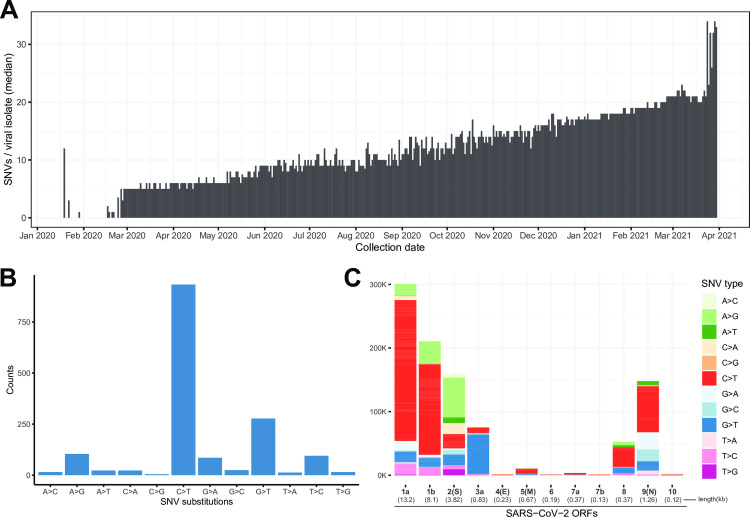
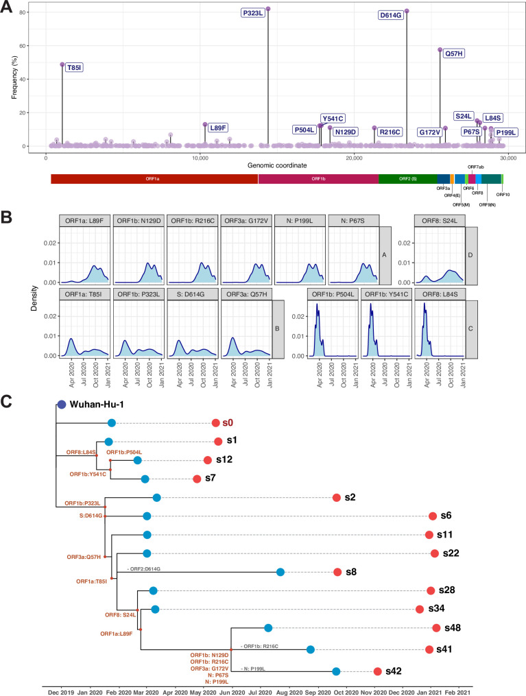
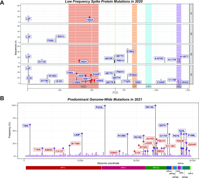
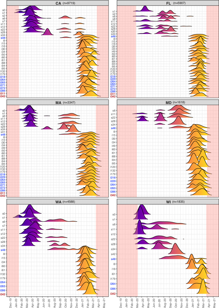
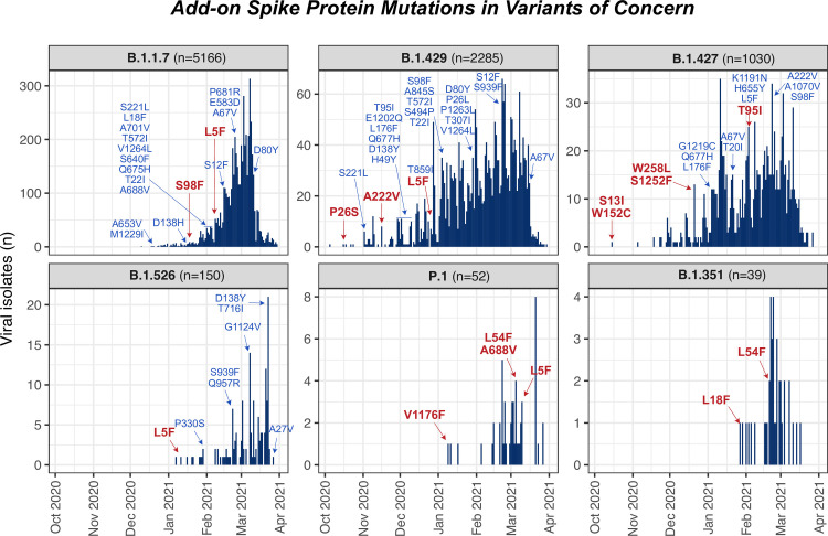
Since the first case of COVID-19 in December 2019 in Wuhan, China, SARS-CoV-2 has spread worldwide and within a year and a half has caused 3.56 million deaths globally. With dramatically increasing infection numbers, and the arrival of new variants with increased infectivity, tracking the evolution of its genome is crucial for effectively controlling the pandemic and informing vaccine platform development. Our study explores evolution of SARS-CoV-2 in a representative cohort of sequences covering the entire genome in the United States, through all of 2020 and early 2021. Strikingly, we detected many accumulating Single Nucleotide Variations (SNVs) encoding amino acid changes in the SARS-CoV-2 genome, with a pattern indicative of RNA editing enzymes as major mutators of SARS-CoV-2 genomes. We report three major variants through October of 2020. These revealed 14 key mutations that were found in various combinations among 14 distinct predominant signatures. These signatures likely represent evolutionary lineages of SARS-CoV-2 in the U.S. and reveal clues to its evolution such as a mutational burst in the summer of 2020 likely leading to a homegrown new variant, and a trend towards higher mutational load among viral isolates, but with occasional mutation loss. The last quartile of 2020 revealed a concerning accumulation of mostly novel low frequency replacement mutations in the Spike protein, and a hypermutable glutamine residue near the putative furin cleavage site. Finally, end of the year data and 2021 revealed the gradual increase to prevalence of known variants of concern, particularly B.1.1.7, that have acquired additional Spike mutations. Overall, our results suggest that predominant viral genomes are dynamically evolving over time, with periods of mutational bursts and unabated mutation accumulation. This high level of existing variation, even at low frequencies and especially in the Spike-encoding region may become problematic when super-spreader events, akin to serial Founder Events in evolution, drive these rare mutations to prominence.
Overview
The claims and statistics of this paper haven’t been checked yet.
Run the check — free, no account needed. It reads the open-access text and checks the sources it cites.
Integrity signals
Registry
Cites
Statistics
Figures
Verification
Claims the paper makes
The claims and statistics of this paper haven’t been checked yet.
Run the check — free, no account needed. It reads the open-access text and checks the sources it cites.
Claims other papers make about this one
Each is checked against this paper’s own text: does it say what the citing paper says it does?
In the citing paper
“However, sequencing data of SARS-CoV-2 from patients revealed persistent accumulation of new mutations over the last two years of the Covid19 pandemic, leading to the emergence of new viral strains that can be more transmissible or virulent5,6 or evade vaccines7–9.”
From The roles of APOBEC-mediated RNA editing in SARS-CoV-2 mutations, replication and fitness (2022) · cites this paper as [6]
Self-check
Percentages, sums and reported differences re-computed from the paper’s own numbers.
The self-check needs the paper’s full text.
Citations
Cites
A dynamic nomenclature proposal for SARS-CoV-2 lineages to assist genomic epidemiology
Rambaut, A., Holmes, E. C., O’Toole, Á., Hill, V., McCrone, J. T., Ruis, C., du Plessis, L., & Pybus, O. G. (2020) · Nature Microbiology · doi:10.1038/s41564-020-0770-5 · Cited by 2,543
Spike mutation D614G alters SARS-CoV-2 fitness
Plante, J. A., Liu, Y., Liu, J., Xia, H., Johnson, B. A., Lokugamage, K. G., Zhang, X., Muruato, A. E., Zou, J., Fontes-Garfias, C. R., et al. (2021) · Nature · doi:10.1038/s41586-020-2895-3 · Cited by 1,558
Persistence and Evolution of SARS-CoV-2 in an Immunocompromised Host
Choi, B., Choudhary, M. C., Regan, J., Sparks, J. A., Padera, R. F., Qiu, X., Solomon, I. H., Kuo, H.-H., Boucau, J., Bowman, K., et al. (2020) · New England Journal of Medicine · doi:10.1056/nejmc2031364 · Cited by 1,246
SARS-CoV-2 evolution during treatment of chronic infection
Kemp, S. A., Collier, D. A., Datir, R. P., Ferreira, I. A. T. M., Gayed, S., Jahun, A., Hosmillo, M., Rees-Spear, C., Mlcochova, P., Lumb, I. U., et al. (2021) · Nature · doi:10.1038/s41586-021-03291-y · Cited by 1,097 · cited 3×
“Further supporting this, are reports of problematic variants arising within individual immunocompromised patients treated with convalescent sera [50] as these escape mutants can clearly arise when subjected to low levels of anti-Spike protein antibody.”
Background
Research works printed but not linked: 70
- C Wang, 2020, A novel coronavirus outbreak of global health concern, The Lancet, 395, 470, 10.1016/S0140-6736(20)30185-9 · DOI · Paper page
- D Cucinotta, 2020, WHO Declares COVID-19 a Pandemic., Acta Bio Medica Atenei Parm, 91, 157
- P V’kovski, 2020, Coronavirus biology and replication: implications for SARS-CoV-2, Nat Rev Microbiol, 1
- FK Yoshimoto, 2020, The Proteins of Severe Acute Respiratory Syndrome Coronavirus-2 (SARS CoV-2 or n-COV19), the Cause of COVID-19., Protein J, 39, 198, 10.1007/s10930-020-09901-4 · DOI
- C Yi, 2020, Key residues of the receptor binding motif in the spike protein of SARS-CoV-2 that interact with ACE2 and neutralizing antibodies, Cell Mol Immunol, 17, 621, 10.1038/s41423-020-0458-z · DOI
- AAT Naqvi, 2020, Insights into SARS-CoV-2 genome, structure, evolution, pathogenesis and therapies: Structural genomics approach., Biochim Biophys Acta BBA—Mol Basis Dis, 1866, 165878, 10.1016/j.bbadis.2020.165878 · DOI
- Y Chen, 2020, Emerging coronaviruses: Genome structure, replication, and pathogenesis, J Med Virol, 92, 418, 10.1002/jmv.25681 · DOI
- SG Fang, 2008, Proteolytic processing of polyproteins 1a and 1ab between non-structural proteins 10 and 11/12 of Coronavirus infectious bronchitis virus is dispensable for viral replication in cultured cells, Virology, 379, 175, 10.1016/j.virol.2008.06.038 · DOI
- H Slanina, Coronavirus replication–transcription complex: Vital and selective NMPylation of a conserved site in nsp9 by the NiRAN-RdRp subunit, Proc Natl Acad Sci
- K Schubert, 2020, SARS-CoV-2 Nsp1 binds the ribosomal mRNA channel to inhibit translation, Nat Struct Mol Biol, 27, 959, 10.1038/s41594-020-0511-8 · DOI
- S Angeletti, 2020, COVID-2019: The role of the nsp2 and nsp3 in its pathogenesis., J Med Virol, 92, 584, 10.1002/jmv.25719 · DOI
- D Benvenuto, 2020, Evolutionary analysis of SARS-CoV-2: how mutation of Non-Structural Protein 6 (NSP6) could affect viral autophagy., J Infect, 81, e24, 10.1016/j.jinf.2020.03.058 · DOI
- RN Kirchdoerfer, 2019, Structure of the SARS-CoV nsp12 polymerase bound to nsp7 and nsp8 co-factors., Nat Commun, 10, 2342, 10.1038/s41467-019-10280-3 · DOI
- NS Ogando, 2020, The Enzymatic Activity of the nsp14 Exoribonuclease Is Critical for Replication of MERS-CoV and SARS-CoV-2, J Virol, 94, 10.1128/JVI.01246-20 · DOI
- Y Ma, 2015, Structural basis and functional analysis of the SARS coronavirus nsp14–nsp10 complex, Proc Natl Acad Sci, 112, 9436, 10.1073/pnas.1508686112 · DOI
- S Klein, 2020, SARS-CoV-2 structure and replication characterized by in situ cryo-electron tomography, Nat Commun, 11, 5885, 10.1038/s41467-020-19619-7 · DOI
- P Flaherty, 2012, Ultrasensitive detection of rare mutations using next-generation targeted resequencing, Nucleic Acids Res, 40, e2, 10.1093/nar/gkr861 · DOI
- R Wang, 2020, Host Immune Response Driving SARS-CoV-2 Evolution., Viruses, 12, 10.3390/v12101095 · DOI · Paper page
- LJ Klimczak, 2020, Similarity between mutation spectra in hypermutated genomes of rubella virus and in SARS-CoV-2 genomes accumulated during the COVID-19 pandemic., PLOS ONE, 15, e0237689, 10.1371/journal.pone.0237689 · DOI · Paper page
- SD Giorgio, 2020, Evidence for host-dependent RNA editing in the transcriptome of SARS-CoV-2., Sci Adv, 6, eabb5813, 10.1126/sciadv.abb5813 · DOI · Paper page
- M-C Liu, 2018, AID/APOBEC-like cytidine deaminases are ancient innate immune mediators in invertebrates., Nat Commun, 9, 1948, 10.1038/s41467-018-04273-x · DOI
- K Nishikura, 2010, Functions and regulation of RNA editing by ADAR deaminases, Annu Rev Biochem, 79, 321, 10.1146/annurev-biochem-060208-105251 · DOI · Paper page
- S Stavrou, 2015, APOBEC3 Proteins in Viral Immunity, J Immunol, 195, 4565, 10.4049/jimmunol.1501504 · DOI
- BR Rosenberg, 2011, Transcriptome-wide sequencing reveals numerous APOBEC1 mRNA-editing targets in transcript 3’ UTRs, Nat Struct Mol Biol, 18, 230, 10.1038/nsmb.1975 · DOI · Paper page
- S Sharma, 2015, APOBEC3A cytidine deaminase induces RNA editing in monocytes and macrophages, Nat Commun, 6, 6881, 10.1038/ncomms7881 · DOI · Paper page
- P Jalili, 2020, Quantification of ongoing APOBEC3A activity in tumor cells by monitoring RNA editing at hotspots, Nat Commun, 11, 2971, 10.1038/s41467-020-16802-8 · DOI
- S Sharma, 2017, Stem-loop structure preference for site-specific RNA editing by APOBEC3A and APOBEC3G., PeerJ, 5, e4136, 10.7717/peerj.4136 · DOI
- WR Gallaher, 2020, A palindromic RNA sequence as a common breakpoint contributor to copy-choice recombination in SARS-COV-2, Arch Virol, 165, 2341, 10.1007/s00705-020-04750-z · DOI
- PJK Libin, 2019, VIRULIGN: fast codon-correct alignment and annotation of viral genomes, Bioinformatics, 35, 1763, 10.1093/bioinformatics/bty851 · DOI
- Á O’Toole, 2021, Tracking the international spread of SARS-CoV-2 lineages B.1.1.7 and B.1.351/501Y-V2., Wellcome Open Res, 6, 121, 10.12688/wellcomeopenres.16661.1 · DOI · Paper page
- BQ Minh, 2020, IQ-TREE 2: New Models and Efficient Methods for Phylogenetic Inference in the Genomic Era, Mol Biol Evol, 37, 1530, 10.1093/molbev/msaa015 · DOI · Paper page
- H Wickham, 2019, Welcome to the Tidyverse., J Open Source Softw, 4, 1686, 10.21105/joss.01686 · DOI · Paper page
- T Galili, 2015, dendextend: an R package for visualizing, adjusting and comparing trees of hierarchical clustering, Bioinforma Oxf Engl, 31, 3718, 10.1093/bioinformatics/btv428 · DOI
- U Bodenhofer, 2015, msa: an R package for multiple sequence alignment, Bioinformatics, 31, 3997, 10.1093/bioinformatics/btv494 · DOI
- L-G Wang, 2020, Treeio: An R Package for Phylogenetic Tree Input and Output with Richly Annotated and Associated Data, Mol Biol Evol, 37, 599, 10.1093/molbev/msz240 · DOI
- G Yu, 2017, ggtree: an r package for visualization and annotation of phylogenetic trees with their covariates and other associated data, Methods Ecol Evol, 8, 28, 10.1111/2041-210X.12628 · DOI
- S Laha, 2020, Characterizations of SARS-CoV-2 mutational profile, spike protein stability and viral transmission, Infect Genet Evol, 85, 104445, 10.1016/j.meegid.2020.104445 · DOI
- Y Toyoshima, 2020, SARS-CoV-2 genomic variations associated with mortality rate of COVID-19, J Hum Genet, 65, 1075, 10.1038/s10038-020-0808-9 · DOI
- AA Pater, 2021, Emergence and Evolution of a Prevalent New SARS-CoV-2 Variant in the United States., BioRxiv, 2021
- YJ Hou, 2020, SARS-CoV-2 D614G variant exhibits efficient replication ex vivo and transmission in vivo, Science, 370, 1464, 10.1126/science.abe8499 · DOI · Paper page
- SS Hassan, 2020, Variability of Accessory Proteins Rules the SARS-CoV-2 Pathogenicity., BioRxiv, 2020
- E Issa, 2020, SARS-CoV-2 and ORF3a: Nonsynonymous Mutations, Functional Domains, and Viral Pathogenesis., MSystems, 5, 10.1128/mSystems.00266-20 · DOI
- R Wang, 2020, Characterizing SARS-CoV-2 mutations in the United States., Res Sq
- SJ Arévalo, 2020, Analysis of the Dynamics and Distribution of SARS-CoV-2 Mutations and its Possible Structural and Functional Implications., BioRxiv, 2020
- O Saha, 2020, Temporal landscape of mutation accumulation in SARS-CoV-2 genomes from Bangladesh: possible implications from the ongoing outbreak in Bangladesh., BioRxiv, 2020
- TC Williams, 2021, SARS-CoV-2 evolution and vaccines: cause for concern?, Lancet Respir Med, 0
- L Verkoczy, 2014, Autoreactivity in HIV-1 broadly neutralizing antibodies: implications for their function & induction by vaccination, Curr Opin HIV AIDS, 9, 224, 10.1097/COH.0000000000000049 · DOI
- B Korber, 2020, Tracking Changes in SARS-CoV-2 Spike: Evidence that D614G Increases Infectivity of the COVID-19 Virus., Cell, 182, 812, 10.1016/j.cell.2020.06.043 · DOI · Paper page
- WB Provine, 2004, Ernst Mayr: Genetics and Speciation., Genetics, 167, 1041, 10.1093/genetics/167.3.1041 · DOI
- SM Clegg, 2002, Genetic consequences of sequential founder events by an island-colonizing bird, Proc Natl Acad Sci, 99, 8127, 10.1073/pnas.102583399 · DOI
- M Shah, 2020, Mutations in the SARS-CoV-2 spike RBD are responsible for stronger ACE2 binding and poor anti-SARS-CoV mAbs cross-neutralization., Comput Struct Biotechnol J, 18, 3402, 10.1016/j.csbj.2020.11.002 · DOI
- SE Galloway, 2021, Emergence of SARS-CoV-2 B.1.1.7 Lineage—United States, December 29, 2020–January 12, 2021., Morb Mortal Wkly Rep, 70, 95, 10.15585/mmwr.mm7003e2 · DOI
- A Bal, 2021, Two-step strategy for the identification of SARS-CoV-2 variant of concern 202012/01 and other variants with spike deletion H69–V70, France, August to December 2020., Eurosurveillance, 26, 2100008
- BB Oude Munnink, 2021, Transmission of SARS-CoV-2 on mink farms between humans and mink and back to humans, Science, 371, 172, 10.1126/science.abe5901 · DOI
- J Lin, Genomic monitoring of SARS-CoV-2 uncovers an Nsp1 deletion variant that modulates type I interferon response, Cell Host Microbe, 2021, S1931312821000457
- X Wang, 2012, The Cellular Antiviral Protein APOBEC3G Interacts with HIV-1 Reverse Transcriptase and Inhibits Its Function during Viral Replication, J Virol, 86, 3777, 10.1128/JVI.06594-11 · DOI
- AM Edwards, 2020, Extracurricular Functions of tRNA Modifications in Microorganisms., Genes, 11, 907, 10.3390/genes11080907 · DOI
- B Meyer, 2020, Identification of the 3-amino-3-carboxypropyl (acp) transferase enzyme responsible for acp3U formation at position 47 in Escherichia coli tRNAs., Nucleic Acids Res, 48, 1435, 10.1093/nar/gkz1191 · DOI
- M Miladi, 2020, The landscape of SARS-CoV-2 RNA modifications, BioRxiv, 2020
- F Poulain, 2020, Footprint of the host restriction factors APOBEC3 on the genome of human viruses, PLOS Pathog, 16, e1008718, 10.1371/journal.ppat.1008718 · DOI
- P Simmonds, 2020, Rampant C→U Hypermutation in the Genomes of SARS-CoV-2 and Other Coronaviruses: Causes and Consequences for Their Short- and Long-Term Evolutionary Trajectories., MSphere, 5, e00408, 10.1128/mSphere.00408-20 · DOI · Paper page
- J Wise, 2021, Covid-19: The E484K mutation and the risks it poses, BMJ, 372, n359, 10.1136/bmj.n359 · DOI
- S Werner, 2020, Machine learning of reverse transcription signatures of variegated polymerases allows mapping and discrimination of methylated purines in limited transcriptomes, Nucleic Acids Res, 48, 3734, 10.1093/nar/gkaa113 · DOI
- W Hoepel, 2021, High titers and low fucosylation of early human anti–SARS-CoV-2 IgG promote inflammation by alveolar macrophages, Sci Transl Med, 13, 10.1126/scitranslmed.abf8654 · DOI
- LC Katzelnick, 2017, Antibody-dependent enhancement of severe dengue disease in humans, Science, 358, 929, 10.1126/science.aan6836 · DOI
- AM Arvin, 2020, A perspective on potential antibody-dependent enhancement of SARS-CoV-2, Nature, 584, 353, 10.1038/s41586-020-2538-8 · DOI · Paper page
- WS Lee, 2020, Antibody-dependent enhancement and SARS-CoV-2 vaccines and therapies., Nat Microbiol, 5, 1185, 10.1038/s41564-020-00789-5 · DOI · Paper page
- Morris SB. Case Series of Multisystem Inflammatory Syndrome in Adults Associated with SARS-CoV-2 Infection—United Kingdom and United States, March–August 2020. MMWR Morb Mortal Wkly Rep 2020;69. https://doi.org/10.15585/mmwr.mm6940e1. · DOI
- M Slatkin, 2012, Serial Founder Effects During Range Expansion: A Spatial Analog of Genetic Drift, Genetics, 191, 171, 10.1534/genetics.112.139022 · DOI
- JMJ Travis, 2007, Deleterious Mutations Can Surf to High Densities on the Wave Front of an Expanding Population, Mol Biol Evol, 24, 2334, 10.1093/molbev/msm167 · DOI
Cited by
Sorted by citations
The roles of APOBEC-mediated RNA editing in SARS-CoV-2 mutations, replication and fitness
Kim, K., Calabrese, P., Wang, S., Qin, C., Rao, Y., Feng, P., & Chen, X. S. (2022) · Scientific Reports · doi:10.1038/s41598-022-19067-x · Cited by 115
Metadata
- Authors
- Tasakis, R. N., Samaras, G., Jamison, A., Lee, M., Paulus, A., Whitehouse, G., Verkoczy, L., Papavasiliou, F. N., & Diaz, M.
- ORCID
- Venue
- PLOS ONE
- Publisher
- Public Library of Science (PLoS)
- Published
- 2021-07-23
- Volume
- 16
- Issue
- 7
- Pages
- e0255169
- ISSN
- 1932-6203
- PMC
- PMC8301627
- Cited by
- 26
- References
- 4
- Language
- en
About these checks
Registry, retraction, Cites and statistics results are computed, not judged, with one exception: a printed reference that no index lists is read once by a model before it counts as a citation error. Claims are extracted by a model and kept only when their quote appears verbatim in the text; each cited source is then checked, with a model’s help, for whether it says what the claim relies on. Those verdicts can be wrong — read the claim’s quote and the cited source before relying on one.
Statistics and arithmetic are recomputed, not judged.
Confidence bands are shown only for fields calibrated on our annotated test set. None is calibrated yet, so no band is shown.
What counts as a claim ›How we measure confidence ›How we check this ›
Are you an author, or do you see a problem with this page? Request a review
