

The DOI matches its Crossref record; 71 of the 71 references it cites were found in a registry or an index.
Aβ mediates F-actin disassembly in dendritic spines leading to cognitive deficits in Alzheimer's disease
Open accessAbstract
Dendritic spine loss is recognized as an early feature of Alzheimer's disease (AD), but the underlying mechanisms are poorly understood. Dendritic spine structure is defined by filamentous actin (F-actin) and we observed depolymerization of synaptosomal F-actin accompanied by increased globular-actin (G-actin) at as early as 1 month of age in a mouse model of AD (APPswe/PS1ΔE9, male mice). This led to recall deficit after contextual fear conditioning (cFC) at 2 months of age in APPswe/PS1ΔE9 male mice, which could be reversed by the actin-polymerizing agent jasplakinolide. Further, the F-actin-depolymerizing agent latrunculin induced recall deficit after cFC in WT mice, indicating the importance of maintaining F-/G-actin equilibrium for optimal behavioral response. Using direct stochastic optical reconstruction microscopy (dSTORM), we show that F-actin depolymerization in spines leads to a breakdown of the nano-organization of outwardly radiating F-actin rods in cortical neurons from APPswe/PS1ΔE9 mice. Our results demonstrate that synaptic dysfunction seen as F-actin disassembly occurs very early, before onset of pathological hallmarks in AD mice, and contributes to behavioral dysfunction, indicating that depolymerization of F-actin is causal and not consequent to decreased spine density. Further, we observed decreased synaptosomal F-actin levels in postmortem brain from mild cognitive impairment and AD patients compared with subjects with normal cognition. F-actin decrease correlated inversely with increasing AD pathology (Braak score, Aβ load, and tangle density) and directly with performance in episodic and working memory tasks, suggesting its role in human disease pathogenesis and progression. SIGNIFICANCE STATEMENT Synaptic dysfunction underlies cognitive deficits in Alzheimer's disease (AD). The cytoskeletal protein actin plays a critical role in maintaining structure and function of synapses. Using cultured neurons and an AD mouse model, we show for the first time that filamentous actin (F-actin) is lost selectively from synapses early in the disease process, long before the onset of classical AD pathology. We also demonstrate that loss of synaptic F-actin contributes directly to memory deficits. Loss of synaptosomal F-actin in human postmortem tissue correlates directly with decreased performance in memory test and inversely with AD pathology. Our data highlight that synaptic cytoarchitectural changes occur early in AD and they may be targeted for the development of therapeutics.
Overview
The claims and statistics of this paper haven’t been checked yet.
Run the check — free, no account needed. It reads the open-access text and checks the sources it cites.
Integrity signals
Registry
Cites
Statistics
Figures
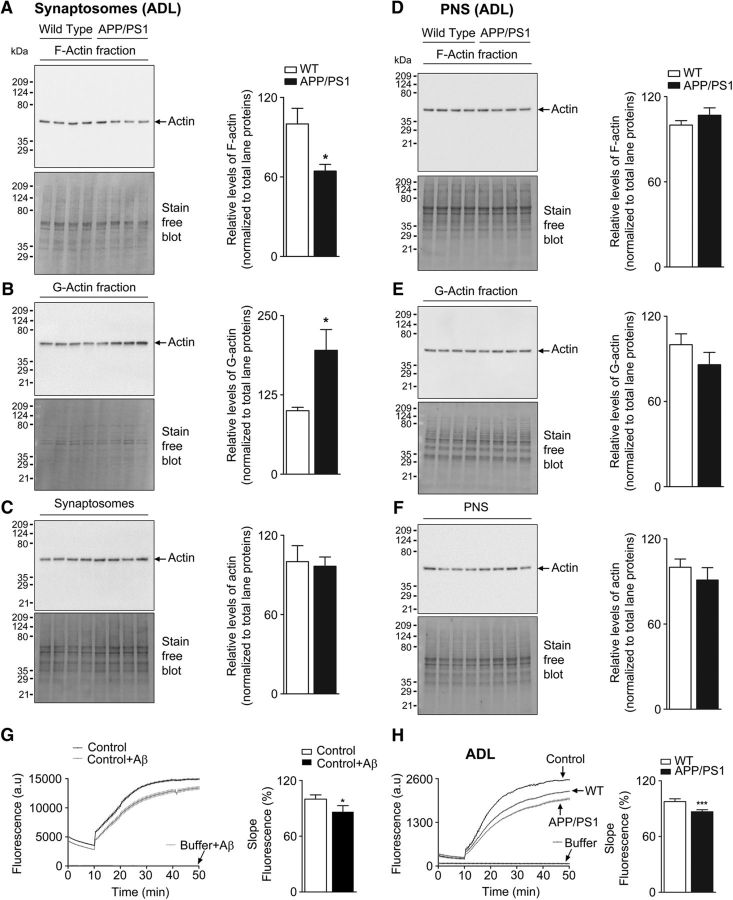
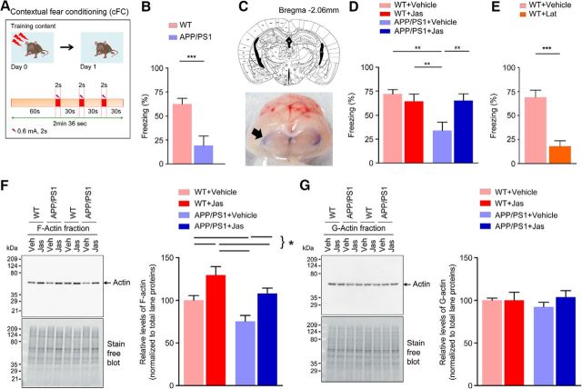
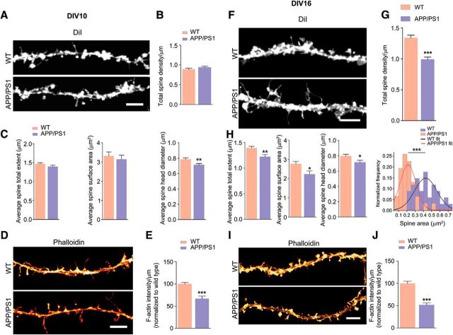
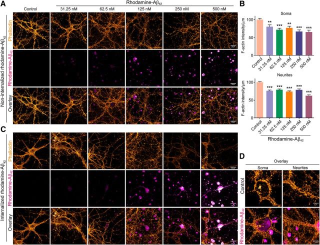
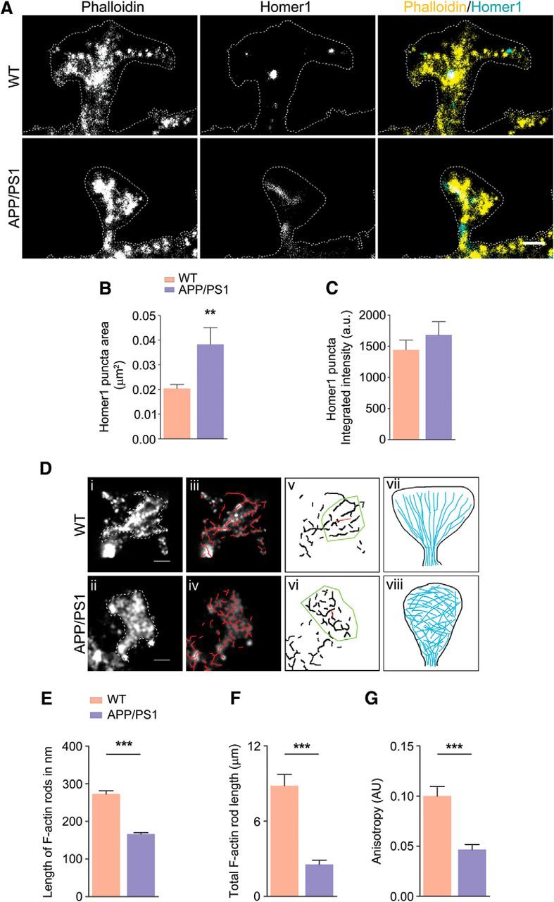
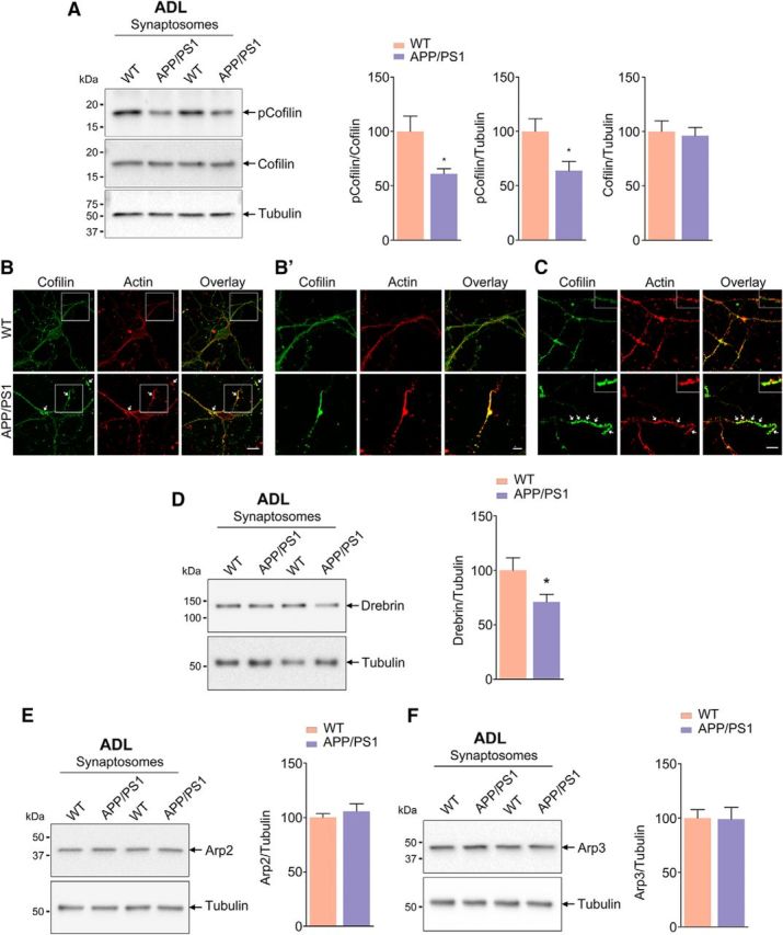
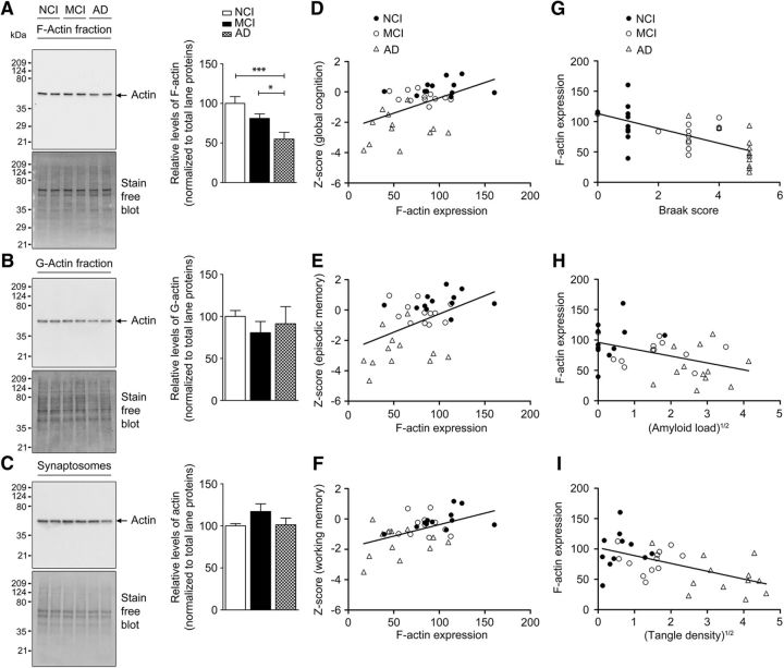
Verification
Claims the paper makes
The claims and statistics of this paper haven’t been checked yet.
Run the check — free, no account needed. It reads the open-access text and checks the sources it cites.
Claims other papers make about this one
Each is checked against this paper’s own text: does it say what the citing paper says it does?
The cited source says what the claim attributes to it.
In the citing paper
“Moreover, even low levels of Aβ can facilitate synaptic F-actin depolymerization and impair dendritic spines at very early stages of the pathology in an APP/presenilin 1 transgenic mouse model (64).”
From Deletion of Abi3 gene locus exacerbates neuropathological features of Alzheimer’s disease in a mouse model of Aβ amyloidosis (2021) · cites this paper as [64]
Self-check
Percentages, sums and reported differences re-computed from the paper’s own numbers.
The self-check needs the paper’s full text.
Citations
Cites
1 retracted
RETRACTED ARTICLE: A specific amyloid-β protein assembly in the brain impairs memory
Lesné, S., Koh, M. T., Kotilinek, L., Kayed, R., Glabe, C. G., Yang, A., Gallagher, M., & Ashe, K. H. (2006) · Nature · doi:10.1038/nature04533 · Cited by 2,769 · cited 1×
“Dendritic spines undergo synaptic-activitydependent modifications such as enlargement and shrinkage/ elimination during LTP (Bosch et al., 2014) or LTD, respectively, and correlate with memory deficits in animal models (Holcomb et al., 1998; Lesné et al., 2006; Shankar et al., 2008).”
BackgroundRetractedThe Mouse Brain in Stereotaxic Coordinates
Paxinos, G. (2001) · Cited by 12,859
Amyloid oligomers exacerbate tau pathology in a mouse model of tauopathy
Selenica, M.-L. B., Brownlow, M., Jimenez, J. P., Lee, D. C., Pena, G., Dickey, C. A., Gordon, M. N., & Morgan, D. (2013) · Neurodegenerative Diseases · doi:10.1159/000337230 · Cited by 10,359
Sub-diffraction-limit imaging by stochastic optical reconstruction microscopy (STORM)
Rust, M., Bates, M., & Zhuang, X. (2006) · Nature Methods · doi:10.1038/nmeth929 · Cited by 7,823
Jack, C. R., Knopman, D. S., Jagust, W. J., Petersen, R. C., Weiner, M. W., Aisen, P. S., Shaw, L. M., Vemuri, P., Wiste, H. J., Weigand, S. D., et al. (2013) · The Lancet Neurology · doi:10.1016/s1474-4422(12)70291-0 · Cited by 4,396
Shankar, G. M., Li, S., Mehta, T. H., Garcia-Munoz, A., Shepardson, N. E., Smith, I., Brett, F. M., Farrell, M. A., Rowan, M. J., Lemere, C. A., et al. (2008) · Nature Medicine · doi:10.1038/nm1782 · Cited by 3,685
An unbiased detector of curvilinear structures
Steger, C. T. (1998) · IEEE Transactions on Pattern Analysis and Machine Intelligence · doi:10.1109/34.659930 · Cited by 1,436
Holcomb, L., Gordon, M. N., McGowan, E., Yu, X., Benkovic, S., Jantzen, P., Wright, K., Saad, I., Mueller, R., Morgan, D., et al. (1998) · Nature Medicine · doi:10.1038/nm0198-097 · Cited by 1,386
Actin, Spectrin, and Associated Proteins Form a Periodic Cytoskeletal Structure in Axons
Xu, K., Zhong, G., & Zhuang, X. (2012) · Science · doi:10.1126/science.1232251 · Cited by 1,269
Direct stochastic optical reconstruction microscopy with standard fluorescent probes
van de Linde, S., Löschberger, A., Klein, T., Heidbreder, M., Wolter, S., Heilemann, M., & Sauer, M. (2011) · Nature Protocols · doi:10.1038/nprot.2011.336 · Cited by 1,104
Cited by
Sorted by citations
Religious Orders Study and Rush Memory and Aging Project
Bennett, D. A., Buchman, A. S., Boyle, P. A., Barnes, L. L., Wilson, R. S., & Schneider, J. A. (2018) · Journal of Alzheimer's Disease · doi:10.3233/jad-179939 · Cited by 1,260
Self-citationDejanovic, B., Huntley, M. A., De Mazière, A., Meilandt, W. J., Wu, T., Srinivasan, K., Jiang, Z., Gandham, V., Friedman, B. A., Ngu, H., et al. (2018) · Neuron · doi:10.1016/j.neuron.2018.10.014 · Cited by 517
Neuroprotective Effects of Quercetin in Alzheimer’s Disease
Khan, H. Z., Ullah, H., Aschner, M., Cheang, W. S., & Akkol, E. K. (2019) · Biomolecules · doi:10.3390/biom10010059 · Cited by 563
Amyloid Beta in Aging and Alzheimer’s Disease
Sehar, U., Rawat, P., Reddy, A. P., Kopel, J. J., & Reddy, P. H. (2022) · International Journal of Molecular Sciences · doi:10.3390/ijms232112924 · Cited by 409
GAP-43 and BASP1 in Axon Regeneration: Implications for the Treatment of Neurodegenerative Diseases
Chung, D., Shum, A. K., & Caraveo, G. (2020) · Frontiers in Cell and Developmental Biology · doi:10.3389/fcell.2020.567537 · Cited by 209
Synapse loss and progress of Alzheimer’s disease -A network model
Kashyap, G., Bapat, D., Das, D., Gowaikar, R., Amritkar, R. E., Rangarajan, G., Ravindranath, V., & Ambika, G. (2019) · Scientific Reports · doi:10.1038/s41598-019-43076-y · Cited by 162
Self-citationTan, C., Lu, N., Wang, C., Chen, D.-Y., Sun, N.-H., Lyu, H., Körbelin, J., Shi, W., Fukunaga, K., Lu, Y., et al. (2019) · Neuron · doi:10.1016/j.neuron.2018.12.036 · Cited by 128
Zhao, M.-Y., Wang, G.-Q., Wang, N.-N., Qiao-yan, Y., Liu, R.-L., & Shi, W. (2019) · Neurological Research · doi:10.1080/01616412.2018.1548747 · Cited by 118
Zhai, L., Shen, H., Sheng, Y., & Guan, Q. (2021) · Journal of Cellular and Molecular Medicine · doi:10.1111/jcmm.16787 · Cited by 115
Super-resolving Microscopy in Neuroscience
Werner, C. D., Sauer, M., & Geis, C. (2021) · Chemical Reviews · doi:10.1021/acs.chemrev.0c01174 · Cited by 113
Ivashko-Pachima, Y., Hadar, A., Grigg, I., Korenková, V., Kapitansky, O., Karmon, G., Gershovits, M., Sayas, C. L., Kooy, R. F., Attems, J., et al. (2021) · Molecular Psychiatry · doi:10.1038/s41380-019-0563-5 · Cited by 107
Understanding the physical basis of memory: Molecular mechanisms of the engram
Luis, C. O. S., & Ryan, T. J. (2022) · Journal of Biological Chemistry · doi:10.1016/j.jbc.2022.101866 · Cited by 114
Ullah, H., Minno, A. D., Santarcangelo, C., Khan, H. Z., & Daglia, M. (2021) · Antioxidants · doi:10.3390/antiox10040546 · Cited by 81
Wei, C., Fan, J. J., Sun, X., Yao, J., Guo, Y., Zhou, B., & Shang, Y. (2020) · Free Radical Biology and Medicine · doi:10.1016/j.freeradbiomed.2020.02.022 · Cited by 73
Effects of sulforaphane in the central nervous system
Huang, C., Wu, J., Chen, D., Jin, J., Wu, Y. H., & Chen, Z. (2019) · European Journal of Pharmacology · doi:10.1016/j.ejphar.2019.03.010 · Cited by 71
Nasiri, E., Alizadeh, A., Roushandeh, A. M., Gazor, R., Hashemi-Firouzi, N., & Golipoor, Z. (2019) · Metabolic Brain Disease · doi:10.1007/s11011-019-00421-4 · Cited by 66
Intracellular Trafficking Mechanisms of Synaptic Dysfunction in Alzheimer’s Disease
Perdigão, C., Barata, M. A., Araújo, M. N. C. D. F., Mirfakhar, F. S., Castanheira, J., & Almeida, C. G. (2020) · Frontiers in Cellular Neuroscience · doi:10.3389/fncel.2020.00072 · Cited by 66
Androuin, A., Potier, B., Nägerl, U. V., Cattaert, D., Danglot, L., Thierry, M., Youssef, I., Triller, A., Duyckaerts, C. J., Hachimi, K. H. E., et al. (2018) · Acta Neuropathologica · doi:10.1007/s00401-018-1847-6 · Cited by 66
Hossain, M. F., Wang, N., Chen, R., Li, S., Roy, J., Uddin, M. G., Li, Z., Lim, L. W., & Song, Y. (2021) · Ageing Research Reviews · doi:10.1016/j.arr.2021.101304 · Cited by 62
Super-resolution microscopy: a closer look at synaptic dysfunction in Alzheimer disease
Padmanabhan, P., Kneynsberg, A., & Götz, J. (2021) · Nature reviews. Neuroscience · doi:10.1038/s41583-021-00531-y · Cited by 59
Karahan, H., Smith, D. C., Kim, B., Dabin, L. C., Al-Amin, M. M., Wijeratne, H. R. S., Pennington, T., Viana di Prisco, G., McCord, B., Lin, P. B.-C., et al. (2021) · Science Advances · doi:10.1126/sciadv.abe3954 · Cited by 53
Anti-Neuroinflammatory Potential of Polyphenols by Inhibiting NF-Κb to Halt Alzheimer's Disease
Uddin, M. S., Hasana, S., Ahmad, J., Hossain, M. F., Rahman, M. M., Behl, T., Rauf, A., Ahmad, A., Hafeez, A., Perveen, A., et al. (2021) · Current Pharmaceutical Design · doi:10.2174/1381612826666201118092422 · Cited by 49
Bello-Medina, P. C., Hernández-Quiroz, F., Pérez-Morales, M., González-Franco, D. A., Cruz-Pauseno, G., Garcı́a-Mena, J., Díaz-Cintra, S. Y., & Pacheco‐López, G. (2021) · Frontiers in Neuroscience · doi:10.3389/fnins.2021.595583 · Cited by 48
Actin Cytoskeleton Role in the Maintenance of Neuronal Morphology and Long-Term Memory
Lamprecht, R. (2021) · Cells · doi:10.3390/cells10071795 · Cited by 48
Borroni, V., & Barrantes, F. J. (2021) · Membranes · doi:10.3390/membranes11090664 · Cited by 49
Kommaddi, R. P., Tomar, D. S., Karunakaran, S., Bapat, D., Nanguneri, S., Ray, A., Schneider, B. L., Nair, D. K., & Ravindranath, V. (2019) · Antioxidants and Redox Signaling · doi:10.1089/ars.2019.7754 · Cited by 44
Self-citationPark, C. E., Cho, Y., Cho, I., Jung, H., Kim, B., Shin, J. H., Choi, S., Kwon, S., Hahn, Y. K., & Chang, J. (2020) · ACS Nano · doi:10.1021/acsnano.0c04915 · Cited by 38
Behnke, J. A., Ye, C., Moberg, K. H., & Zheng, J. Q. (2021) · STAR Protocols · doi:10.1016/j.xpro.2021.100689 · Cited by 41
Synaptic aging disrupts synaptic morphology and function in cerebellar Purkinje cells
Wang, L., Deng, J., Fan, W., Yan, M., Sun, Y.-Z., & Deng, J.-X. (2018) · Neural Regeneration Research · doi:10.4103/1673-5374.233445 · Cited by 37
Samanta, S., Rajasekhar, K., Ramesh, M., Murugan, N. A., Alam, S., Shah, D., Clement, J. P., & Govindaraju, T. (2021) · Advanced Therapeutics · doi:10.1002/adtp.202000225 · Cited by 36
Kreis, P., Gallrein, C., Puente, E. R., Mack, T. G. A., Kroon, C., Dinkel, V., Willmes, C. G., Murk, K., Dieck, S. T., Schuman, E. M., et al. (2019) · Nature Communications · doi:10.1038/s41467-019-08420-w · Cited by 35
Hulshof, L. A., Frajmund, L. A., van Nuijs, D., van der Heijden, D. C. N., Middeldorp, J., & Hol, E. M. (2022) · Neurobiology of Aging · doi:10.1016/j.neurobiolaging.2021.12.009 · Cited by 39
Role of actin in organelle trafficking in neurons
Venkatesh, K., Mathew, A., & Koushika, S. P. (2019) · Cytoskeleton · doi:10.1002/cm.21580 · Cited by 38
Real-time nanoscale organization of amyloid precursor protein
Kedia, S., Ramakrishna, P., Netrakanti, P. R., Jose, M., Sibarita, J., Nadkarni, S., & Nair, D. K. (2020) · Nanoscale · doi:10.1039/d0nr00052c · Cited by 33
Self-citationCdc42GAP deficiency contributes to the Alzheimer’s disease phenotype
Zhu, M., Xiao, B., Xue, T., Qin, S., Ding, J., Wu, Y. H., Tang, Q., Huang, M., Zhao, N., Ye, Y., et al. (2023) · Brain · doi:10.1093/brain/awad184 · Cited by 32
Synaptosome as a tool in Alzheimer’s disease research
Ahmad, F., & Liu, P. (2020) · Brain Research · doi:10.1016/j.brainres.2020.147009 · Cited by 33
Nargeh, H., Aliabadi, F., Ajami, M., & Pazoki–Toroudi, H. (2021) · Journal of Agricultural and Food Chemistry · doi:10.1021/acs.jafc.1c00923 · Cited by 32
Bello-Medina, P. C., González-Franco, D. A., Vargas-Rodríguez, I., & Díaz-Cintra, S. Y. (2021) · Neurología (English Edition) · doi:10.1016/j.nrleng.2019.06.008 · Cited by 35
Epigenetic treatment of behavioral and physiological deficits in a tauopathy mouse model
Wang, W., Cao, Q., Tan, T., Yang, F., Williams, J. B., & Yan, Z. (2021) · Aging Cell · doi:10.1111/acel.13456 · Cited by 30
Bello-Medina, P. C., Corona-Cervantes, K., Zavala-Torres, N. G., González, A., Pérez-Morales, M., González-Franco, D. A., Gómez, A., García-Mena, J., Díaz-Cintra, S. Y., & Pacheco‐López, G. (2022) · International Journal of Molecular Sciences · doi:10.3390/ijms23158209 · Cited by 33
Three-dimensional real time imaging of amyloid β aggregation on living cells
Kuragano, M., Yamashita, R., Chikai, Y., Kitamura, R., & Tokuraku, K. (2020) · Scientific Reports · doi:10.1038/s41598-020-66129-z · Cited by 30
Kommaddi, R. P., Gowaikar, R., Haseena, P. A., Diwakar, L., Singh, K. P., & Mondal, A. (2024) · Journal of Biological Chemistry · doi:10.1016/j.jbc.2023.105619 · Cited by 29
Self-citationAltered Actin Dynamics in Cell Migration of GNE Mutant Cells
Devi, S. S., Yadav, R., & Arya, R. (2021) · Frontiers in Cell and Developmental Biology · doi:10.3389/fcell.2021.603742 · Cited by 27
Mitigation of synaptic and memory impairments via F-actin stabilization in Alzheimer’s disease
Haseena, P. A., Basavaraju, N., Chandran, M., Jaleel, A., Bennett, D. A., & Kommaddi, R. P. (2024) · Alzheimer s Research & Therapy · doi:10.1186/s13195-024-01558-w · Cited by 26
Self-citationBello-Medina, P. C., González-Franco, D. A., Vargas-Rodríguez, I., & Díaz-Cintra, S. Y. (2019) · Neurología · doi:10.1016/j.nrl.2019.06.002 · Cited by 25
The role of molecular diffusion within dendritic spines in synaptic function
Obashi, K., Taraska, J. W., & Okabe, S. (2021) · The Journal of General Physiology · doi:10.1085/jgp.202012814 · Cited by 24
The power of super-resolution microscopy in modern biomedical science
Sun, N., Jia, Y., Bai, S., Li, Q., Dai, L., & Li, J. (2023) · Advances in Colloid and Interface Science · doi:10.1016/j.cis.2023.102880 · Cited by 23
Mahaman, Y. A. R., Huang, F., Embaye, K. S., Wang, X., & Zhu, F. (2021) · Frontiers in Cell and Developmental Biology · doi:10.3389/fcell.2021.680118 · Cited by 22
Ansari, M. A., Al‐Jarallah, A., & Babiker, F. A. (2023) · Cells · doi:10.3390/cells12131728 · Cited by 21
Rarinca, V., Nicoară, M., Ureche, D., & Ciobica, A. S. (2023) · Antioxidants · doi:10.3390/antiox12071418 · Cited by 21
Kim, H.-J., Kim, B., Kim, H.-S., & Cho, J. (2020) · Molecular Brain · doi:10.1186/s13041-020-0565-x · Cited by 21
Novel approaches to increase synaptic resilience as potential treatments for Alzheimer’s disease
Pham, A. Q., & Dore, K. (2023) · Seminars in Cell & Developmental Biology · doi:10.1016/j.semcdb.2022.03.032 · Cited by 21
Gong, J., Jin, Z., Chen, H., He, J., Zhang, Y., & Yang, X. (2023) · Advanced Drug Delivery Reviews · doi:10.1016/j.addr.2023.114791 · Cited by 21
Chatterjee, M., Kwon, J., Benedict, J., Kamceva, M., Kurup, P. K., & Lombroso, P. J. (2021) · Experimental Brain Research · doi:10.1007/s00221-020-06028-x · Cited by 21
INPP5D/SHIP1: Expression, Regulation and Roles in Alzheimer’s Disease Pathophysiology
Olufunmilayo, E. O., & Holsinger, R. M. D. (2023) · Genes · doi:10.3390/genes14101845 · Cited by 19
Early hippocampal hyperexcitability and synaptic reorganization in mouse models of amyloidosis
Ray, A., Loghinov, I., Ravindranath, V., & Barth, A. L. (2024) · iScience · doi:10.1016/j.isci.2024.110629 · Cited by 17
Self-citationc-Abl Deficiency Provides Synaptic Resiliency Against Aβ-Oligomers
Gutiérrez, D. A., Vargas, L. M., Chandía-Cristi, A., de la Fuente, C., Leal, N. R., & Alvarez, A. R. (2019) · Frontiers in Cellular Neuroscience · doi:10.3389/fncel.2019.00526 · Cited by 19
LIMK, Cofilin 1 and actin dynamics involvement in fear memory processing
Medina, C., de la Fuente, V., Dieck, S. T., Nassim-Assir, B., Dalmay, T., Bartnik, I., Lunardi, P. S., Alvares, L. D. O., Schuman, E. M., Letzkus, J. J., et al. (2020) · Neurobiology of Learning and Memory · doi:10.1016/j.nlm.2020.107275 · Cited by 18
Impaired Synaptic Plasticity Mechanisms in Alzheimer’s Disease
Ebrahimi, R., Golzari, Z., Heidari‐Foroozan, M., Khosravi, A., Sharghi, S. G., Saleh, M., Salarvandian, S., & Esmaeilpour, K. (2025) · Metabolic Brain Disease · doi:10.1007/s11011-025-01712-9 · Cited by 21
Yan, M., Meng, L., Dai, L., Zhang, X., Chen, G., Zheng, Y., Zha, Y., Zeng, Y., & Zhang, Z. (2020) · Biochemical and Biophysical Research Communications · doi:10.1016/j.bbrc.2020.06.101 · Cited by 18
Guptarak, J., Scaduto, P., Tumurbaatar, B., Zhang, W. R., Jupiter, D. C., Taglialatela, G., & Fracassi, A. (2024) · Alzheimer s & Dementia · doi:10.1002/alz.13900 · Cited by 17
Nanguneri, S., Pramod, R. T., Efimova, N., Das, D., Jose, M., Svitkina, T., & Nair, D. K. (2019) · eNeuro · doi:10.1523/eneuro.0425-18.2019 · Cited by 17
Self-citationKalirin‐7 prevents dendritic spine dysgenesis induced by amyloid beta‐derived oligomers
Xie, Z., Shapiro, L. P., Cahill, M. E., Russell, T. A., Lacor, P. N., Klein, W. L., & Penzes, P. (2018) · European Journal of Neuroscience · doi:10.1111/ejn.14311 · Cited by 16
Wu, N., Meng, S., Li, Z., Fang, J., Cheng, Q., Kong, T., & Liu, Z. (2024) · Small · doi:10.1002/smll.202307603 · Cited by 16
Cui, Z., Guo, Z., Wei, L., Zou, X., Zhu, Z., Liu, Y., Wang, J., Chen, L., Wang, D., & Ke, Z. (2022) · Pain · doi:10.1097/j.pain.0000000000002648 · Cited by 17
Kommaddi, R. P., Verma, A., Terrera, G. M., Tiwari, V., Chithanathan, K., Diwakar, L., Gowaikar, R., Karunakaran, S., Malo, P. K., Graff‐Radford, N. R., et al. (2023) · Translational Psychiatry · doi:10.1038/s41398-023-02411-8 · Cited by 18
Self-citationMorphological Features of Human Dendritic Spines
Renner, J., & Rasia‐Filho, A. A. (2023) · Advances in neurobiology · doi:10.1007/978-3-031-36159-3_9 · Cited by 18
Unraveling Early Signs of Navigational Impairment in APPswe/PS1dE9 Mice Using Morris Water Maze
Karunakaran, S. (2020) · Frontiers in Neuroscience · doi:10.3389/fnins.2020.568200 · Cited by 16
Self-citationSuper-resolution imaging of the neuronal cytoskeleton
Butler-Hallissey, C., & Leterrier, C. (2024) · npj Imaging · doi:10.1038/s44303-024-00054-y · Cited by 13
A Palette of Fluorescent Aβ42 Peptides Labelled at a Range of Surface-Exposed Sites
Thacker, D., Bless, M., Barghouth, M., Zhang, E., & Linse, S. S. (2022) · International Journal of Molecular Sciences · doi:10.3390/ijms23031655 · Cited by 13
Busquets, O., Parcerisas, A., Verdaguer, E., Ettcheto, M., Camins, A., Beas‐Zárate, C., Castro-Torres, R. D., & Auladell, C. (2020) · Journal of Alzheimer s Disease · doi:10.3233/jad-201053 · Cited by 14
Tacke, C., DiStefano, P. S., Lindsay, R. M., Metzdorf, K., Zagrebelsky, M., & Körte, M. (2022) · Frontiers in Molecular Neuroscience · doi:10.3389/fnmol.2022.945348 · Cited by 12
Ammassari‐Teule, M. (2020) · Frontiers in Synaptic Neuroscience · doi:10.3389/fnsyn.2020.566615 · Cited by 12
Lopes, C. R., Silva, J. S., Santos, J., Rodrigues, M. S., Madeira, D., Oliveira, A., Moreira-de-Sá, A., Lourenço, V. S., Gonçalves, F. Q., Silva, H. B. L., et al. (2023) · International Journal of Molecular Sciences · doi:10.3390/ijms24086968 · Cited by 10
Cofilin: Molecular and Cellular Functions and Its Role in the Functioning of the Nervous System
Kovaleva, T. F., Maksimova, N. S., Жуков, И. Ю., Pershin, V. I., Mukhina, I. V., & Gainullin, M. (2019) · Neurochemical Journal · doi:10.1134/s1819712419010124 · Cited by 10
Yadav, R., Oswalia, J., Ghosh, A., & Arya, R. (2022) · NeuroMolecular Medicine · doi:10.1007/s12017-022-08711-4 · Cited by 10
Scholes, H. M., Cryar, A., Kerr, F., Sutherland, D., Gethings, L. A., Vissers, J. P. C., Lees, J., Orengo, C. A., Partridge, L., & Thalassinos, K. (2020) · Scientific Reports · doi:10.1038/s41598-020-74748-9 · Cited by 13
Sarkar, S., Gharami, K., Mondal, A., Padmanabhan, K., Paidi, R. K., Srikumar, B. N., & Biswas, S. C. (2025) · Acta Neuropathologica Communications · doi:10.1186/s40478-025-02060-6 · Cited by 9
Kawaguchi, Y., Matsubayashi, J., Kawakami, Y., Nishida, R., Kurihara, Y., & Takei, K. (2022) · Molecular Medicine · doi:10.1186/s10020-022-00581-7 · Cited by 9
Kirimtat, A., Krejcar, O., Marešová, P., Selamat, A., & Kuča, K. (2024) · IEEE Access · doi:10.1109/access.2024.3484928 · Cited by 8
VANL-100 Attenuates Beta-Amyloid-Induced Toxicity in SH-SY5Y Cells
Collins, A. E., Saleh, T. M., & Kalisch, B. E. (2022) · International Journal of Molecular Sciences · doi:10.3390/ijms24010442 · Cited by 10
Karbowski, J., & Urban, P. (2023) · Scientific Reports · doi:10.1038/s41598-023-49321-9 · Cited by 9
Lin, C., Yang, S., Huang, Y.-J., & Lane, H. (2019) · Scientific Reports · doi:10.1038/s41598-019-55414-1 · Cited by 7
Li, K., Jiang, Y., Li, G., Liu, T., & Yang, Z. R. (2020) · ACS Chemical Neuroscience · doi:10.1021/acschemneuro.0c00574 · Cited by 8
Actin Cytoskeleton at the Synapse: An Alzheimer's Disease Perspective
Haseena, P. A., Basavaraju, N., Gupta, A., & Kommaddi, R. P. (2025) · Cytoskeleton · doi:10.1002/cm.21993 · Cited by 9
Self-citationThe double‐edged sword effect of HDAC6 in Aβ toxicities
Cheng, K., Hwang, Y., & Chiang, H. (2021) · The FASEB Journal · doi:10.1096/fj.202101061r · Cited by 8
Early β adrenoceptor dependent time window for fear memory persistence in APPswe/PS1dE9 mice
Karunakaran, S. (2021) · Scientific Reports · doi:10.1038/s41598-020-79487-5 · Cited by 7
Self-citationMaeda, T., Shimamori, K., Kurita, H., Tokuraku, K., & Kuragano, M. (2024) · Experimental Cell Research · doi:10.1016/j.yexcr.2024.113958 · Cited by 7
The emerging role of disulfidptosis in Alzheimer's disease
Zhou, Q., Zheng, N., Chen, Z., Xie, L., Yang, X., Sun, Q., Lin, J., Li, B., & Li, L. (2025) · European Journal of Pharmacology · doi:10.1016/j.ejphar.2025.178085 · Cited by 10
Medvedeva, M. V., Serebryakova, M. V., Matyushenko, A. M., Nefedova, V. V., Muronetz, V. I., & Schmalhausen, E. V. (2024) · Archives of Biochemistry and Biophysics · doi:10.1016/j.abb.2024.110189 · Cited by 6
Moghaddam, H. S., & Aarabi, M. H. (2018) · Journal of Neuroscience · doi:10.1523/jneurosci.0844-18.2018 · Cited by 7
Protective Effects of Statins against Alzheimer Disease
Rezakhani, L., Salimi, Z., Zarei, F., Moradpour, F., Khazaei, M. R., Khazaei, M., & Pourjalili, M. (2023) · Ewha Medical Journal · doi:10.12771/emj.2023.e17 · Cited by 6
Resilience mechanisms underlying Alzheimer’s disease
Chew, C. S., Jy, L., Ng, K. Y., Koh, R. Y., & Chye, S. M. (2025) · Metabolic Brain Disease · doi:10.1007/s11011-024-01507-4 · Cited by 6
Zhu, X., Zhang, Y., Yan, X., Zhao, Y., Shi, L., Sun, Z., Kang, M. H., Zong, Y., Li, Q., & Xu, Z. (2025) · Neuroscience Letters · doi:10.1016/j.neulet.2025.138329 · Cited by 5
Reaction–Diffusion Model-Based Research on Formation Mechanism of Neuron Dendritic Spine Patterns
Jia, Y., Zhao, Q., Yin, H., Guo, S., Sun, M., Yang, Z. R., & Zhao, X. (2021) · Frontiers in Neurorobotics · doi:10.3389/fnbot.2021.563682 · Cited by 5
Kot, K., Kot, Y. G., Kurbanov, R., Андріяш, Г. С., Tigunova, O., Blume, Y. B., & Shulga, S. (2024) · Frontiers in Neuroscience · doi:10.3389/fnins.2024.1325287 · Cited by 5
Fluorescent probes for neuroscience: imaging ex vivo brain tissue sections
Schwehr, B. J., Hartnell, D., Ellison, G., Hindes, M. T., Milford, B., Dallerba, E., Hickey, S. M., Pfeffer, F. M., Brooks, D. A., Massi, M., et al. (2024) · The Analyst · doi:10.1039/d4an00663a · Cited by 5
Chemical Stimulation of Rodent and Human Cortical Synaptosomes: Implications in Neurodegeneration
Ahmad, F., Yu, J. H., Lladó, A., & Liu, P. (2021) · Cells · doi:10.3390/cells10051174 · Cited by 6
Characterization of a Human Neuronal Culture System for the Study of Cofilin–Actin Rod Pathology
Tahtamouni, L. H., Alderfer, S. A., Kuhn, T. B., Minamide, L. S., Chanda, S., Ruff, M. R., & Bamburg, J. R. (2023) · Biomedicines · doi:10.3390/biomedicines11112942 · Cited by 5
Therapeutic potential of isothiocyanates by targeting the NRF2 pathway
García‐Yagüe, Á. J., Cazalla, E., & Cuadrado, A. (2026) · Free Radical Biology and Medicine · doi:10.1016/j.freeradbiomed.2026.03.029 · Cited by 5
Metadata
- Authors
- Kommaddi, R. P., Das, D., Karunakaran, S., Nanguneri, S., Bapat, D., Ray, A., Shaw, E., Bennett, D. A., Nair, D., & Ravindranath, V.
- Affiliations
- Indian Institute of Science Bangalore (IN)
- Rush University Medical Center (US)
- Corresponding author
- Ravindranath, Vijayalakshmi
- ORCID
- Venue
- The Journal of Neuroscience
- Publisher
- Society for Neuroscience
- Published
- 2017-12-15
- Volume
- 38
- Issue
- 5
- Pages
- 1085-1099
- ISSN
- 0270-6474 (ISSN-L)
- 1529-2401
- PMC
- PMC5792472
- OpenAlex
- W2774657197
- Cited by
- 135
- References
- 71
- Citations by year
- 2026: 3 · 2025: 22 · 2024: 13 · 2023: 16 · 2022: 13 · 2021: 25 · 2020: 17 · 2019: 20 · 2018: 6
- FWCI
- 8.70
- Type
- journal article
- Language
- en
- Open access
- hybrid
- doi.org
- Locations
- Journal of Neuroscience (publishedVersion, cc-by)
- Europe PMC (PubMed Central) (publishedVersion, cc-by)
- PubMed Central (publishedVersion, cc-by)
- PubMed (publishedVersion)
- NOT FOUND REPOSITORY (Indian Institute of Science Bangalore) (acceptedVersion)
- Funders
- Tata Trusts
- Science and Engineering Research Board (PDF/2016/003226)
- National Institute on Aging (R01AG017917, R01AG015819, P30AG010161, RF1AG015819)
- Topics
- Life Sciences › Neuroscience › Cellular and Molecular Neuroscience › Alzheimer's disease research and treatments
- Life Sciences › Neuroscience › Cellular and Molecular Neuroscience › Neuroscience and Neuropharmacology Research
- Health Sciences › Medicine › Psychiatry and Mental health › Dementia and Cognitive Impairment Research
- Keywords
- amyloid-beta, F-actin depolymerization, dendritic spines, actin cytoskeleton, synaptic dysfunction, APPswe/PS1ΔE9 mice, contextual fear conditioning, memory impairment, jasplakinolide, latrunculin, actin polymerization, postmortem brain, working memory
- MeSH terms
- Actins, Aging, Alzheimer Disease, Animals, Autopsy, Cognition Disorders, Conditioning, Classical, Fear, Female, Humans, Male, Mice, Inbred C57BL, Mice, Transgenic, Mental Recall, Synaptosomes, Amyloid beta-Peptides, Dendritic Spines, Actin Depolymerizing Factors, Mice, Cognitive Dysfunction, Primary Cell Culture
About these checks
Registry, retraction, Cites and statistics results are computed, not judged, with one exception: a printed reference that no index lists is read once by a model before it counts as a citation error. Claims are extracted by a model and kept only when their quote appears verbatim in the text; each cited source is then checked, with a model’s help, for whether it says what the claim relies on. Those verdicts can be wrong — read the claim’s quote and the cited source before relying on one.
Statistics and arithmetic are recomputed, not judged.
Confidence bands are shown only for fields calibrated on our annotated test set. None is calibrated yet, so no band is shown.
What counts as a claim ›How we measure confidence ›How we check this ›
Are you an author, or do you see a problem with this page? Request a review
