

The DOI matches its Crossref record; 11 of the 11 references it cites were found in a registry or an index.
CX3CL1 Is Neuroprotective in Permanent Focal Cerebral Ischemia in Rodents
Abstract
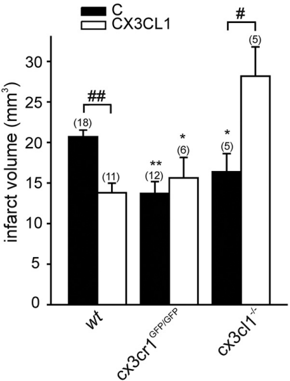
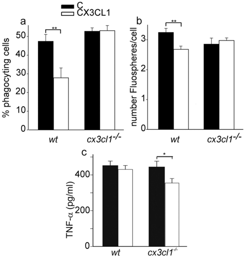
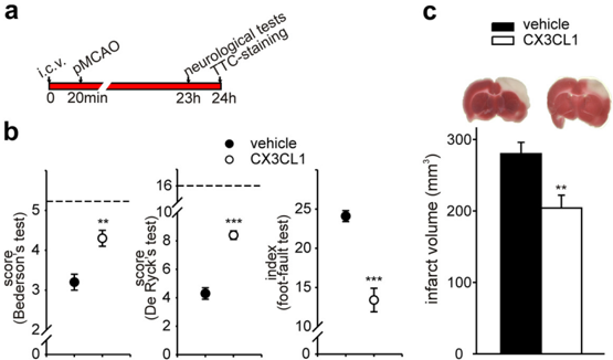
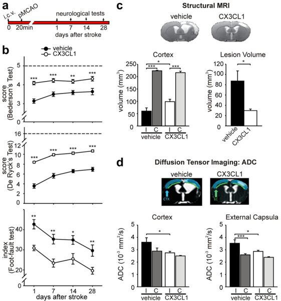
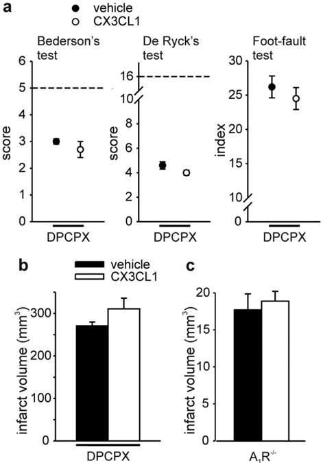
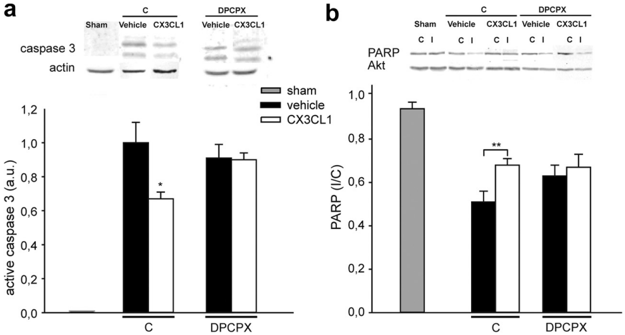
The chemokine CX3CL1 and its receptor CX3CR1 are constitutively expressed in the nervous system. In this study, we used in vivo murine models of permanent middle cerebral artery occlusion (pMCAO) to investigate the protective potential of CX3CL1. We report that exogenous CX3CL1 reduced ischemia-induced cerebral infarct size, neurological deficits, and caspase-3 activation. CX3CL1-induced neuroprotective effects were long lasting, being observed up to 50 d after pMCAO in rats. The neuroprotective action of CX3CL1 in different models of brain injuries is mediated by its inhibitory activity on microglia and, in vitro , requires the activation of adenosine receptor 1 (A 1 R). We show that, in the presence of the A 1 R antagonist 1,3-dipropyl-8-cyclopentylxanthine and in A 1 R −/− mice, the neuroprotective effect of CX3CL1 on pMCAO was abolished, indicating the critical importance of the adenosine system in CX3CL1 protection also in vivo . In apparent contrast with the above reported data but in agreement with previous findings, cx3cl1 −/− and cx3cr1 GFP/GFP mice, respectively, deficient in CX3CL1 or CX3CR1, had less severe brain injury on pMCAO, and the administration of exogenous CX3CL1 increased brain damage in cx3cl1 −/− ischemic mice. We also report that CX3CL1 induced a different phagocytic activity in wild type and cx3cl1 −/− microglia in vitro during cotreatment with the medium conditioned by neurons damaged by oxygen-glucose deprivation. Together, these data suggest that acute administration of CX3CL1 reduces ischemic damage via an adenosine-dependent mechanism and that the absence of constitutive CX3CL1–CX3CR1 signaling changes the outcome of microglia-mediated effects during CX3CL1 administration to ischemic brain.
Overview
The claims and statistics of this paper haven’t been checked yet.
Run the check — free, no account needed. It reads the open-access text and checks the sources it cites.
Integrity signals
Registry
Cites
Statistics
Figures
Verification
Claims the paper makes
The claims and statistics of this paper haven’t been checked yet.
Run the check — free, no account needed. It reads the open-access text and checks the sources it cites.
Claims other papers make about this one
Each is checked against this paper’s own text: does it say what the citing paper says it does?
In the citing paper
“Cipriani et al. [55] found that CX3CL1 could reduce infarct volume and improve behavioral outcomes for middle cerebral artery occlusion rats.”
From CX3CL1/CX3CR1 axis attenuates early brain injury via promoting the delivery of exosomal microRNA-124 from neuron to microglia after subarachnoid hemorrhage (2020) · cites this paper as [55]
Self-check
Percentages, sums and reported differences re-computed from the paper’s own numbers.
The self-check needs the paper’s full text.
Citations
Cites
Microglia-mediated neurotoxicity: uncovering the molecular mechanisms
Block, M. L., Zecca, L., & Hong, J.-S. (2007) · Nature Reviews Neuroscience · doi:10.1038/nrn2038 · Cited by 4,110
Control of microglial neurotoxicity by the fractalkine receptor
Cardona, A. E., Pioro, E. P., Sasse, M. E., Kostenko, V., Cardona, S. M., Dijkstra, I. M., Huang, D., Kidd, G., Dombrowski, S., Dutta, R., et al. (2006) · Nature Neuroscience · doi:10.1038/nn1715 · Cited by 1,531
Regulation of Tau Pathology by the Microglial Fractalkine Receptor
Bhaskar, K., Konerth, M., Kokiko-Cochran, O. N., Cardona, A., Ransohoff, R. M., & Lamb, B. T. (2010) · Neuron · doi:10.1016/j.neuron.2010.08.023 · Cited by 639 · cited 5×
“, 2010) but also with the enhanced microtubule-associated protein tau phosphorylation (Bhaskar et al., 2010), suggesting opposite effects at different phases of the disease.”
BackgroundInfluentialCaspase signalling controls microglia activation and neurotoxicity
Burguillos, M. A., Deierborg, T., Kavanagh, E., Persson, A., Hajji, N., Garcia-Quintanilla, A., Cano, J., Brundin, P., Englund, E., Venero, J. L., et al. (2011) · Nature · doi:10.1038/nature09788 · Cited by 585
Microglial Cx3cr1 knockout prevents neuron loss in a mouse model of Alzheimer's disease
Fuhrmann, M., Bittner, T., Jung, C. K. E., Burgold, S., Page, R. M., Mitteregger, G., Haass, C., LaFerla, F. M., Kretzschmar, H., & Herms, J. (2010) · Nature Neuroscience · doi:10.1038/nn.2511 · Cited by 562
Lee, S., Varvel, N. H., Konerth, M. E., Xu, G., Cardona, A. E., Ransohoff, R. M., & Lamb, B. T. (2010) · The American Journal of Pathology · doi:10.2353/ajpath.2010.100265 · Cited by 522 · cited 5×
“It is reported that CX3CL1 reduces the phagocytic activity of microglia (Lee et al., 2010).”
BackgroundInfluentialInhibition of Microglial Phagocytosis Is Sufficient To Prevent Inflammatory Neuronal Death
Neher, J. J., Neniskyte, U., Zhao, J.-W., Bal-Price, A., Tolkovsky, A. M., & Brown, G. C. (2011) · The Journal of Immunology · doi:10.4049/jimmunol.1003600 · Cited by 373
Liu, Z., Condello, C., Schain, A., Harb, R., & Grutzendler, J. (2010) · The Journal of Neuroscience · doi:10.1523/jneurosci.4403-10.2010 · Cited by 275 · cited 2×
“In different models of neuroinflammatory diseases, CX3CR1 deficiency correlated with enhanced microglia phagocytosis (Lee et al., 2010; Liu et al., 2010) and altered production of inflammatory cytokines (Cardona et al.”
BackgroundCX3CL1 reduces neurotoxicity and microglial activation in a rat model of Parkinson’s disease
Pabon, M. M., Bachstetter, A. D., Hudson, C. E., Gemma, C., & Bickford, P. C. (2011) · Journal of Neuroinflammation · doi:10.1186/1742-2094-8-9 · Cited by 241
Mice deficient in fractalkine are less susceptible to cerebral ischemia-reperfusion injury
Soriano, S. G., Amaravadi, L. S., Wang, Y. F., Zhou, H., Yu, G. X., Tonra, J. R., Fairchild-Huntress, V., Fang, Q., Dunmore, J. H., Huszar, D., et al. (2002) · Journal of Neuroimmunology · doi:10.1016/s0165-5728(02)00033-4 · Cited by 222
Lyons, A., Lynch, A. M., Downer, E. J., Hanley, R., O’Sullivan, J. B., Smith, A., & Lynch, M. A. (2009) · Journal of Neurochemistry · doi:10.1111/j.1471-4159.2009.06253.x · Cited by 202
Research works printed but not linked: 38
- 10.1161/01.STR.23.9.1337 · DOI
- 10.1016/j.tins.2007.08.007 · DOI · Paper page
- 10.1161/STROKEAHA.108.531632 · DOI
- Chapman, 2000, Fractalkine cleavage from neuronal membranes represents an acute event in the inflammatory response to excitotoxic brain damage, J Neurosci, 20, 1
- 10.1128/MCB.21.9.3159-3165.2001 · DOI
- 10.1007/s11302-005-0649-1 · DOI
- 10.1038/jcbfm.2008.64 · DOI · Paper page
- 10.1016/S0166-2236(99)01401-0 · DOI · Paper page
- 10.1038/sj.cdd.4402132 · DOI
- 10.1073/pnas.95.18.10896 · DOI · Paper page
- 10.1038/sj.jcbfm.9600362 · DOI
- 10.1073/pnas.161292398 · DOI
- 10.1128/MCB.20.11.4106-4114.2000 · DOI · Paper page
- 10.1007/s00210-009-0477-x · DOI
- 10.1002/glia.20610 · DOI
- 10.1523/JNEUROSCI.5360-06.2007 · DOI · Paper page
- 10.1523/JNEUROSCI.5505-08.2009 · DOI
- 10.1038/npp.2010.26 · DOI
- 10.4049/jimmunol.180.11.7590 · DOI · Paper page
- 10.1016/j.jneuroim.2005.03.023 · DOI · Paper page
- 10.1016/S0006-8993(03)02867-1 · DOI
- 10.1038/sj.bmt.1701300 · DOI
- Namura, 1998, Activation and cleavage of caspase-3 in apoptosis induced by experimental cerebral ischemia, J Neurosci, 18, 3659, 10.1523/JNEUROSCI.18-10-03659.1998 · DOI
- 10.1371/journal.pone.0011746 · DOI
- 10.1074/jbc.M110.169839 · DOI
- 10.1111/j.1460-9568.2004.03564.x · DOI
- 10.1111/j.1471-4159.2006.03823.x · DOI
- 10.1016/0006-8993(95)01153-6 · DOI
- 10.1038/jcbfm.2008.121 · DOI
- 10.1124/jpet.106.105064 · DOI
- 10.1046/j.1460-9568.2002.02007.x · DOI
- Villa, 2007, The interleukin-8 (IL(/CXCL8) receptor inhibitor reparixin improves neurological deficits and reduces long-term inflammation in permanent and transient cerebral ischemia in rats, Mol Med, 13, 125, 10.2119/2007-00008.Villa · DOI
- 10.1016/0014-2999(96)00101-X · DOI
- 10.1016/j.neulet.2005.02.019 · DOI
- 10.1097/01.WCB.0000122742.72175.1B · DOI
- 10.1073/pnas.95.26.15769 · DOI · Paper page
- 10.1016/j.brainres.2009.06.068 · DOI
- 10.1002/(SICI)1098-1136(20000215)29:4<305::AID-GLIA2>3.0.CO;2-V · DOI
Cited by
Sorted by citations
Microglia, seen from the CX3CR1 angle
Wolf, Y., Yona, S., Kim, K.-W., & Jung, S. (2013) · Frontiers in Cellular Neuroscience · doi:10.3389/fncel.2013.00026 · Cited by 316
Microglia in Health and Disease
Ransohoff, R. M., & Khoury, J. E. (2016) · Cold Spring Harbor Perspectives in Biology · doi:10.1101/cshperspect.a020560 · Cited by 303
Modulating neurotoxicity through CX3CL1/CX3CR1 signaling
Limatola, C., & Ransohoff, R. M. (2014) · Frontiers in Cellular Neuroscience · doi:10.3389/fncel.2014.00229 · Cited by 210
Self-citationThe brain’s best friend: microglial neurotoxicity revisited
Hellwig, S., Heinrich, A., & Biber, K. (2013) · Frontiers in Cellular Neuroscience · doi:10.3389/fncel.2013.00071 · Cited by 126
Time-dependent effects of CX3CR1 in a mouse model of mild traumatic brain injury
Febinger, H. Y., Thomasy, H. E., Pavlova, M. N., Ringgold, K. M., Barf, P. R., George, A. M., Grillo, J. N., Bachstetter, A. D., Garcia, J. A., Cardona, A. E., et al. (2015) · Journal of Neuroinflammation · doi:10.1186/s12974-015-0386-5 · Cited by 110
“For instance, in a model of cerebral ischemia and spinal cord injury, CX3CR1 mice showed neuroprotection and improved functional recovery [23–26].”
Metadata
- Authors
- Cipriani, R., Villa, P., Chece, G., Lauro, C., Paladini, A., Micotti, E., Perego, C., De Simoni, M.-G., Fredholm, B. B., Eusebi, F., & Limatola, C.
- Venue
- The Journal of Neuroscience
- Publisher
- Society for Neuroscience
- Published
- 2011-11-09
- Volume
- 31
- Issue
- 45
- Pages
- 16327-16335
- ISSN
- 0270-6474
- 1529-2401
- Cited by
- 166
- References
- 11
- Language
- en
About these checks
Registry, retraction, Cites and statistics results are computed, not judged, with one exception: a printed reference that no index lists is read once by a model before it counts as a citation error. Claims are extracted by a model and kept only when their quote appears verbatim in the text; each cited source is then checked, with a model’s help, for whether it says what the claim relies on. Those verdicts can be wrong — read the claim’s quote and the cited source before relying on one.
Statistics and arithmetic are recomputed, not judged.
Confidence bands are shown only for fields calibrated on our annotated test set. None is calibrated yet, so no band is shown.
What counts as a claim ›How we measure confidence ›How we check this ›
Are you an author, or do you see a problem with this page? Request a review
