

The DOI matches its Crossref record; 58 of the 58 references it cites were found in a registry or an index.
Wnt-7a Induces Presynaptic Colocalization of α7-Nicotinic Acetylcholine Receptors and Adenomatous Polyposis Coli in Hippocampal Neurons
Open accessAbstract
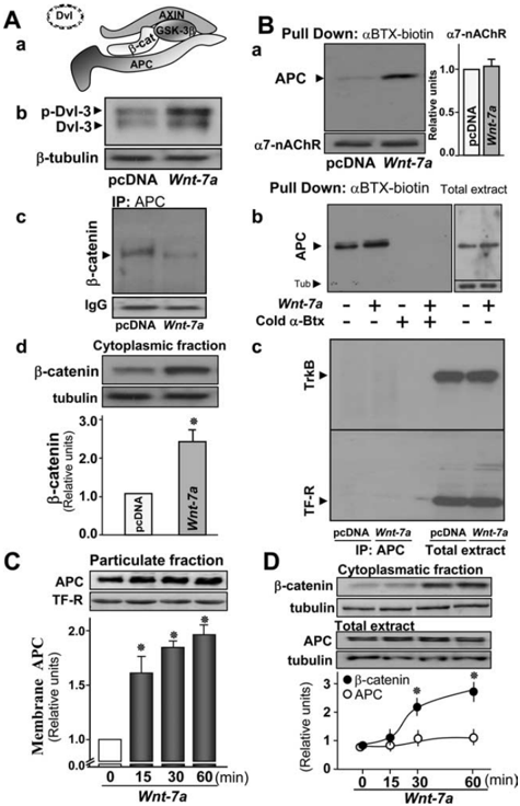
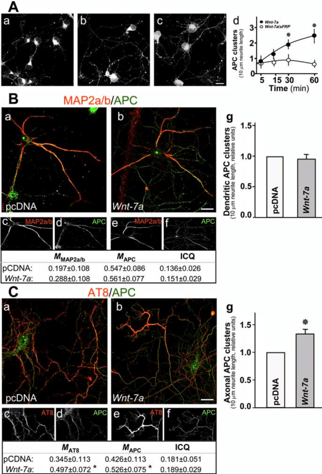
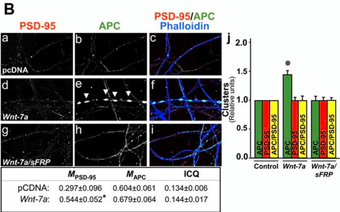
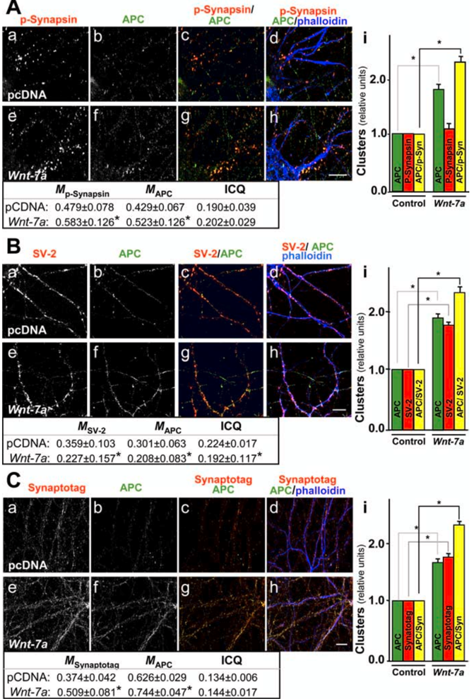
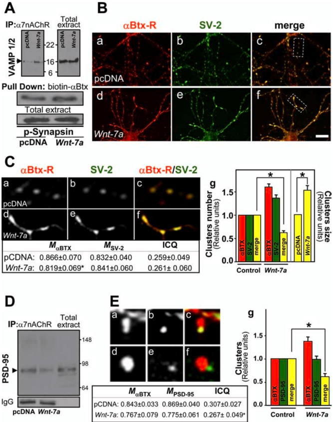
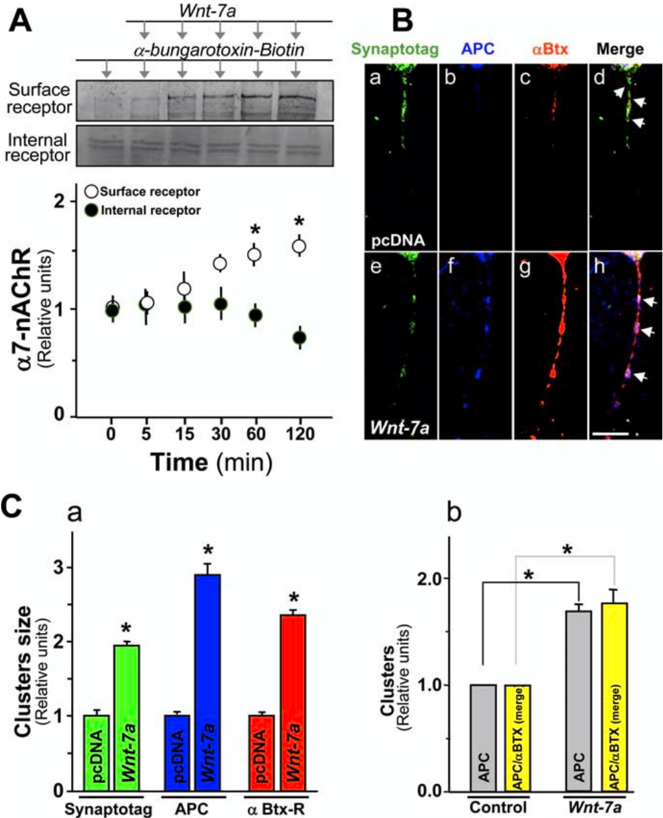
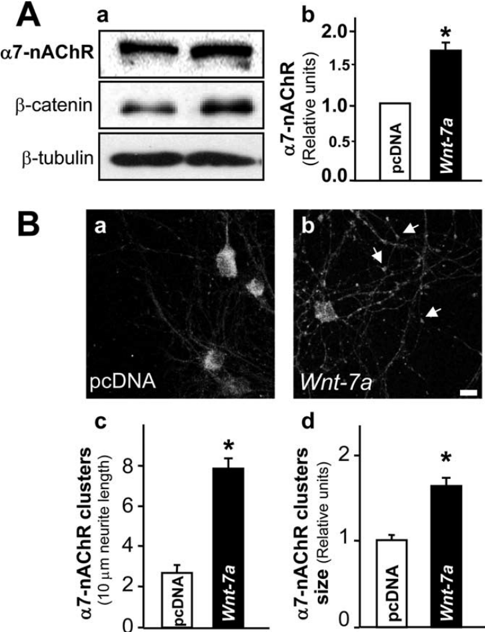
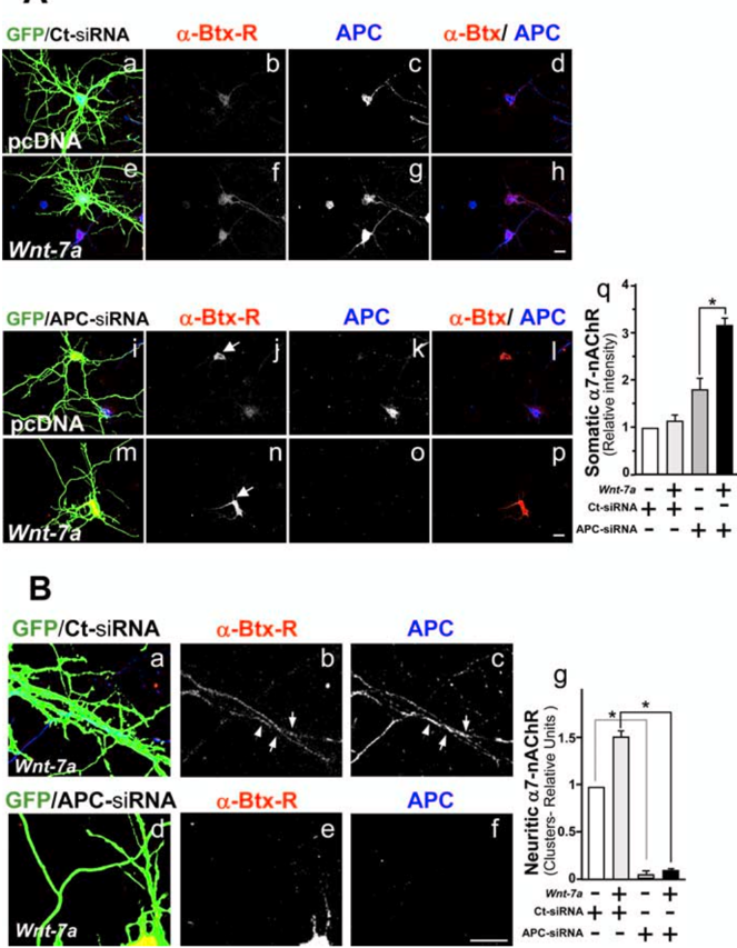
Nicotinic acetylcholine receptors (nAChRs) contribute significantly to hippocampal function. α7-nAChRs are present in presynaptic sites in hippocampal neurons and may influence transmitter release, but the factors that determine their presynaptic localization are unknown. We report here that Wnt-7a, a ligand active in the canonical Wnt signaling pathway, induces dissociation of the adenomatous polyposis coli (APC) protein from the β-catenin cytoplasmic complex and the interaction of APC with α7-nAChRs in hippocampal neurons. Interestingly,Wnt-7a induces the relocalization of APC to membranes, clustering of APC in neurites, and coclustering of APC with different, presynaptic protein markers.Wnt-7a also increases the number and size of coclusters of α7-nAChRs and APC in presynaptic terminals. These short-term changes in α7-nAChRs occur in the few minutes after ligand exposure and involve translocation to the plasma membrane without affecting total receptor levels. Longer-term exposure to Wnt-7a increases nAChR α7 subunit levels in an APC-independent manner and increases clusters of α7-nAChRs in neurites via an APC-dependent process. Together, these results demonstrate that stimulation through the canonical Wnt pathway regulates the presynaptic localization of APC and α7-nAChRs with APC serving as an intermediary in the α7-nAChR relocalization process. Modulation by Wnt signaling may be essential for α7-nAChR expression and function in synapses.
Overview
The claims and statistics of this paper haven’t been checked yet.
Run the check — free, no account needed. It reads the open-access text and checks the sources it cites.
Integrity signals
Registry
Cites
Statistics
Figures
Verification
Claims the paper makes
The claims and statistics of this paper haven’t been checked yet.
Run the check — free, no account needed. It reads the open-access text and checks the sources it cites.
Claims other papers make about this one
Each is checked against this paper’s own text: does it say what the citing paper says it does?
In the citing paper
“nonetheless, Wnt2 and other proteins traditionally associated with neural development may have important roles in post-critical period regulation of neuronal function (Farias et al., 2007; Moon et al., 2004; Wayman et al., 2006).”
From Unifying Views of Autism Spectrum Disorders: A Consideration of Autoregulatory Feedback Loops (2016) · cites this paper as Farias et al., 2007
Self-check
Percentages, sums and reported differences re-computed from the paper’s own numbers.
The self-check needs the paper’s full text.
Citations
Cites
Alzheimer's Disease: Genes, Proteins, and Therapy
Selkoe, D. J. (2001) · Physiological Reviews · doi:10.1152/physrev.2001.81.2.741 · Cited by 6,063
Alzheimer's Disease: A Disorder of Cortical Cholinergic Innervation
Coyle, J. T., Price, D. L., & DeLong, M. R. (1983) · Science · doi:10.1126/science.6338589 · Cited by 3,052
β‐catenin is a target for the ubiquitin–proteasome pathway
Aberle, H., Bauer, A., Stappert, J., Kispert, A., & Kemler, R. (1997) · The EMBO Journal · doi:10.1093/emboj/16.13.3797 · Cited by 2,586
Measurement of co‐localization of objects in dual‐colour confocal images
Manders, E. M. M., Verbeek, F. J., & Aten, J. A. (1993) · Journal of Microscopy · doi:10.1111/j.1365-2818.1993.tb03313.x · Cited by 2,132
WNT and β-catenin signalling: diseases and therapies
Moon, R. T., Kohn, A. D., Ferrari, G. V. D., & Kaykas, A. (2004) · Nature Reviews Genetics · doi:10.1038/nrg1427 · Cited by 1,763
Wnt signalling regulates adult hippocampal neurogenesis
Lie, D. C., Colamarino, S. A., Song, H., Désiré, L., Mira, H., Consiglio, A., Lein, E. S., Jessberger, S., Lansford, H., Dearie, A. R., et al. (2005) · Nature · doi:10.1038/nature04108 · Cited by 1,544
Lustig, B., Jerchow, B., Sachs, M., Weiler, S., Pietsch, T., Karsten, U., van de Wetering, M., Clevers, H., Schlag, P., Birchmeier, W., et al. (2002) · Molecular and Cellular Biology · doi:10.1128/mcb.22.4.1184-1193.2002 · Cited by 1,198
The Promise and Perils of Wnt Signaling Through β-Catenin
Moon, R. T., Bowerman, B. A., Boutros, M., & Perrimon, N. (2002) · Science · doi:10.1126/science.1071549 · Cited by 970
Synaptic Targeting by Alzheimer's-Related Amyloid β Oligomers
Lacor, P. N., Buniel, M. C., Chang, L., Fernandez, S. J., Gong, Y., Viola, K. L., Lambert, M. P., Velasco, P. T., Bigio, E. H., Finch, C. E., et al. (2004) · The Journal of Neuroscience · doi:10.1523/jneurosci.3432-04.2004 · Cited by 954
Hippocampal synaptic transmission enhanced by low concentrations of nicotine
Gray, R. G. F., Rajan, A. S., Radcliffe, K. A., Yakehiro, M., & Dani, J. A. (1996) · Nature · doi:10.1038/383713a0 · Cited by 947
Nusse, R., & VARMUS, H. E. (1992) · Cell · doi:10.1016/0092-8674(92)90630-u · Cited by 814
Nicotinic Receptors in the Development and Modulation of CNS Synapses
Role, L. W., & Berg, D. K. (1996) · Neuron · doi:10.1016/s0896-6273(00)80134-8 · Cited by 762
Li, Q., Lau, A., Morris, T. J., Guo, L. E., Fordyce, C. B., & Stanley, E. F. (2004) · Journal of Neuroscience · doi:10.1523/jneurosci.0346-04.2004 · Cited by 708
Axonal Remodeling and Synaptic Differentiation in the Cerebellum Is Regulated by WNT-7a Signaling
Hall, A. C., Lucas, F. R., & Salinas, P. C. (2000) · Cell · doi:10.1016/s0092-8674(00)80689-3 · Cited by 699
Nicotinic acetylcholine involvement in cognitive function in animals
Levin, E. D., & Simon, B. B. (1998) · Psychopharmacology · doi:10.1007/s002130050667 · Cited by 675
Zhou, F., Zhou, J., Dedhar, S., Wu, Y.-H., & Snider, W. D. (2004) · Neuron · doi:10.1016/j.neuron.2004.05.011 · Cited by 540
Destabilization of β-catenin by mutations in presenilin-1 potentiates neuronal apoptosis
Zhang, Z., Hartmann, H., Do, V. M., Abramowski, D., Sturchler, C., Staufenbiel, M., Sommer, B., van de Wetering, M., Clevers, H. C., Säftig, P., et al. (1998) · Nature · doi:10.1038/27208 · Cited by 515
Wnt signaling through Dishevelled, Rac and JNK regulates dendritic development
Rosso, S. B., Sussman, D., Wynshaw-Boris, A., & Salinas, P. C. (2005) · Nature Neuroscience · doi:10.1038/nn1374 · Cited by 485 · cited 2×
“, 2006) were prepared as described previously (Hall et al., 2000; Rosso et al., 2004; Quintanilla et al., 2005; Seto and Bellen, 2006).”
MethodCalcium modulation and high calcium permeability of neuronal nicotinic acetylcholine receptors
Vernino, S. A., Amador, M., Luetje, C. W., Patrick, J., & Dani, J. A. (1992) · Neuron · doi:10.1016/0896-6273(92)90114-s · Cited by 463
Binding of APC to the Human Homolog of the Drosophila Discs Large Tumor Suppressor Protein
Matsumine, A., Ogai, A., Senda, T., Okumura, N., Satoh, K., Baeg, G., KAWAHARA, T., Kobayashi, S., Okada, M., Toyoshima, K., et al. (1996) · Science · doi:10.1126/science.272.5264.1020 · Cited by 421
Ultrastructural Distribution of the α7 Nicotinic Acetylcholine Receptor Subunit in Rat Hippocampus
Fabian‐Fine, R., Skehel, P., Errington, M. L., Davies, H. A., Sher, E., Stewart, M. G., & Fine, A. (2001) · Journal of Neuroscience · doi:10.1523/jneurosci.21-20-07993.2001 · Cited by 364
Molecular organization of the postsynaptic specialization
Sheng, M. (2001) · Proceedings of the National Academy of Sciences · doi:10.1073/pnas.111146298 · Cited by 343
Ferrari, G. V. D., Chacón, M. A., Barría, M. I., Garrido, J., Godoy, J. A., Olivares, G. H., Reyes, A. E., Alvarez, A. R., Bronfman, M., & Inestrosa, N. C. (2003) · Molecular Psychiatry · doi:10.1038/sj.mp.4001208 · Cited by 333
Self-citationMAP2 is localized to the dendrites of hippocampal neurons which develop in culture
Cáceres, A., Banker, G. A., Steward, O., Binder, L. I., & Payne, M. R. (1984) · Developmental Brain Research · doi:10.1016/0165-3806(84)90167-6 · Cited by 320
Roles of Wnt proteins in neural development and maintenance
Patapoutian, A., & Reichardt, L. F. (2000) · Current Opinion in Neurobiology · doi:10.1016/s0959-4388(00)00100-8 · Cited by 319
Wnt signaling function in Alzheimer’s disease
Ferrari, G. V. D., & Inestrosa, N. C. (2000) · Brain Research Reviews · doi:10.1016/s0165-0173(00)00021-7 · Cited by 289
Self-citationNicotinic Stimulation Produces Multiple Forms of Increased Glutamatergic Synaptic Transmission
Radcliffe, K. A., & Dani, J. A. (1998) · The Journal of Neuroscience · doi:10.1523/jneurosci.18-18-07075.1998 · Cited by 271
Alkondon, M., & Albuquerque, E. X. (2001) · Journal of Neurophysiology · doi:10.1152/jn.2001.86.6.3043 · Cited by 238
Ahmad‐Annuar, A., Ciani, L., Simeonidis, I., Herreros, J., Fredj, N. B., Rosso, S. B., Hall, A. C., Brickley, S. G., & Salinas, P. C. (2006) · The Journal of Cell Biology · doi:10.1083/jcb.200511054 · Cited by 233
Kem, W. R. (2000) · Behavioural Brain Research · doi:10.1016/s0166-4328(00)00211-4 · Cited by 233
Wnt-3a overcomes β-amyloid toxicity in rat hippocampal neurons
Alvarez, A. R., Godoy, J. A., Müllendorff, K., Olivares, G. H., Bronfman, M., & Inestrosa, N. C. (2004) · Experimental Cell Research · doi:10.1016/j.yexcr.2004.02.028 · Cited by 212
Self-citationNicotinic α7 receptors: Synaptic options and downstream signaling in neurons
Berg, D. K., & Conroy, W. G. (2002) · Journal of Neurobiology · doi:10.1002/neu.10116 · Cited by 198
González‐Sancho, J. M., Brennan, K. R., Castelo‐Soccio, L. A., & Brown, A. M. C. (2004) · Molecular and Cellular Biology · doi:10.1128/mcb.24.11.4757-4768.2004 · Cited by 184
Protein kinase C inhibits amyloid β‐peptide neurotoxicity by acting on members of the Wnt pathway
Garrido, J., Godoy, J. A., Alvarez, A. R., Bronfman, M., & Inestrosa, N. C. (2002) · The FASEB Journal · doi:10.1096/fj.02-0327fje · Cited by 164
Self-citationInternalization is required for proper Wingless signaling in Drosophila melanogaster
Seto, E., & Bellen, H. J. (2006) · The Journal of Cell Biology · doi:10.1083/jcb.200510123 · Cited by 157
The genetics of sensory gating deficits in schizophrenia
Freedman, R., Olincy, A., Ross, R. G., Waldo, M. C., Stevens, K. E., Adler, L. E., & Leonard, S. (2003) · Current Psychiatry Reports · doi:10.1007/s11920-003-0032-2 · Cited by 153
Casein Kinase Iε Modulates the Signaling Specificities of Dishevelled
Cong, F., Schweizer, L., & VARMUS, H. E. (2004) · Molecular and Cellular Biology · doi:10.1128/mcb.24.5.2000-2011.2004 · Cited by 143
Kawai, H., Zago, W., & Berg, D. K. (2002) · The Journal of Neuroscience · doi:10.1523/jneurosci.22-18-07903.2002 · Cited by 140
Signal transduction during amyloid-β-peptide neurotoxicity: role in Alzheimer disease
Fuentealba, R. A., Farı́as, G. G., Scheu, J., Bronfman, M., Marzolo, M., & Inestrosa, N. C. (2004) · Brain Research Reviews · doi:10.1016/j.brainresrev.2004.07.018 · Cited by 126
Self-citationSchulte, G., Bryja, V., Rawal, N., Castelo‐Branco, G., Sousa, K. M., & Arenas, E. (2005) · Journal of Neurochemistry · doi:10.1111/j.1471-4159.2004.03022.x · Cited by 125
Caricasole, A., Copani, A., Caruso, A., Caraci, F., Iacovelli, L., Sortino, M. A., Terstappen, G. C., & Nicoletti, F. (2003) · Trends in Pharmacological Sciences · doi:10.1016/s0165-6147(03)00100-7 · Cited by 124
Pharmacotherapy for Alzheimer's disease: progress and prospects
Palmer, A. M. (2002) · Trends in Pharmacological Sciences · doi:10.1016/s0165-6147(02)02056-4 · Cited by 118
Quintanilla, R. A., Muñoz, F. J., Metcalfe, M., Hitschfeld, M., Olivares, G. H., Godoy, J. A., & Inestrosa, N. C. (2005) · Journal of Biological Chemistry · doi:10.1074/jbc.m411936200 · Cited by 114
Self-citationRegulation of acetylcholine receptor clustering by the tumor suppressor APC
Wang, J., Zheng, J., Zhang, L., Zhou, G., Braun, J., Yao, Y., & Wang, Z.-Z. (2003) · Nature Neuroscience · doi:10.1038/nn1128 · Cited by 110
Conroy, W. G., & Berg, D. K. (1998) · Molecular Pharmacology · doi:10.1124/mol.53.3.392 · Cited by 110
Neurite outgrowth involves adenomatous polyposis coli protein and β-catenin
Votin, V., Nelson, W. J., & Barth, A. I. M. (2005) · Journal of Cell Science · doi:10.1242/jcs.02679 · Cited by 101
Banerjee, C., Nyengaard, J., Wevers, A., de Vos, R. A., Steur, E. N. J. J., Lindstrom, J., Pilz, K., Nowacki, S., Bloch, W., & Schröder, H. (2000) · Neurobiology of Disease · doi:10.1006/nbdi.2000.0317 · Cited by 100
Herber, D. L., Severance, E. G., Cuevas, J., Morgan, D., & Gordon, M. N. (2004) · Journal of Histochemistry & Cytochemistry · doi:10.1177/002215540405201013 · Cited by 93
Borroni, V., Borroni, V., Baier, C. J., Lang, T., Bonini, I. C., White, M. M., Garbus, I., & Barrantes, F. J. (2006) · Molecular Membrane Biology · doi:10.1080/09687860600903387 · Cited by 92
Self-citationZhao, L., Kuo, Y.-P., George, A. A., Peng, J., Purandare, M. S., Schroeder, K. M., Lukas, R. J., & Wu, J. (2003) · Journal of Pharmacology and Experimental Therapeutics · doi:10.1124/jpet.103.048777 · Cited by 84
Self-citationTrans-Golgi network delivery of synaptic proteins in synaptogenesis
Sytnyk, V., Leshchyns’ka, I., Dityatev, A. E., & Schachner, M. (2003) · Journal of Cell Science · doi:10.1242/jcs.00956 · Cited by 82
The adenomatous polyposis coli protein: in the limelight out at the edge
Dikovskaya, D., Zumbrunn, J., Penman, G. A., & Näthke, I. S. (2001) · Trends in Cell Biology · doi:10.1016/s0962-8924(01)02069-4 · Cited by 80
Neuronal Nicotinic Synapse Assembly Requires the Adenomatous Polyposis Coli Tumor Suppressor Protein
Temburni, M. K., Rosenberg, M. M., Pathak, N. H., McConnell, R. E., & Jacob, M. H. (2004) · Journal of Neuroscience · doi:10.1523/jneurosci.1826-04.2004 · Cited by 66
Farías, G. G., Godoy, J. A., Hernández, F., Avila, J., Fisher, A., & Inestrosa, N. C. (2004) · Neurobiology of Disease · doi:10.1016/j.nbd.2004.07.016 · Cited by 59
Self-citationNeuronal localization of the adenomatous polyposis coli tumor suppressor protein
Brakeman, J. S. F., Gu, S. H., Wang, X. B., Dolin, G., & Baraban, J. M. (1999) · Neuroscience · doi:10.1016/s0306-4522(98)00605-8 · Cited by 56
Penman, G. A., Leung, L., & Näthke, I. S. (2005) · Journal of Cell Science · doi:10.1242/jcs.02589 · Cited by 40
Zago, W. M., Massey, K. A., & Berg, D. K. (2006) · Molecular and Cellular Neuroscience · doi:10.1016/j.mcn.2005.11.009 · Cited by 24
Khorchid, A., Larocca, J. N., & Almazán, G. (1999) · Journal of Neuroscience Research · doi:10.1002/(sici)1097-4547(19991215)58:6 · Cited by 20
Cited by
Sorted by citations
Emerging roles of Wnts in the adult nervous system
Inestrosa, N. C., & Arenas, E. (2009) · Nature reviews. Neuroscience · doi:10.1038/nrn2755 · Cited by 622
Self-citationWnt/Ca2+ signaling pathway: a brief overview
De, A. (2011) · Acta Biochimica et Biophysica Sinica · doi:10.1093/abbs/gmr079 · Cited by 565
Wnt signaling in the nervous system and in Alzheimer's disease
Inestrosa, N. C., & Varela-Nallar, L. (2014) · Journal of Molecular Cell Biology · doi:10.1093/jmcb/mjt051 · Cited by 324
Self-citationWnt signaling in neural circuit assembly
Salinas, P. C., & Zou, Y. (2008) · Annual Review of Neuroscience · doi:10.1146/annurev.neuro.31.060407.125649 · Cited by 274
WNT signaling in neuronal maturation and synaptogenesis
Rosso, S. B., & Inestrosa, N. C. (2013) · Frontiers in Cellular Neuroscience · doi:10.3389/fncel.2013.00103 · Cited by 272
Self-citation
Metadata
- Authors
- Farías, G. G., Vallés, A. S., Colombres, M., Godoy, J. A., Toledo, E. M., Lukas, R. J., Barrantes, F. J., & Inestrosa, N. C.
- Affiliations
- Pontificia Universidad Católica de Chile (CL)
- Barrow Neurological Institute (US)
- Instituto de Investigaciones Bioquímicas de Bahía Blanca, CONICET-UNS (AR)
- Venue
- The Journal of Neuroscience
- Publisher
- Society for Neuroscience
- Published
- 2007-05-16
- Volume
- 27
- Issue
- 20
- Pages
- 5313-5325
- ISSN
- 0270-6474 (ISSN-L)
- 1529-2401
- PMC
- PMC6672358
- OpenAlex
- W2081376704
- Cited by
- 108
- References
- 58
- Citations by year
- 2026: 1 · 2025: 1 · 2024: 2 · 2023: 1 · 2022: 4 · 2021: 7 · 2020: 6 · 2019: 4 · 2018: 4 · 2017: 3 · 2016: 4 · 2015: 5 · 2014: 8 · 2013: 17 · 2012: 8
- FWCI
- 3.31
- Type
- journal article
- Language
- en
- Open access
- hybrid
- doi.org
- Licence
- Locations
- Journal of Neuroscience (publishedVersion, cc-by-nc-sa)
- PubMed Central (publishedVersion)
- PubMed (publishedVersion)
- LA Referencia (Red Federada de Repositorios Institucionales de Publicaciones Científicas) (submittedVersion, cc-by-nc-sa)
- Americanae (AECID Library) (submittedVersion, cc-by-nc-sa)
- Funders
- Barrow Neurological Foundation
- Consejo Nacional de Investigaciones Científicas y Técnicas
- Universidad Nacional del Sur
- Arizona Biomedical Research Commission
- National Institutes of Health (DA015389)
- Comisión Nacional de Investigación Científica y Tecnológica
- National Institute on Drug Abuse (R01DA015389, DA015389)
- National Institute of Neurological Disorders and Stroke (NS040417, R01NS040417)
- Topics
- Life Sciences › Biochemistry, Genetics and Molecular Biology › Molecular Biology › Nicotinic Acetylcholine Receptors Study
- Life Sciences › Neuroscience › Cellular and Molecular Neuroscience › Neuroscience and Neuropharmacology Research
- Life Sciences › Biochemistry, Genetics and Molecular Biology › Molecular Biology › Wnt/β-catenin signaling in development and cancer
- Keywords
- Wnt7a, alpha7 nicotinic acetylcholine receptor, adenomatous polyposis coli, presynaptic localization, hippocampal neurons, Wnt/β-catenin signaling, β-catenin, synaptic transmission, plasma membrane translocation
- MeSH terms
- Animals, Cell Line, Cells, Cultured, Hippocampus, Humans, Mice, Inbred C57BL, Neurons, Proto-Oncogene Proteins, Receptors, Nicotinic, Signal Transduction, Rats, Sprague-Dawley, Presynaptic Terminals, Adenomatous Polyposis Coli Protein, Wnt Proteins, beta Catenin, Mice, Rats, alpha7 Nicotinic Acetylcholine Receptor
About these checks
Registry, retraction, Cites and statistics results are computed, not judged, with one exception: a printed reference that no index lists is read once by a model before it counts as a citation error. Claims are extracted by a model and kept only when their quote appears verbatim in the text; each cited source is then checked, with a model’s help, for whether it says what the claim relies on. Those verdicts can be wrong — read the claim’s quote and the cited source before relying on one.
Statistics and arithmetic are recomputed, not judged.
Confidence bands are shown only for fields calibrated on our annotated test set. None is calibrated yet, so no band is shown.
What counts as a claim ›How we measure confidence ›How we check this ›
Are you an author, or do you see a problem with this page? Request a review
