

The DOI matches its Crossref record; 46 of the 46 references it cites were found in a registry or an index.
Parvalbumin Is a Mobile Presynaptic Ca2+Buffer in the Calyx of Held that Accelerates the Decay of Ca2+and Short-Term Facilitation
Open accessAbstract
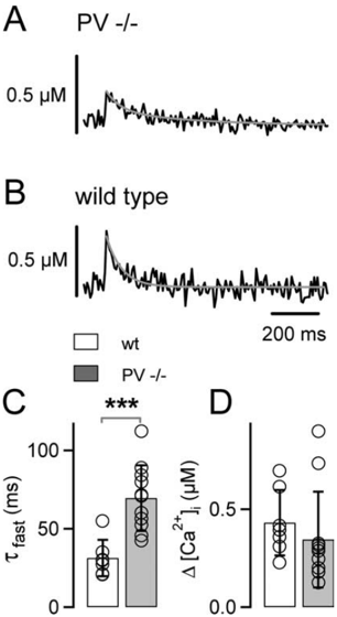
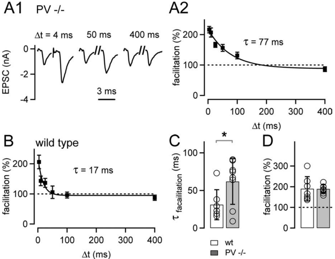
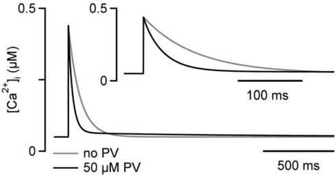
Presynaptic Ca 2+signaling plays a crucial role in short-term plasticity of synaptic transmission. Here, we studied the role of mobile endogenous presynaptic Ca 2+buffer(s) in modulating paired-pulse facilitation at a large excitatory nerve terminal in the auditory brainstem, the calyx of Held. To do so, we assessed the effect of presynaptic whole-cell recording, which should lead to the diffusional loss of endogenous mobile Ca 2+buffers, on paired-pulse facilitation and on intracellular Ca 2+concentration ([Ca 2+]i) transients evoked by action potentials. In unperturbed calyces briefly preloaded with the Ca 2+indicator fura-6F, the [Ca 2+]i transient decayed surprisingly fast (τ fast, ∼30 ms). Presynaptic whole-cell recordings made without additional Ca 2+buffers slowed the decay kinetics of [Ca 2+]i and paired-pulse facilitation (twofold to threefold), but the amplitude of the [Ca 2+]i transient was changed only marginally. The fast [Ca 2+]i decay was restored by adding the slow Ca 2+buffer EGTA (50–100 μ m) or parvalbumin (100 μ m), a Ca 2+-binding protein with slow Ca 2+-binding kinetics, to the presynaptic pipette solution. In contrast, the fast Ca 2+buffer fura-2 strongly reduced the amplitude of the [Ca 2+]i transient and slowed its decay, suggesting that the mobile endogenous buffer in calyces of Held has slow, rather than fast, binding kinetics. In parvalbumin knock-out mice, the decay of [Ca 2+]i and facilitation was slowed approximately twofold compared with wild-type mice, similar to what is observed during whole-cell recordings in rat calyces of Held. Thus, in young calyces of Held, a mobile Ca 2+buffer with slow binding kinetics, primarily represented by parvalbumin, accelerates the decay of spatially averaged [Ca 2+]i and paired-pulse facilitation.
Overview
The claims and statistics of this paper haven’t been checked yet.
Run the check — free, no account needed. It reads the open-access text and checks the sources it cites.
Integrity signals
Registry
Cites
Statistics
Figures
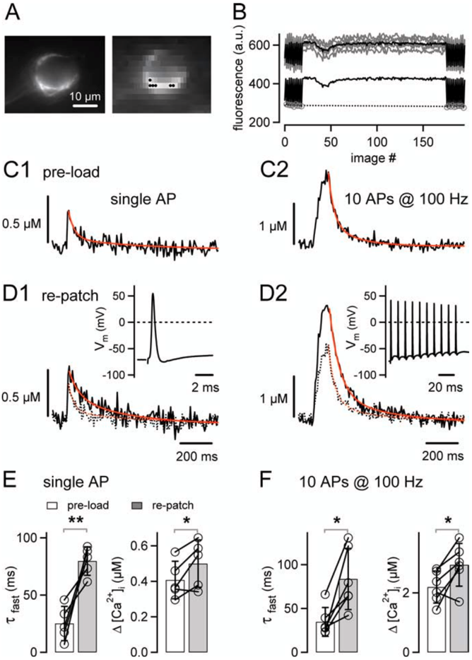
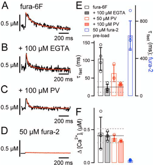
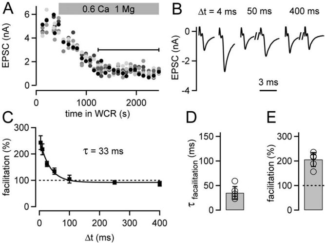
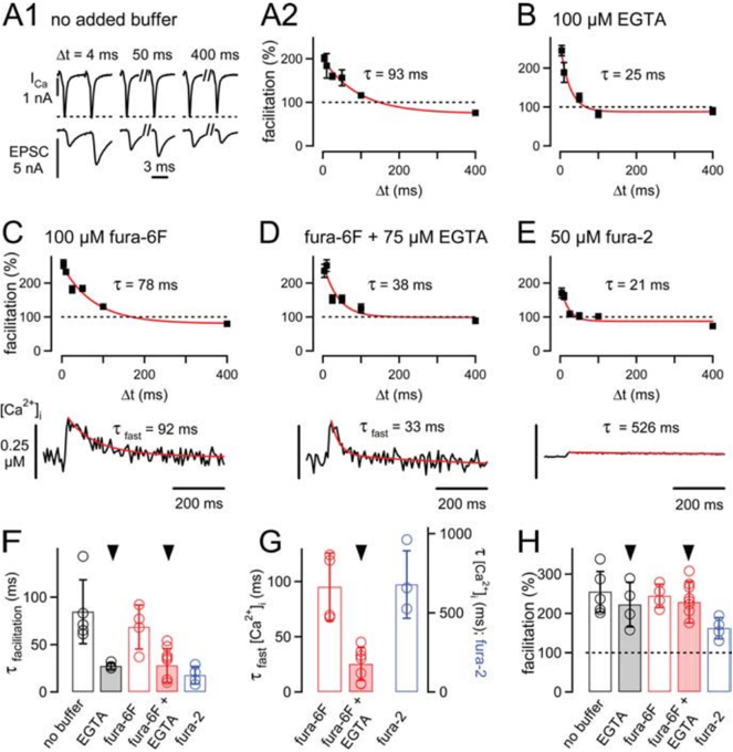
Verification
Claims the paper makes
The claims and statistics of this paper haven’t been checked yet.
Run the check — free, no account needed. It reads the open-access text and checks the sources it cites.
Claims other papers make about this one
Each is checked against this paper’s own text: does it say what the citing paper says it does?
In the citing paper
“The absence of PV in PV-/- Pvalb neurons does not affect basal synaptic transmission, but enhances facilitation [49, 50] and shortens delayed transmitter release [51].”
Self-check
Percentages, sums and reported differences re-computed from the paper’s own numbers.
The self-check needs the paper’s full text.
Citations
Cites
A new generation of Ca2+ indicators with greatly improved fluorescence properties.
Grynkiewicz, G., Poenie, M., & Tsien, R. Y. (1985) · Journal of Biological Chemistry · doi:10.1016/s0021-9258(19)83641-4 · Cited by 21,562
Polazzi, E., Mengoni, I., Caprini, M., Peña‐Altamira, E., Kurtys, E., & Monti, B. (2012) · Neurosignals · doi:10.1159/000337115 · Cited by 9,023
Short-Term Synaptic Plasticity
Zucker, R. S., & Regehr, W. G. (2002) · Annual Review of Physiology · doi:10.1146/annurev.physiol.64.092501.114547 · Cited by 5,068
Calbindin D-28k and parvalbumin in the rat nervous system
Celio, M. R. (1990) · Neuroscience · doi:10.1016/0306-4522(90)90091-h · Cited by 2,076
Kv3 channels: voltage-gated K+ channels designed for high-frequency repetitive firing
Rudy, B., & McBain, C. J. (2001) · Trends in Neurosciences · doi:10.1016/s0166-2236(00)01892-0 · Cited by 781
Calcium gradients and buffers in bovine chromaffin cells.
Neher, E., & Augustine, G. J. (1992) · The Journal of Physiology · doi:10.1113/jphysiol.1992.sp019127 · Cited by 753
Calcium influx and transmitter release in a fast CNS synapse
Borst, J. G. G., & Sakmann, B. J. (1996) · Nature · doi:10.1038/383431a0 · Cited by 632
Schneggenburger, R., Meyer, A. C., & Neher, E. (1999) · Neuron · doi:10.1016/s0896-6273(00)80789-8 · Cited by 625
Self-citationDeterminants of the Time Course of Facilitation at the Granule Cell to Purkinje Cell Synapse
Atluri, P. P., & Regehr, W. G. (1996) · Journal of Neuroscience · doi:10.1523/jneurosci.16-18-05661.1996 · Cited by 513
Pre‐ and postsynaptic whole‐cell recordings in the medial nucleus of the trapezoid body of the rat.
Borst, J. G. G., Helmchen, F., & Sakmann, B. J. (1995) · The Journal of Physiology · doi:10.1113/jphysiol.1995.sp021095 · Cited by 427
Cited by
Sorted by citations
Multiple Roles of Calcium Ions in the Regulation of Neurotransmitter Release
Neher, E., & Sakaba, T. (2008) · Neuron · doi:10.1016/j.neuron.2008.08.019 · Cited by 966
Calcium Channel Regulation and Presynaptic Plasticity
Catterall, W. A., & Few, A. P. (2008) · Neuron · doi:10.1016/j.neuron.2008.09.005 · Cited by 647
González‐Burgos, G., & Lewis, D. A. (2008) · Schizophrenia Bulletin · doi:10.1093/schbul/sbn070 · Cited by 592
Nanodomain coupling between Ca2+ channels and sensors of exocytosis at fast mammalian synapses
Eggermann, E., Bucurenciu, I., Goswami, S. P., & Jonas, P. (2012) · Nature Reviews Neuroscience · doi:10.1038/nrn3125 · Cited by 500
Short-Term Presynaptic Plasticity
Regehr, W. G. (2012) · Cold Spring Harbor Perspectives in Biology · doi:10.1101/cshperspect.a005702 · Cited by 482
Calcium Channels, Synaptic Plasticity, and Neuropsychiatric Disease
Nanou, E., & Catterall, W. A. (2018) · Neuron · doi:10.1016/j.neuron.2018.03.017 · Cited by 492
GABAergic Interneurons Shape the Functional Maturation of the Cortex
Le Magueresse, C., & Monyer, H. (2013) · Neuron · doi:10.1016/j.neuron.2013.01.011 · Cited by 425
Cytosolic Ca2+ BuffersPreprint
Schwaller, B. (2010) · Cold Spring Harbor Perspectives in Biology · doi:10.1101/cshperspect.a004051 · Cited by 387
Self-citationNeuroplasticity of Neocortical Circuits in Schizophrenia
Lewis, D. A., & González‐Burgos, G. (2007) · Neuropsychopharmacology · doi:10.1038/sj.npp.1301563 · Cited by 372
Bucurenciu, I., Kulik, A., Schwaller, B., Frotscher, M., & Jonas, P. (2008) · Neuron · doi:10.1016/j.neuron.2007.12.026 · Cited by 274
Self-citationThe continuing disappearance of “pure” Ca2+ buffers
Schwaller, B. (2008) · Cellular and Molecular Life Sciences · doi:10.1007/s00018-008-8564-6 · Cited by 257
Self-citationGABA Neuron Alterations, Cortical Circuit Dysfunction and Cognitive Deficits in Schizophrenia
González‐Burgos, G., Fish, K. N., & Lewis, D. A. (2011) · Neural Plasticity · doi:10.1155/2011/723184 · Cited by 250
Filice, F., Vörckel, K. J., Sungur, A. Ö., Wöhr, M., & Schwaller, B. (2016) · Molecular Brain · doi:10.1186/s13041-016-0192-8 · Cited by 240
Self-citationOxidative Stress‐Related Mechanisms in Schizophrenia Pathogenesis and New Treatment Perspectives
Ermakov, E. A., Dmitrieva, E. M., Parshukova, D. A., Kazantseva, D. V., Vasilieva, A. R., & Smirnova, L. P. (2021) · Oxidative Medicine and Cellular Longevity · doi:10.1155/2021/8881770 · Cited by 256
The Calyx of Held Synapse: From Model Synapse to Auditory Relay
Borst, J. G. G., & van Hoeve, J. S. S. (2011) · Annual Review of Physiology · doi:10.1146/annurev-physiol-020911-153236 · Cited by 225
A General Model of Synaptic Transmission and Short-Term Plasticity
Pan, B., & Zucker, R. S. (2009) · Neuron · doi:10.1016/j.neuron.2009.03.025 · Cited by 195
Calcium Signaling in Dendritic SpinesPreprint
Higley, M. J., & Sabatini, B. L. (2012) · Cold Spring Harbor Perspectives in Biology · doi:10.1101/cshperspect.a005686 · Cited by 187
Calcium Channels and Short-term Synaptic Plasticity
Catterall, W. A., Leal, K., & Nanou, E. (2013) · Journal of Biological Chemistry · doi:10.1074/jbc.r112.411645 · Cited by 153
Physiology of intracellular calcium buffering
Eisner, D. A., Neher, E., Taschenberger, H., & Smith, G. L. (2023) · Physiological Reviews · doi:10.1152/physrev.00042.2022 · Cited by 162
Hoftman, G. D., & Lewis, D. A. (2011) · Schizophrenia Bulletin · doi:10.1093/schbul/sbr029 · Cited by 128
The Parvalbumin Hypothesis of Autism Spectrum Disorder
Filice, F., Janickova, L., Henzi, T., Bilella, A., & Schwaller, B. (2020) · Frontiers in Cellular Neuroscience · doi:10.3389/fncel.2020.577525 · Cited by 118
Self-citationBrenowitz, S. D., & Regehr, W. G. (2007) · Journal of Neuroscience · doi:10.1523/jneurosci.1064-07.2007 · Cited by 114
Calcium-Binding Proteins as Determinants of Central Nervous System Neuronal Vulnerability to Disease
Fairless, R., Williams, S. K., & Diem, R. (2019) · International Journal of Molecular Sciences · doi:10.3390/ijms20092146 · Cited by 110
Actin-dependent rapid recruitment of reluctant synaptic vesicles into a fast-releasing vesicle pool
Lee, J. S., Ho, W., & Lee, S. (2012) · Proceedings of the National Academy of Sciences · doi:10.1073/pnas.1114072109 · Cited by 102
Relative Roles of Different Mechanisms of Depression at the Mouse Endbulb of Held
Yang, H., & Xu‐Friedman, M. A. (2008) · Journal of Neurophysiology · doi:10.1152/jn.01293.2007 · Cited by 101
Sommeijer, J.-P., & Levelt, C. N. (2012) · PLoS ONE · doi:10.1371/journal.pone.0035323 · Cited by 100
Regulation of transmitter release by Ca2+ and synaptotagmin: insights from a large CNS synapse
Kochubey, O., Lou, X., & Schneggenburger, R. (2011) · Trends in Neurosciences · doi:10.1016/j.tins.2011.02.006 · Cited by 92
Self-citationCytosolic Ca 2+ Buffers Are Inherently Ca 2+ Signal ModulatorsPreprint
Schwaller, B. (2019) · Cold Spring Harbor Perspectives in Biology · doi:10.1101/cshperspect.a035543 · Cited by 94
Self-citationJiang, M., Zhu, J., Liu, Y., Yang, M., Tian, C., Jiang, S., Wang, Y., Guo, H., Wang, K., & Shu, Y. (2012) · PLoS Biology · doi:10.1371/journal.pbio.1001324 · Cited by 89
Orduz, D., Bischop, D. P., Schwaller, B., Schiffmann, S. N., & Gall, D. (2013) · The Journal of Physiology · doi:10.1113/jphysiol.2012.250795 · Cited by 88
Self-citationLin, K., Taschenberger, H., & Neher, E. (2022) · Proceedings of the National Academy of Sciences · doi:10.1073/pnas.2207987119 · Cited by 84
Efficient Ca2+ buffering in fast‐spiking basket cells of rat hippocampus
Aponte, Y., Bischofberger, J., & Jónás, P. (2008) · The Journal of Physiology · doi:10.1113/jphysiol.2007.147298 · Cited by 81
Bornschein, G., Arendt, O., Hallermann, S., Brachtendorf, S., Eilers, J., & Schmidt, H. (2013) · The Journal of Physiology · doi:10.1113/jphysiol.2013.254128 · Cited by 84
Lazarus, M. S., & Huang, Z. J. (2011) · Journal of Neurophysiology · doi:10.1152/jn.00729.2010 · Cited by 77
Schwaller, B. (2012) · Biochimica et Biophysica Acta (BBA) - General Subjects · doi:10.1016/j.bbagen.2011.11.008 · Cited by 75
Self-citationThe chandelier neuron in schizophrenia
Lewis, D. A. (2010) · Developmental Neurobiology · doi:10.1002/dneu.20825 · Cited by 76
Müller, M., Felmy, F., & Schneggenburger, R. (2008) · The Journal of Physiology · doi:10.1113/jphysiol.2008.155838 · Cited by 71
Self-citationChang, S., Reim, K., Pedersen, M., Neher, E., Brose, N., & Taschenberger, H. (2015) · Journal of Neuroscience · doi:10.1523/jneurosci.4841-14.2015 · Cited by 66
Müller, M., Goutman, J., Kochubey, O., & Schneggenburger, R. (2010) · Journal of Neuroscience · doi:10.1523/jneurosci.4378-09.2010 · Cited by 67
Self-citationAltered Cortical GABA Neurotransmission in Schizophrenia: Insights into Novel Therapeutic Strategies
Stan, A. D., & Lewis, D. A. (2012) · Current Pharmaceutical Biotechnology · doi:10.2174/138920112800784925 · Cited by 64
Developmental Changes in Short-Term Plasticity at the Rat Calyx of Held Synapse
Crins, T., Rusu, S., Rodríguez-Contreras, A., & Borst, J. G. (2011) · Journal of Neuroscience · doi:10.1523/jneurosci.1995-11.2011 · Cited by 59
Wang, L., Fedchyshyn, M. J., & Yang, Y. (2009) · Molecular Brain · doi:10.1186/1756-6606-2-36 · Cited by 60
Reduced endogenous Ca 2+ buffering speeds active zone Ca 2+ signaling
Delvendahl, I., Jablonski, L., Baade, C., Matveev, V., Neher, E., & Hallermann, S. (2015) · Proceedings of the National Academy of Sciences · doi:10.1073/pnas.1508419112 · Cited by 63
Mechanisms of short‐term plasticity at neuromuscular active zones of Drosophila
Hallermann, S., Heckmann, M., & Kittel, R. J. (2010) · HFSP journal · doi:10.2976/1.3338710 · Cited by 58
Keller, D. X., Babai, N. Z., Kochubey, O., Han, Y., Markram, H., Schürmann, F., & Schneggenburger, R. (2015) · PLoS Computational Biology · doi:10.1371/journal.pcbi.1004253 · Cited by 61
Self-citationFast resupply of synaptic vesicles requires synaptotagmin-3
Weingarten, D. J., Shrestha, A., Juda-Nelson, K., Kissiwaa, S. A., Spruston, E., & Jackman, S. L. (2022) · Nature · doi:10.1038/s41586-022-05337-1 · Cited by 55
Guilmi, M. N. D., Wang, T., Inchauspe, C. G., Forsythe, I. D., Ferrari, M. D., van den Maagdenberg, A. M. J. M., Borst, J. G. G., & Uchitel, O. D. (2014) · Journal of Neuroscience · doi:10.1523/jneurosci.2526-13.2014 · Cited by 53
Cortical Interneuron Subtypes Vary in Their Axonal Action Potential Properties
Casale, A. E., Foust, A. J., Bal, T., & McCormick, D. A. (2015) · Journal of Neuroscience · doi:10.1523/jneurosci.1467-13.2015 · Cited by 53
Lipstein, N., Chang, S., Lin, K., López-Murcia, F. J., Neher, E., Taschenberger, H., & Brose, N. (2021) · Neuron · doi:10.1016/j.neuron.2021.09.054 · Cited by 53
Neuroplasticity of excitatory and inhibitory cortical circuits in schizophrenia
Lewis, D. A. (2009) · Dialogues in Clinical Neuroscience · doi:10.31887/dcns.2009.11.3/dalewis · Cited by 56
Curley, A. A., Eggan, S. M., Lazarus, M. S., Huang, Z. J., Volk, D. W., & Lewis, D. A. (2013) · Neurobiology of Disease · doi:10.1016/j.nbd.2012.10.018 · Cited by 51
Mechanisms underlying short‐term modulation of transmitter release by presynaptic depolarization
Hori, T., & Takahashi, T. (2009) · The Journal of Physiology · doi:10.1113/jphysiol.2009.168765 · Cited by 51
Synaptic plasticity in the auditory system: a review
Friauf, E., Fischer, A. U., & Fuhr, M. F. (2015) · Cell and Tissue Research · doi:10.1007/s00441-015-2176-x · Cited by 49
The Cerebellar Mossy Fiber Synapse as a Model for High-Frequency Transmission in the Mammalian CNS
Delvendahl, I., & Hallermann, S. (2016) · Trends in Neurosciences · doi:10.1016/j.tins.2016.09.006 · Cited by 52
Kosaka, T., & Kosaka, K. (2007) · Neuroscience Research · doi:10.1016/j.neures.2007.09.008 · Cited by 48
Mochida, S. (2019) · International Journal of Molecular Sciences · doi:10.3390/ijms20092217 · Cited by 49
Kumar, P., Goettemoeller, A. M., Espinosa‐García, C., Tobin, B. R., Tfaily, A., Nelson, R. S., Natu, A., Dammer, E. B., Santiago, J. V., Malepati, S., et al. (2024) · Nature Communications · doi:10.1038/s41467-024-47028-7 · Cited by 53
A Single-Compartment Model of Calcium Dynamics in Nerve Terminals and DendritesPreprint
Helmchen, F., & Tank, D. W. (2015) · Cold Spring Harbor Protocols · doi:10.1101/pdb.top085910 · Cited by 47
Acute Complexin Knockout Abates Spontaneous and Evoked Transmitter Release
López-Murcia, F. J., Reim, K., Jahn, O., Taschenberger, H., & Brose, N. (2019) · Cell Reports · doi:10.1016/j.celrep.2019.02.030 · Cited by 45
The Calyx of Held in the auditory system: structure, function, and development
Baydyuk, M., Xu, J.-H., & Wu, L. (2016) · Hearing Research · doi:10.1016/j.heares.2016.03.009 · Cited by 33
Turovsky, E. A., Turovskaya, M. V., Kononov, A. V., & Зинченко, В. П. (2013) · Experimental Neurology · doi:10.1016/j.expneurol.2013.09.006 · Cited by 35
A theory of synaptic transmission
Wang, B., & Dudko, O. K. (2021) · eLife · doi:10.7554/elife.73585 · Cited by 38
Decreased parvalbumin mRNA expression in dorsolateral prefrontal cortex in Parkinson′s disease
Lanoue, A. C., Blatt, G. J., & Soghomonian, J. (2013) · Brain Research · doi:10.1016/j.brainres.2013.07.025 · Cited by 36
Woehler, A., Lin, K., & Neher, E. (2014) · The Journal of Physiology · doi:10.1113/jphysiol.2014.281097 · Cited by 31
Leão, R. M., & von Gersdorff, H. (2009) · Brazilian Journal of Medical and Biological Research · doi:10.1590/s0100-879x2009000100014 · Cited by 32
Zhang, X., Xu, L., Zhu, X., Tang, Z.-H., Dong, Y.-B., Yu, Z., Shang, Q. S., Wang, Z., & Shen, H. (2023) · Nature Communications · doi:10.1038/s41467-023-43930-8 · Cited by 29
Translating neuronal activity at the synapse: presynaptic calcium sensors in short-term plasticity
de Jong, A. P. H., & Fioravante, D. (2014) · Frontiers in Cellular Neuroscience · doi:10.3389/fncel.2014.00356 · Cited by 28
Nakamura, Y., Reva, M., & DiGregorio, D. A. (2018) · Journal of Neuroscience · doi:10.1523/jneurosci.2061-17.2018 · Cited by 30
Hennig, M. H., Postlethwaite, M., Forsythe, I. D., & Graham, B. P. (2008) · The Journal of Physiology · doi:10.1113/jphysiol.2008.152124 · Cited by 29
Shepard, R. M., Heslin, K. A., & Coutellier, L. D. (2016) · Neurobiology of Disease · doi:10.1016/j.nbd.2016.12.012 · Cited by 27
Short-term synaptic plasticity and intensity coding
MacLeod, K. M. (2011) · Hearing Research · doi:10.1016/j.heares.2011.03.001 · Cited by 29
Yao, L., & Sakaba, T. (2010) · Journal of Neurophysiology · doi:10.1152/jn.00685.2010 · Cited by 26
Makowiecki, K., Garrett, A. R., Harvey, A. R., & Rodger, J. (2018) · Scientific Reports · doi:10.1038/s41598-018-23979-y · Cited by 26
Neurotransmitter Release Site Replenishment and Presynaptic Plasticity
Mochida, S. (2020) · International Journal of Molecular Sciences · doi:10.3390/ijms22010327 · Cited by 26
Neher, E., & Taschenberger, H. (2020) · Neuroscience · doi:10.1016/j.neuroscience.2020.10.012 · Cited by 15
Ca 2+ regulation of glutamate release from inner hair cells of hearing mice
Tobón, L. M. J., & Moser, T. (2023) · Proceedings of the National Academy of Sciences · doi:10.1073/pnas.2311539120 · Cited by 25
Gilmanov, I. R., Samigullin, D. V., Vyskočil, F., Nikolsky, E. E., & Bukharaeva, E. A. (2008) · Journal of Computational Neuroscience · doi:10.1007/s10827-008-0079-5 · Cited by 24
Wichmann, C. (2015) · Hearing Research · doi:10.1016/j.heares.2015.07.008 · Cited by 21
Burgess, J. R., & Raven, E. L. (2009) · Advances in inorganic chemistry · doi:10.1016/s0898-8838(09)00205-0 · Cited by 21
Sehring, I. M., Klotz, C., Beisson, J., & Plattner, H. (2008) · Cell Calcium · doi:10.1016/j.ceca.2008.06.004 · Cited by 21
New insights into short-term synaptic facilitation at the frog neuromuscular junction
Ma, J., Kelly, L. S., Ingram, J., Price, T. J., Meriney, S. D., & Dittrich, M. (2014) · Journal of Neurophysiology · doi:10.1152/jn.00198.2014 · Cited by 20
Iremonger, K. J., Benediktsson, A. M., & Bains, J. S. (2010) · Frontiers in Neuroendocrinology · doi:10.1016/j.yfrne.2010.03.002 · Cited by 20
Babai, N. Z., Kochubey, O., Keller, D. X., & Schneggenburger, R. (2014) · Journal of Neuroscience · doi:10.1523/jneurosci.1990-14.2014 · Cited by 20
Self-citationLin, K., Erazo-Fischer, E., & Taschenberger, H. (2012) · Journal of Neuroscience · doi:10.1523/jneurosci.3838-11.2012 · Cited by 19
Janickova, L., & Schwaller, B. (2020) · Frontiers in Cellular Neuroscience · doi:10.3389/fncel.2020.571216 · Cited by 19
Self-citationLiao, C., & Lien, C. (2009) · Neuroscience · doi:10.1016/j.neuroscience.2009.09.052 · Cited by 19
Lin, K., Taschenberger, H., & Neher, E. (2016) · The Journal of Physiology · doi:10.1113/jp272773 · Cited by 18
Presynaptic GCaMP expression decreases vesicle release probability at the calyx of Held
Singh, M., Lujan, B., & Renden, R. (2018) · Synapse · doi:10.1002/syn.22040 · Cited by 17
Komaki, A., Shahidi, ., Sarihi, A., Hasanein, P., Lashgari, R., Haghparast, A., Salehi, I., & Kourosh‐Arami, M. (2013) · Europe PMC (PubMed Central) · Cited by 18
Szabolcsi, V., & Celio, M. R. (2014) · Glia · doi:10.1002/glia.22768 · Cited by 18
Schwaller, B. (2010) · Elsevier eBooks · doi:10.1016/b978-0-12-374145-5.00120-0 · Cited by 17
Self-citationЗинченко, В. П., Turovskaya, M. V., Teplov, I. Y., Berezhnov, A. V., & Turovsky, E. A. (2016) · BIOPHYSICS · doi:10.1134/s0006350916010280 · Cited by 17
Details of Ca2+ dynamics matter
Neher, E. (2008) · The Journal of Physiology · doi:10.1113/jphysiol.2008.153080 · Cited by 16
Samigullin, D. V., Fatikhov, N. F., Khaziev, E. F., Skorinkin, A. I., Nikolsky, E. E., & Bukharaeva, E. A. (2015) · Frontiers in Synaptic Neuroscience · doi:10.3389/fnsyn.2014.00029 · Cited by 15
Transients in global Ca 2+ concentration induced by electrical activity in a giant nerve terminal
Neher, E., & Taschenberger, H. (2013) · The Journal of Physiology · doi:10.1113/jphysiol.2012.248617 · Cited by 15
Zachařová, G., & Paleček, J. (2009) · Physiological Research · doi:10.33549/physiolres.931738 · Cited by 14
Diffusion of Ca2+ from Small Boutons en Passant into the Axon Shapes AP-Evoked Ca2+ Transients
Tran, V., & Stricker, C. (2018) · Biophysical Journal · doi:10.1016/j.bpj.2018.07.018 · Cited by 13
Loss of KCC2 in GABAergic Neurons Causes Seizures and an Imbalance of Cortical Interneurons
Zavalin, K. A., Hassan, A., Fu, C., Delpire, E. J., & Lagrange, A. H. (2022) · Frontiers in Molecular Neuroscience · doi:10.3389/fnmol.2022.826427 · Cited by 14
Gandhi, T., Liu, C., Adeyelu, T. T., Canepa, C. R., & Lee, C. C. (2023) · Frontiers in Behavioral Neuroscience · doi:10.3389/fnbeh.2023.1114789 · Cited by 13
López-Murcia, F. J., Lin, K., Berns, M. M. M., Ranjan, M., Lipstein, N., Neher, E., Brose, N., Reim, K., & Taschenberger, H. (2024) · Proceedings of the National Academy of Sciences of the United States of America · doi:10.1073/pnas.2320505121 · Cited by 13
Metadata
- Authors
- Müller, M., Felmy, F., Schwaller, B., & Schneggenburger, R.
- Affiliations
- École Polytechnique Fédérale de Lausanne (CH)
- Ludwig-Maximilians-Universität München (DE)
- University of Fribourg (CH)
- Venue
- The Journal of Neuroscience
- Publisher
- Society for Neuroscience
- Published
- 2007-02-28
- Volume
- 27
- Issue
- 9
- Pages
- 2261-2271
- ISSN
- 0270-6474 (ISSN-L)
- 1529-2401
- PMC
- PMC6673482
- OpenAlex
- W1975952480
- Cited by
- 155
- References
- 46
- Citations by year
- 2026: 4 · 2025: 4 · 2024: 3 · 2023: 7 · 2022: 4 · 2021: 7 · 2020: 4 · 2019: 5 · 2018: 7 · 2017: 1 · 2016: 8 · 2015: 9 · 2014: 11 · 2013: 10 · 2012: 8
- FWCI
- 5.97
- Type
- journal article
- Language
- en
- Open access
- hybrid
- doi.org
- Licence
- Locations
- Journal of Neuroscience (publishedVersion, cc-by-nc-sa)
- Europe PMC (PubMed Central) (publishedVersion)
- PubMed Central (publishedVersion)
- PubMed (publishedVersion)
- Zurich Open Repository and Archive (University of Zurich) (submittedVersion)
- Zurich Open Repository and Archive (University of Zurich)
- Funders
- National Science Foundation
- Deutsche Forschungsgemeinschaft (TP B1)
- Schweizerischer Nationalfonds zur Förderung der Wissenschaftlichen Forschung (100400, 3100A0)
- Topics
- Life Sciences › Neuroscience › Cellular and Molecular Neuroscience › Neuroscience and Neuropharmacology Research
- Life Sciences › Biochemistry, Genetics and Molecular Biology › Molecular Biology › Ion channel regulation and function
- Life Sciences › Neuroscience › Sensory Systems › Ion Channels and Receptors
- Keywords
- parvalbumin, calyx of Held, paired-pulse facilitation, short-term plasticity, calcium transients, fura-2, EGTA, auditory brainstem, excitatory synapses
- MeSH terms
- Animals, Brain Stem, Calcium, Calcium-Binding Proteins, Mice, Inbred C57BL, Synaptic Transmission, Neuronal Plasticity, Parvalbumins, Rats, Wistar, Receptors, Presynaptic, Mice, Knockout, Patch-Clamp Techniques, Mice, Rats, In Vitro Techniques
About these checks
Registry, retraction, Cites and statistics results are computed, not judged, with one exception: a printed reference that no index lists is read once by a model before it counts as a citation error. Claims are extracted by a model and kept only when their quote appears verbatim in the text; each cited source is then checked, with a model’s help, for whether it says what the claim relies on. Those verdicts can be wrong — read the claim’s quote and the cited source before relying on one.
Statistics and arithmetic are recomputed, not judged.
Confidence bands are shown only for fields calibrated on our annotated test set. None is calibrated yet, so no band is shown.
What counts as a claim ›How we measure confidence ›How we check this ›
Are you an author, or do you see a problem with this page? Request a review
