

The DOI matches its Crossref record; 66 of the 67 references it cites were found in a registry or an index.
Pharmacological reversal of synaptic and network pathology in human MECP2‐KO neurons and cortical organoids
Open accessAbstract
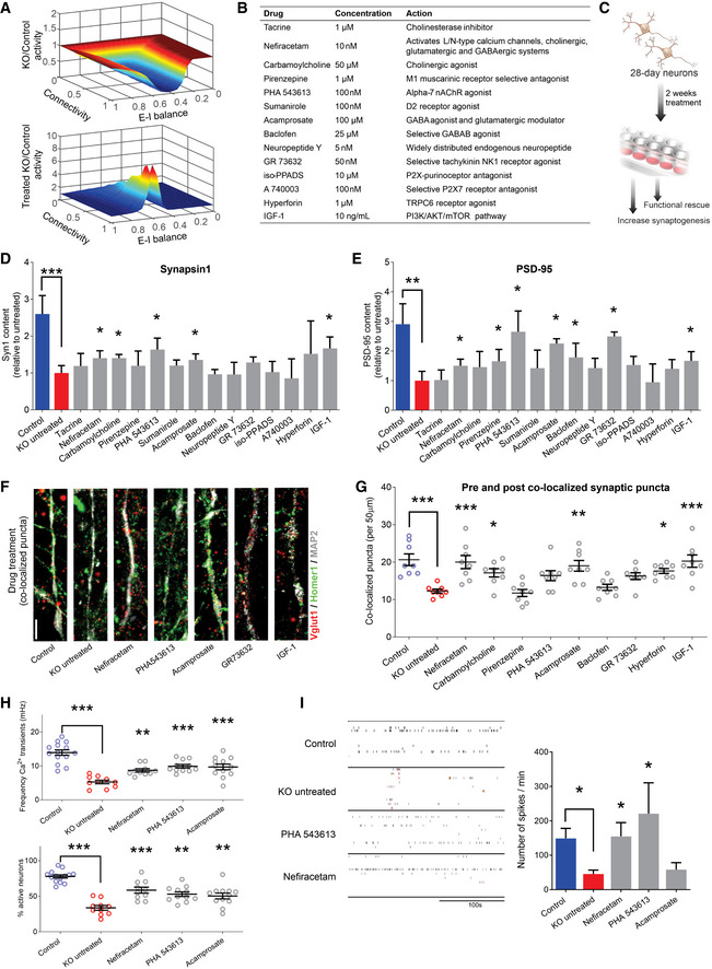
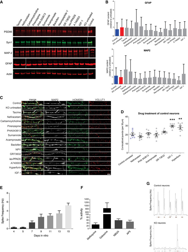
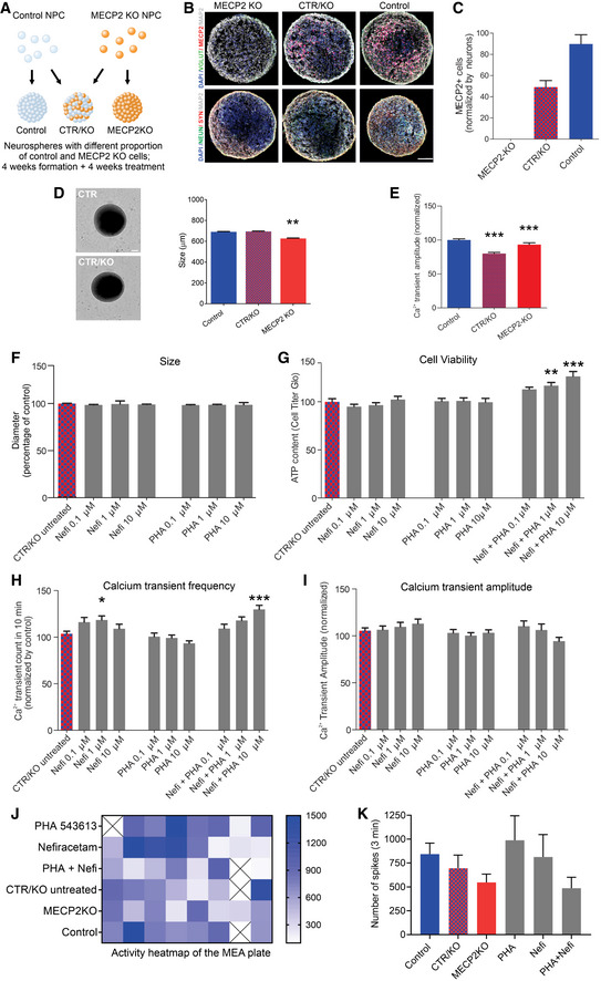
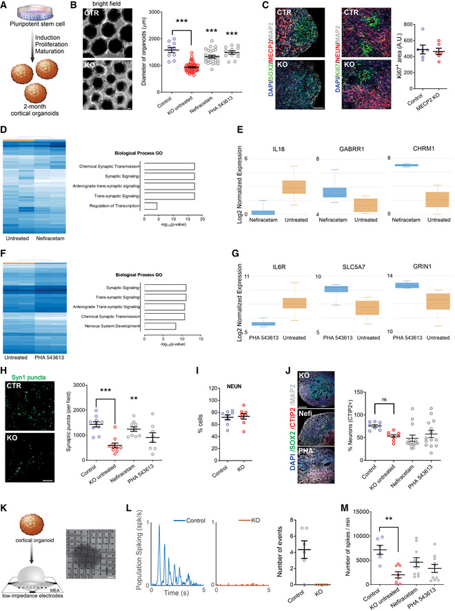
Duplication or deficiency of the X‐linked MECP2 gene reliably produces profound neurodevelopmental impairment. MECP2 mutations are almost universally responsible for Rett syndrome (RTT), and particular mutations and cellular mosaicism of MECP2 may underlie the spectrum of RTT symptomatic severity. No clinically approved treatments for RTT are currently available, but human pluripotent stem cell technology offers a platform to identify neuropathology and test candidate therapeutics. Using a strategic series of increasingly complex human stem cell‐derived technologies, including human neurons, MECP2 ‐mosaic neurospheres to model RTT female brain mosaicism, and cortical organoids, we identified synaptic dysregulation downstream from knockout of MECP2 and screened select pharmacological compounds for their ability to treat this dysfunction. Two lead compounds, Nefiracetam and PHA 543613, specifically reversed MECP2‐ knockout cytologic neuropathology. The capacity of these compounds to reverse neuropathologic phenotypes and networks in human models supports clinical studies for neurodevelopmental disorders in which MeCP2 deficiency is the predominant etiology.
Overview
The claims and statistics of this paper haven’t been checked yet.
Run the check — free, no account needed. It reads the open-access text and checks the sources it cites.
Integrity signals
Registry
Cites
Statistics
Figures
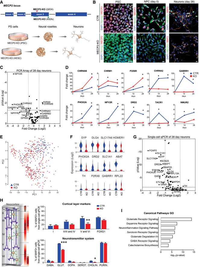
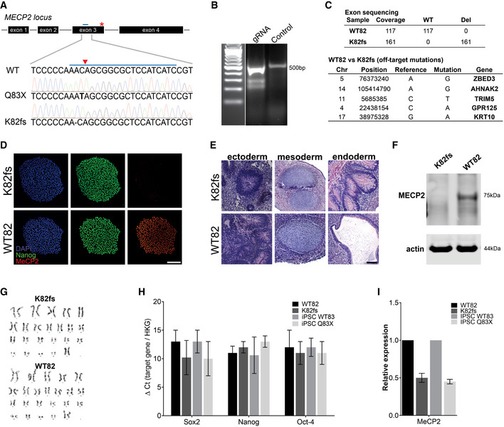
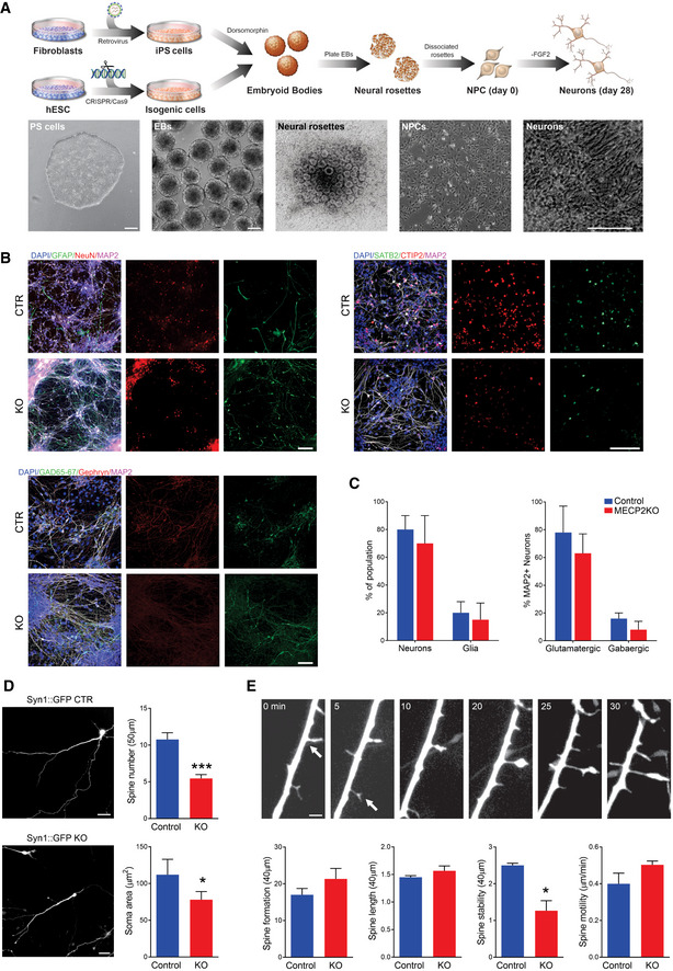
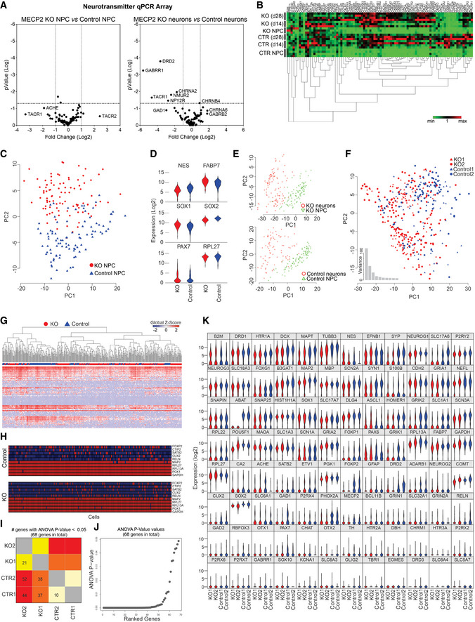
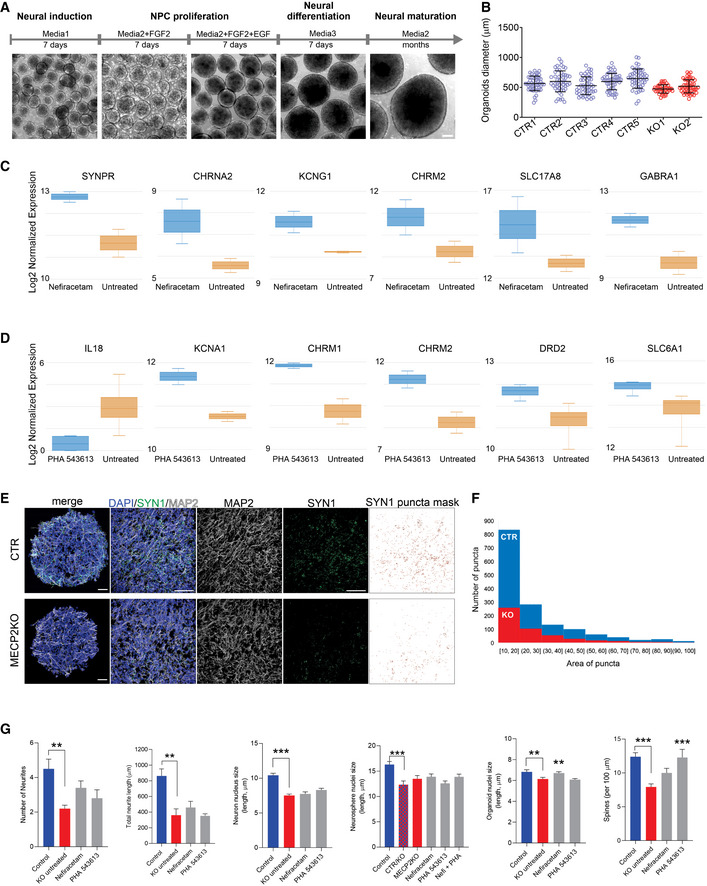
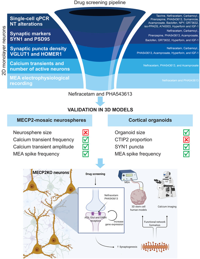
Verification
Claims the paper makes
The claims and statistics of this paper haven’t been checked yet.
Run the check — free, no account needed. It reads the open-access text and checks the sources it cites.
Claims other papers make about this one
Each is checked against this paper’s own text: does it say what the citing paper says it does?
The check wasn’t confident either way, so it gives no verdict — never disputed.
In the citing paper
“whereas Nefiracetam has a broader effect, targeting both L- and N-type calcium channels.”
From CYFIP1 governs the development of cortical axons by modulating calcium availability (2025) · cites this paper as [74]
Experimental — accuracy not yet measured
Self-check
Percentages, sums and reported differences re-computed from the paper’s own numbers.
The self-check needs the paper’s full text.
Citations
Cites
Cytoscape: A software Environment for integrated models of biomolecular interaction networks
Shannon, P., Markiel, A., Ozier, O., Baliga, N. S., Wang, J. T., Ramage, D., Amin, N., Schwikowski, B., & Ideker, T. (2003) · Genome Research · doi:10.1101/gr.1239303 · Cited by 51,888
Shrestha, S., & Offer, S. M. (2016) · Medical Epigenetics · doi:10.1159/000444713 · Cited by 8,951
Rett syndrome is caused by mutations in X-linked MECP2, encoding methyl-CpG-binding protein 2
Amir, R. E., Van den Veyver, I. B., Wan, M., Tran, C. Q., Francke, U., & Zoghbi, H. Y. (1999) · Nature Genetics · doi:10.1038/13810 · Cited by 4,990
Chaos in Neuronal Networks with Balanced Excitatory and Inhibitory Activity
van Vreeswijk, C., & Sompolinsky, H. (1996) · Science · doi:10.1126/science.274.5293.1724 · Cited by 1,831
MeCP2, a Key Contributor to Neurological Disease, Activates and Represses Transcription
Chahrour, M., Jung, S. Y., Shaw, C., Zhou, X., Wong, S. T. C., Qin, J., & Zoghbi, H. Y. (2008) · Science · doi:10.1126/science.1153252 · Cited by 1,823
WebGestalt: an integrated system for exploring gene sets in various biological contexts
Zhang, B., Kirov, S. A., & Snoddy, J. R. (2005) · Nucleic Acids Research · doi:10.1093/nar/gki475 · Cited by 1,803
Conserved cell types with divergent features in human versus mouse cortex
Hodge, R. D., Bakken, T. E., Miller, J. A., Smith, K. A., Barkan, E. R., Graybuck, L. T., Close, J. L., Long, B., Johansen, N., Penn, O., et al. (2019) · Nature · doi:10.1038/s41586-019-1506-7 · Cited by 1,775 · cited 1×
“Notably, the most different systems are glutamatergic, serotoninergic, and cholinergic (Hodge et al, 2019; Sjostedt et al, 2020).”
BackgroundAcetylcholine as a Neuromodulator: Cholinergic Signaling Shapes Nervous System Function and Behavior
Picciotto, M. R., Higley, M. J., & Mineur, Y. S. (2012) · Neuron · doi:10.1016/j.neuron.2012.08.036 · Cited by 1,404
Marchetto, M. C. N., Carromeu, C., Acab, A., Yu, D., Yeo, G. W., Mu, Y., Chen, G., Gage, F. H., & Muotri, A. R. (2010) · Cell · doi:10.1016/j.cell.2010.10.016 · Cited by 1,329
Self-citationSomatic coding mutations in human induced pluripotent stem cells
Gore, A., Li, Z., Fung, H.-L., Young, J. E., Agarwal, S., Antosiewicz-Bourget, J., Canto, I., Giorgetti, A., Israel, M. A., Kiskinis, E., et al. (2011) · Nature · doi:10.1038/nature09805 · Cited by 1,273
Cited by
Sorted by citations
Human cerebral organoids — a new tool for clinical neurology research
Eichmüller, O. L., & Knoblich, J. A. (2022) · Nature Reviews Neurology · doi:10.1038/s41582-022-00723-9 · Cited by 319
Uzquiano, A., Kedaigle, A. J., Pigoni, M., Paulsen, B. D. S., Adiconis, X., Kim, K., Faits, T., Nagaraja, S., Antón-Bolaños, N., Gerhardinger, C., et al. (2022) · Cell · doi:10.1016/j.cell.2022.09.010 · Cited by 257
New epilepsy therapies in development
Klein, P., Kaminski, R. M., Koepp, M. J., & Löscher, W. (2024) · Nature Reviews Drug Discovery · doi:10.1038/s41573-024-00981-w · Cited by 166
Modelling human brain development and disease with organoids
Birtele, M., Lancaster, M. A., & Quadrato, G. (2024) · Nature Reviews Molecular Cell Biology · doi:10.1038/s41580-024-00804-1 · Cited by 177
McCready, F. P., Gordillo-Sampedro, S., Pradeepan, K. S., Martínez-Trujillo, J., & Ellis, J. (2022) · Biology · doi:10.3390/biology11020316 · Cited by 61
A beginner’s guide on the use of brain organoids for neuroscientists: a systematic review
Mulder, L., Depla, J. A., Sridhar, A., Wolthers, K. C., Pajkrt, D., & de Sá, R. V. (2023) · Stem Cell Research & Therapy · doi:10.1186/s13287-023-03302-x · Cited by 67
Impact of alcohol exposure on neural development and network formation in human cortical organoids
Adams, J. W., Negraes, P. D., Truong, J., Tran, T. H., Szeto, R. A., Guerra, B. S., Herai, R. H., Teodorof‐Diedrich, C., Spector, S. A., Campo, M. D., et al. (2022) · Molecular Psychiatry · doi:10.1038/s41380-022-01862-7 · Cited by 59
Self-citationHuman 3D brain organoids: steering the demolecularization of brain and neurological diseases
Adlakha, Y. K. (2023) · Cell Death Discovery · doi:10.1038/s41420-023-01523-w · Cited by 63
Porciúncula, L. O., Goto‐Silva, L., Ledur, P. F., & Rehen, S. K. (2021) · Frontiers in Neuroscience · doi:10.3389/fnins.2021.674563 · Cited by 54
Acharya, P., Choi, N. Y., Shrestha, S., Jeong, S., & Lee, M. (2023) · Biotechnology and Bioengineering · doi:10.1002/bit.28606 · Cited by 50
Yıldırım, M., Delepine, C., Feldman, D., Pham, V. A., Chou, S., Ip, J. P. K., Nott, A., Tsai, L., Ming, G., So, P. T. C., et al. (2022) · eLife · doi:10.7554/elife.78079 · Cited by 45
Stem Cell–Based Organoid Models of Neurodevelopmental Disorders
Wang, L., Owusu-Hammond, C., Sievert, D., & Gleeson, J. G. (2023) · Biological Psychiatry · doi:10.1016/j.biopsych.2023.01.012 · Cited by 44
Suzuki, I., Matsuda, N., Han, X. B., Noji, S., Shibata, M., Nagafuku, N., & Ishibashi, Y. (2023) · Advanced Science · doi:10.1002/advs.202207732 · Cited by 48
Cerina, M., Piastra, M. C., & Frega, M. (2023) · Progress in Biomedical Engineering · doi:10.1088/2516-1091/acce12 · Cited by 44
Generation of ‘semi-guided’ cortical organoids with complex neural oscillations
Fitzgerald, M. Q., Chu, T., Puppo, F., Blanch, R., Chillón, M., Subramaniam, S., & Muotri, A. R. (2024) · Nature Protocols · doi:10.1038/s41596-024-00994-0 · Cited by 45
Self-citationLevy, R. J., & Pașca, S. P. (2022) · Biological Psychiatry · doi:10.1016/j.biopsych.2022.11.017 · Cited by 45
Mok, R. S. F., Zhang, W., Sheikh, T. I., Pradeepan, K. S., Fernandes, I. R., DeJong, L. C., Benigno, G., Hildebrandt, M. R., Mufteev, M., Rodrigues, D. C., et al. (2022) · Translational Psychiatry · doi:10.1038/s41398-022-02216-1 · Cited by 35
Self-citationExploring the complexity of MECP2 function in Rett syndrome
Liu, Y., Whitfield, T. W., Bell, G. W., Guo, R., Flamier, A., Young, R. A., & Jaenisch, R. (2025) · Nature reviews. Neuroscience · doi:10.1038/s41583-025-00926-1 · Cited by 39
Human pluripotent stem cell (hPSC) and organoid models of autism: opportunities and limitations
Kilpatrick, S., Irwin, C. L., & Singh, K. K. (2023) · Translational Psychiatry · doi:10.1038/s41398-023-02510-6 · Cited by 34
Cerebral Organoids as an Experimental Platform for Human Neurogenomics
Nowakowski, T. J., & Salama, S. R. (2022) · Cells · doi:10.3390/cells11182803 · Cited by 27
Brain Organoids: Studying Human Brain Development and Diseases in a Dish
Xu, J., & Wen, Z. (2021) · Stem Cells International · doi:10.1155/2021/5902824 · Cited by 25
Bhattacharya, A., Choi, W. W. Y., Muffat, J., & Li, Y. (2021) · Journal of Molecular Biology · doi:10.1016/j.jmb.2021.167386 · Cited by 24
Modeling Autism Spectrum Disorders with Induced Pluripotent Stem Cell-Derived Brain Organoids
Santos, J. L. D. S., Araújo, C. D. A., Rocha, C. A. G., Costa‐Ferro, Z. S. M., & Souza, B. S. D. F. (2023) · Biomolecules · doi:10.3390/biom13020260 · Cited by 29
Brain Organoids and Assembloids—From Disease Modeling to Drug Discovery
Ajongbolo, A. O., & Langhans, S. A. (2025) · Cells · doi:10.3390/cells14110842 · Cited by 25
Deciphering the physiopathology of neurodevelopmental disorders using brain organoids
Dionne, O., Sabatié, S., & Laurent, B. (2024) · Brain · doi:10.1093/brain/awae281 · Cited by 22
Yokoi, R., Shibata, M., Odawara, A., Ishibashi, Y., Nagafuku, N., Matsuda, N., & Suzuki, I. (2021) · Biochemistry and Biophysics Reports · doi:10.1016/j.bbrep.2021.101148 · Cited by 20
Wu, W., Yao, H., Negraes, P. D., Wang, J., Trujillo, C. A., de Souza, J. S., Muotri, A. R., & Haddad, G. G. (2022) · Neurobiology of Disease · doi:10.1016/j.nbd.2022.105882 · Cited by 19
Self-citationGuerreiro, S. R., & Maciel, P. (2023) · Cells · doi:10.3390/cells12040538 · Cited by 20
iPSC toolbox for understanding and repairing disrupted brain circuits in autism
Chiola, S., Edgar, N. U., & Shcheglovitov, A. (2021) · Molecular Psychiatry · doi:10.1038/s41380-021-01288-7 · Cited by 18
Chromatin dynamics in human brain development and disease
Valencia, A. M., & Pașca, S. P. (2021) · Trends in Cell Biology · doi:10.1016/j.tcb.2021.09.001 · Cited by 17
Akol, I., Gather, F., & Vogel, T. (2022) · International Journal of Molecular Sciences · doi:10.3390/ijms23020954 · Cited by 20
Cerebral organoids as an in vitro model to study autism spectrum disorders
Rabeling, A., & Goolam, M. (2022) · Gene Therapy · doi:10.1038/s41434-022-00356-z · Cited by 17
Doorn, N., van Hugte, E., Ciptasari, U., Mordelt, A., Meijer, H. G. E., Schubert, D., Frega, M., Kasri, N. N., & van Putten, M. J. A. M. (2023) · Stem Cell Reports · doi:10.1016/j.stemcr.2023.06.003 · Cited by 16
Swan, M., dos Santos, R. P., & Witte, F. (2022) · Quantum Reports · doi:10.3390/quantum4010008 · Cited by 16
Xu, Y., Liu, P., Yan, Z.-Z., Mi, T., Wang, Y., Li, Q., Teng, Z., & Liu, C. (2022) · Stem Cell Research & Therapy · doi:10.1186/s13287-022-03216-0 · Cited by 16
Sandhu, A., Kumar, A., Rawat, K., Gautam, V., Sharma, A., & Saha, L. (2023) · World Journal of Clinical Cases · doi:10.12998/wjcc.v11.i14.3114 · Cited by 17
Bajikar, S. S., Sztainberg, Y., Trostle, A. J., Tirumala, H. P., Wan, Y., Harrop, C. L., Bengtsson, J. D., Carvalho, C. M. B., Pehlivan, D., Suter, B., et al. (2024) · Human Molecular Genetics · doi:10.1093/hmg/ddae135 · Cited by 16
Microglial involvement in autism spectrum disorder: insights from human data and iPSC models
Michels, S., Mali, A. S., Jäntti, H., Rezaie, M., & Malm, T. (2025) · Brain Behavior and Immunity · doi:10.1016/j.bbi.2025.106071 · Cited by 16
Alterations of presynaptic proteins in autism spectrum disorder
Yeo, X. Y., Lim, Y. T., Chae, W. R., Park, C., Park, H., & Jung, S. (2022) · Frontiers in Molecular Neuroscience · doi:10.3389/fnmol.2022.1062878 · Cited by 16
Gomes, A. R., Fernandes, T. G., Cabral, J. M. S., & Diogo, M. M. (2021) · International Journal of Molecular Sciences · doi:10.3390/ijms22073751 · Cited by 15
Rubio, A. D., Hamilton, L. L., Bausch, M., Jin, M., Papetti, A. V., Jiang, P., & Yelamanchili, S. V. (2025) · Journal of Neuroimmune Pharmacology · doi:10.1007/s11481-025-10181-x · Cited by 17
Soares, E. S., Obiols-Guardia, A., Jorge-Torres, O. C., Oliveira-Mateos, C., Soler, M., Ramesh-Kumar, D., Setién, F., van Rossum, D., Pascual‐Alonso, A., Xiol, C., et al. (2021) · Molecular Therapy — Nucleic Acids · doi:10.1016/j.omtn.2021.12.030 · Cited by 14
Hung, P., Wu, K. L. H., Chen, C., Siu, K.-K., Hsin, Y., Wang, L., & Wang, F. (2021) · International Journal of Molecular Sciences · doi:10.3390/ijms22137174 · Cited by 15
Brain Organoids to Evaluate Cellular Therapies
García-Delgado, A. B., Campos‐Cuerva, R., Rosell‐Valle, C., Martín-López, M., Casado, C., Ferrari, D., Márquez-Rivas, J., Sánchez‐Pernaute, R., & Fernández‐Muñoz, B. (2022) · Animals · doi:10.3390/ani12223150 · Cited by 14
Human microglial cells as a therapeutic target in a neurodevelopmental disease model
Mesci, P., LaRock, C. N., Jeziorski, J. J., Nakashima, H., Chermont, N., Ferrasa, A., Herai, R. H., Ozaki, T., Saleh, A., Snethlage, C. E., et al. (2024) · Stem Cell Reports · doi:10.1016/j.stemcr.2024.06.013 · Cited by 15
Self-citationBerryer, M. H., Rizki, G., Nathanson, A. J., Klein, J. A., Trendafilova, D., Susco, S. G., Lam, D., Messana, A., Holton, K. M., Karhohs, K. W., et al. (2023) · eLife · doi:10.7554/elife.80168 · Cited by 14
Recent advances in stem cells and gene editing: Drug discovery and therapeutics
Bayarsaikhan, D., Bayarsaikhan, G., & Lee, B. (2021) · Progress in molecular biology and translational science · doi:10.1016/bs.pmbts.2021.01.019 · Cited by 12
Muzzi, L., Lisa, D. D., Falappa, M., Pepe, S., Maccione, A., Pastorino, L., Martinoia, S., & Frega, M. (2022) · Bioengineering · doi:10.3390/bioengineering10040449 · Cited by 14
Assessing the Utility of Organoid Intelligence: Scientific and Ethical Perspectives
Nestor, M. W., & Wilson, R. L. (2025) · Organoids · doi:10.3390/organoids4020009 · Cited by 11
Parmentier, T., James, F., Hewitson, E., Bailey, C. D. C., Werry, N., Sheridan, S., Perlis, R., Perreault, M., Gaitero, L., Lalonde, J., et al. (2022) · Scientific Reports · doi:10.1038/s41598-022-13071-x · Cited by 9
Tsuda, B., Pate, S. C., Tye, K. M., Siegelmann, H. T., & B, T. J. S. A. (2021) · bioRxiv (Cold Spring Harbor Laboratory) · doi:10.1101/2021.05.31.446462 · Cited by 9
Self-citationAdams, J. W., Vinokur, A., de Souza, J. S., Austria, C., Guerra, B. S., Herai, R. H., Wahlin, K. J., & Muotri, A. R. (2024) · Cell Reports · doi:10.1016/j.celrep.2024.113867 · Cited by 8
Self-citationSoares, E. S., Velasco, C. D., Tarrasón, A., Soler, M., Srinivas, T., Setién, F., Oliveira-Mateos, C., Casado-Peláez, M., Martinez‐Verbo, L., Armstrong, J., et al. (2025) · Nucleic Acids Research · doi:10.1093/nar/gkaf074 · Cited by 9
Impact of intermittent lead exposure on hominid brain evolution
Joannes‐Boyau, R., de Souza, J. S., Arora, M., Austin, C. E., Westaway, K., Moffat, I., Wang, W., Liao, W., Zhang, Y., Adams, J. W., et al. (2025) · Science Advances · doi:10.1126/sciadv.adr1524 · Cited by 9
Self-citationMicrophysiological models of the central nervous system with fluid flow
Zilberman, A., & Cornelison, R. C. (2021) · Brain Research Bulletin · doi:10.1016/j.brainresbull.2021.05.015 · Cited by 7
Adeyeye, A. O., Mirsadeghi, S., Gutierrez, M. M. F., & Hsieh, J. (2024) · Epilepsy & Behavior · doi:10.1016/j.yebeh.2024.109982 · Cited by 7
Neurodevelopmental disorders: 2022 update
Sabariego-Navarro, M., Fernández‐Blanco, Á., Sierra, C., & Dierssen, M. (2022) · Free Neuropathology · doi:10.17879/freeneuropathology-2022-3801 · Cited by 10
Three-dimensional human brain organoids: A next-generation model to decode Alzheimer’s disease
Kumari, S., Ashvin, Dhapola, R., Sharma, P., Paidlewar, M., Vellingiri, B., Medhi, B., & HariKrishnaReddy, D. (2025) · Ageing Research Reviews · doi:10.1016/j.arr.2025.102928 · Cited by 6
MeCP2 regulates telencephalic development in human cerebral organoids
Tenreiro, M. F., Mohana-Borges, R., Sánchez-Sánchez, S. M., Dias, Â. R. M., Blanch, R., & Muotri, A. R. (2025) · Cell Reports · doi:10.1016/j.celrep.2025.116670 · Cited by 8
Self-citationYokoi, R., Nagafuku, N., Ishibashi, Y., Matsuda, N., & Suzuki, I. (2023) · Organoids · doi:10.3390/organoids2040014 · Cited by 8
Tenreiro, M. F., & Muotri, A. R. (2026) · Developmental Cell · doi:10.1016/j.devcel.2026.02.018 · Cited by 6
Self-citationThom, R. P., Warren, T. L., Khan, S., Muhle, R., Wang, P. P., Brennand, K., Zürcher, N. R., Veenstra-VanderWeele, J. M., & Hoffman, E. J. (2025) · Journal of Child and Adolescent Psychopharmacology · doi:10.1089/cap.2025.0023 · Cited by 5
Differentiation Defect Into GABAergic Neurons in Cerebral Organoids From Autism Patients
Hali, S., Yao, X., Hao, G., Jin, Z., Fu, K., Li, Y., Wang, L., Yoo, H., La, H., Park, C., et al. (2025) · CNS Neuroscience & Therapeutics · doi:10.1111/cns.70449 · Cited by 5
Huber, A., Sarne, V., Beribisky, A. V., Ackerbauer, D., Derdak, S. V., Madritsch, S., Etzler, J., Huck, S., Scholze, P., Görgülü, I., et al. (2024) · Stem Cells and Development · doi:10.1089/scd.2023.0233 · Cited by 5
Aberrant Cortical Layer Development of Brain Organoids Derived from Noonan Syndrome-iPSCs
Kim, B., Koh, Y., Do, H., Ju, Y., Choi, J. B., Cho, G., Yoo, H., Lee, B. H., Han, J., Park, J.-E., et al. (2022) · International Journal of Molecular Sciences · doi:10.3390/ijms232213861 · Cited by 7
Dormant viral pathways underlie space-induced neural senescencePreprint
Martins, A. M. A., Nakashima, H., Macia, Á., Umeyama, T., Miyashita, S., Nascimento, L. L., Coelho, L. B. V., Mesci, P., Viirre, E., Suarez, N., et al. (2025) · bioRxiv (Cold Spring Harbor Laboratory) · doi:10.1101/2025.11.02.686043 · Cited by 6
Self-citationMARTINS, A. M. A., Biagi, D., Tsu, B. L., Fischer, J. D. S. D. G., Coelho, L. B. V., Carvalho, P. C., & Muotri, A. R. (2026) · Scientific Data · doi:10.1038/s41597-026-06881-5 · Cited by 4
Self-citationJeong, Y., Kim, M. W., Lee, S., Park, S., Jeong, K. S., Lee, Y. H., Lee, S., Chung, H., Kim, J., & Kim, C. (2025) · Biomedicine & Pharmacotherapy · doi:10.1016/j.biopha.2025.117821 · Cited by 4
Human pluripotent stem cell (hPSC)-derived models for autism spectrum disorder drug discovery
Nie, L., Irwin, C. L., Geahchan, S., & Singh, K. K. (2024) · Expert Opinion on Drug Discovery · doi:10.1080/17460441.2024.2416484 · Cited by 4
Yang, Z., Teaney, N. A., Buttermore, E. D., Şahin, M., & Saber, W. A. (2025) · Frontiers in Neuroscience · doi:10.3389/fnins.2024.1524577 · Cited by 4
Modeling forebrain regional development and connectivity by human brain organoids
Choe, M. S., Lo, C., & Park, I. (2025) · Current Opinion in Genetics & Development · doi:10.1016/j.gde.2025.102324 · Cited by 5
Pluripotent stem cells-based neural organoids for modelling human brain development and diseases
Tong, L., Tian, P., Wang, R., Niu, S., Wang, R., Ye, Y., Wu, Y., Zhang, W., Wang, Y., Foggetti, A., et al. (2025) · Cell & Bioscience · doi:10.1186/s13578-025-01515-6 · Cited by 3
Shams, S., Cronell, P., Landin, J., Pietri, T., Gimdal, A. E., Kettunen, P., & Westberg, L. (2025) · BMC Neuroscience · doi:10.1186/s12868-025-00946-8 · Cited by 3
How copy number variations shape brain developmental disorders: Unraveling the synaptic puzzle
Wang, T., TRABZONLU, K., Jones, E. F., & Syed, Y. A. (2025) · Psychiatry and Clinical Neurosciences · doi:10.1111/pcn.70009 · Cited by 2
Human Brain Models of Intellectual Disability: Experimental Advances and Novelties
Merckx, N., & Esch, H. V. (2022) · International Journal of Molecular Sciences · doi:10.3390/ijms23126476 · Cited by 2
Yakimov, A. M., Евгеньевна, Т. Е., Paramonova, A. I., Vasilieva, A. A., Константинович, Р. Ф., Дмитриевна, Р. А., & Дмитренко, Д. В. (2025) · Epilepsy and Paroxysmal Conditions · doi:10.17749/2077-8333/epi.par.con.2025.211 · Cited by 2
Mok, R. S., Zhang, W., Sheikh, T. I., Pradeepan, K., Fernandes, I. R., DeJong, L. C., Benigno, G., Hildebrandt, M. R., Mufteev, M., Rodrigues, D. C., et al. (2020) · bioRxiv (Cold Spring Harbor Laboratory) · doi:10.1101/2020.07.12.189621 · Cited by 2
Self-citationSuzuki, I., Matsuda, N., Han, X. B., Noji, S., Shibata, M., Nagafuku, N., & Ishibashi, Y. (2022) · bioRxiv (Cold Spring Harbor Laboratory) · doi:10.1101/2022.11.09.515884 · Cited by 2
CYFIP1 governs the development of cortical axons by modulating calcium availability
Ricci, C., Midroit, M. J., Caicci, F., Achsel, T., Domínguez-Iturza, N., & Bagni, C. (2025) · Nature Communications · doi:10.1038/s41467-025-65801-0 · Cited by 1
It, F. E., Faivre, L. O., Thauvin‐Robinet, C., Vitobello, A., & Duplomb, L. (2024) · médecine/sciences · doi:10.1051/medsci/2024100 · Cited by 1
Miranda, J. P., & Solá, S. (2021) · Frontiers in Cell and Developmental Biology · doi:10.3389/fcell.2021.720554 · Cited by 1
Yıldırım, M., Delepine, C., Feldman, D., Pham, V. A., Chou, S., Ip, J. P. K., Nott, A., Tsai, L., Ming, G., So, P. T. C., et al. (2022) · bioRxiv (Cold Spring Harbor Laboratory) · doi:10.1101/2022.02.28.482282 · Cited by 1
Latour, B. L., Gunnewiek, T. M. K., & Kasri, N. N. (2022) · Elsevier eBooks · doi:10.1016/b978-0-12-822277-5.00010-9
Wang, S., van Hugte, E., Ciptasari, U., & Kasri, N. N. (2022) · Neuromethods · doi:10.1007/978-1-0716-2569-9_7
Xu, Y., Liu, P., Yan, Z.-Z., Mi, T., Wang, Y., Li, Q., Teng, Z., & Liu, C. (2023) · Figshare · doi:10.6084/m9.figshare.22605065
Human Cerebral Organoids in Pillar/Perfusion Plates for Modeling Neurodevelopmental Disorders
Acharya, P. (2024) · University of North Texas Digital Library (University of North Texas) · doi:10.12794/metadc2332637
Goebel, S., Cordova-Martinez, D., Verselis, V. K., & Francesconi, A. (2024) · bioRxiv (Cold Spring Harbor Laboratory) · doi:10.1101/2024.11.01.621616
Callegari, F., Fabbri, R., Barabino, V., Massobrio, P., Magliaro, C., & Brofiga, M. (2025) · APL Bioengineering · doi:10.1063/5.0288838
Role of brain organoids in Rett syndrome
Mohan, G., & Vishnupriya, P. (2025) · Elsevier eBooks · doi:10.1016/b978-0-443-29898-1.00014-9
Use of brain organoids for Parkinson's disease and Alzheimer's diseases
Kumari, S., Sharma, P., Dhapola, R., Ashvin, Chaudhary, B. N., Vellingiri, B., & HariKrishnaReddy, D. (2025) · Elsevier eBooks · doi:10.1016/b978-0-443-29898-1.00015-0
Genetic Manipulation in Organoid Models and Their Applications in Central Nervous System Pathologies
Rustamov, A., Kocahasan, M., Rumman, B., Rahbarghazi, R., & Sokullu, E. (2025) · Molecular Neurobiology · doi:10.1007/s12035-025-05425-z
Mapping human neurodevelopment—Brain organoids meet lineage tracing
Liaci, C., & Quadrato, G. (2025) · FEBS Letters · doi:10.1002/1873-3468.70250 · Cited by 1
Dunn, A. W., Sit, T. P. H., Feord, R. C., Soltani, A., Yin, Y., Turner, R., Loureiro, I., Eglen, S. J., Paulsen, O., & Mierau, S. B. (2025) · bioRxiv (Cold Spring Harbor Laboratory) · doi:10.64898/2025.12.17.694950
Xu, Y.-J., Liu, P.-P., Yan, Z.-Z., 米廷伟, Wang, Y.-Y., Li, Q., 腾兆乾, & Liu, C.-M. (2023) · Figshare · doi:10.6084/m9.figshare.22605062
Xu, Y.-J., Liu, P.-P., Yan, Z.-Z., 米廷伟, Wang, Y.-Y., Li, Q., 腾兆乾, & Liu, C.-M. (2023) · Figshare · doi:10.6084/m9.figshare.22605059
Advances in Stem Cell Research for MECP2-Related Disorders
Akaba, Y., Takahashi, S., & Tsujimura, K. (2025) · Stem cell biology and regenerative medicine · doi:10.1007/978-3-031-94101-6_2
Zhang, X., Jiang, Y., Wang, H., & Hu, R. (2026) · Elsevier eBooks · doi:10.1016/b978-0-443-40423-8.00008-7
Hommersom, M. P., Puvogel, S., Scheefhals, N., Carpentiero, E., Bouma, M. J., van Beusekom, E., Dillen, L., van de Warrenburg, B. P. C., Kasri, N. N., & van Bokhoven, H. (2026) · Stem Cell Reports · doi:10.1016/j.stemcr.2025.102783
Neuromodulators Generate Multiple Context-Relevant Behaviors in Recurrent Neural Networks
Tsuda, B., Pate, S. C., Tye, K. M., Siegelmann, H. T., & B, T. J. S. A. (2026) · Neural Computation · doi:10.1162/neco.a.1489 · Cited by 11
Self-citationSyndrome de Rett et implication des organes périphériques : de la physiopathologie à la thérapie
Borloz, E. (2021) · theses.fr (ABES) · doi:10.70675/6910a242za2edz4509zbc5aze548081377dd
Metadata
- Authors
- Trujillo, C. A., Adams, J. W., Negraes, P. D., Carromeu, C., Tejwani, L., Acab, A., Tsuda, B., Thomas, C. A., Sodhi, N., Fichter, K. M., Romero, S., Zanella, F., Sejnowski, T. J., Ulrich, H., & Muotri, A. R.
- Affiliations
- University of California San Diego (US)
- Salk Institute for Biological Studies (US)
- Universidade de São Paulo (BR)
- Corresponding author
- Trujillo, Cleber A
- Muotri, Alysson R
- ORCID
- Venue
- EMBO Molecular Medicine
- Published
- 2020-12-08
- Volume
- 13
- Issue
- 1
- Pages
- e12523-e12523
- ISSN
- 1757-4676 (ISSN-L)
- 1757-4684
- PMC
- PMC7799367
- OpenAlex
- W3110698055
- Cited by
- 101
- References
- 67
- Citations by year
- 2026: 6 · 2025: 27 · 2024: 13 · 2023: 16 · 2022: 23 · 2021: 14 · 2020: 2
- FWCI
- 8.21
- Type
- journal article
- Language
- en
- Open access
- gold
- doi.org
- Locations
- EMBO Molecular Medicine (publishedVersion, cc-by)
- PubMed Central (publishedVersion, cc-by)
- PubMed (publishedVersion)
- eScholarship (California Digital Library) (submittedVersion, cc-by)
- DOAJ (DOAJ: Directory of Open Access Journals) (submittedVersion, cc-by-sa)
- eScholarship (California Digital Library) (submittedVersion)
- Funders
- HHS | NIH | National Institute of Mental Health (R01MH094753, R01MH103134, P01 NICHD033113)
- California Institute for Regenerative Medicine (TR2‐01814, TR4‐06747)
- HHS | National Institutes of Health (1‐DP2‐OD006495‐01, U19MH107367)
- HHS | NIH | National Institute of General Medical Sciences (5T32GM007198)
- UCSD Center for Academic Research and Training in Anthropogeny
- Brain & Behavior Research Foundation (NARSAD Independent Investigator Grant)
- HHS | NIH | National Institute on Alcohol Abuse and Alcoholism (K01AA026911)
- Loulou Foundation
- Sao Paulo Research Foundation (2018/07366‐4)
- Topics
- Life Sciences › Biochemistry, Genetics and Molecular Biology › Genetics › Genetics and Neurodevelopmental Disorders
- Life Sciences › Neuroscience › Cellular and Molecular Neuroscience › Neuroscience and Neuropharmacology Research
- Life Sciences › Neuroscience › Developmental Neuroscience › Neurogenesis and neuroplasticity mechanisms
- Keywords
- MeCP2, Rett syndrome, cortical organoids, synaptic dysfunction, neurodevelopmental disorders, nefiracetam, drug screening, X-linked disorders
- MeSH terms
- Female, Humans, Neurons, Organoids, Phenotype, Pyrrolidinones, Quinuclidines, Rett Syndrome, Bridged Bicyclo Compounds, Heterocyclic, Methyl-CpG-Binding Protein 2, Gene Knockout Techniques
About these checks
Registry, retraction, Cites and statistics results are computed, not judged, with one exception: a printed reference that no index lists is read once by a model before it counts as a citation error. Claims are extracted by a model and kept only when their quote appears verbatim in the text; each cited source is then checked, with a model’s help, for whether it says what the claim relies on. Those verdicts can be wrong — read the claim’s quote and the cited source before relying on one.
Statistics and arithmetic are recomputed, not judged.
Confidence bands are shown only for fields calibrated on our annotated test set. None is calibrated yet, so no band is shown.
What counts as a claim ›How we measure confidence ›How we check this ›
Are you an author, or do you see a problem with this page? Request a review
