

Large-scale image-based profiling of single-cell phenotypes in arrayed CRISPR-Cas9 gene perturbation screens
Open accessThe DOI matches its Crossref record; 35 of the 35 references it cites were found in a registry or an index.
Abstract
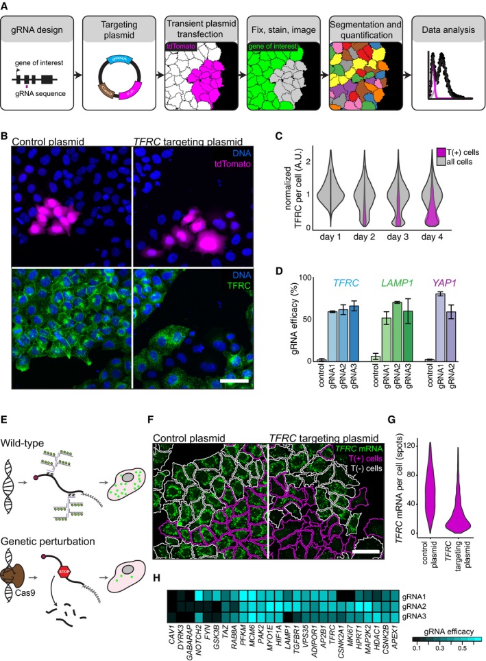
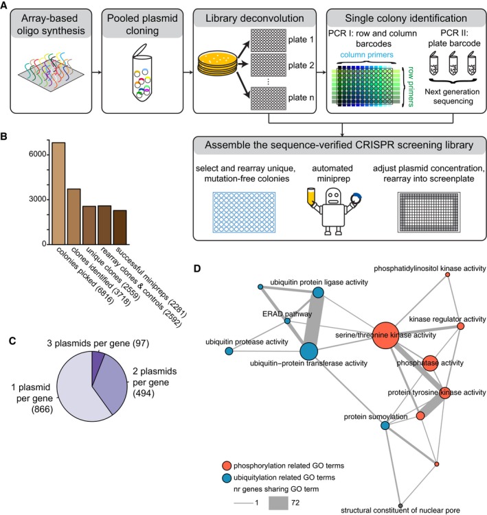
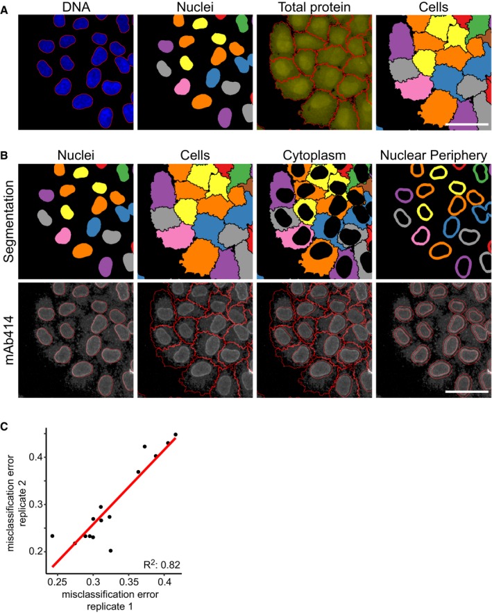
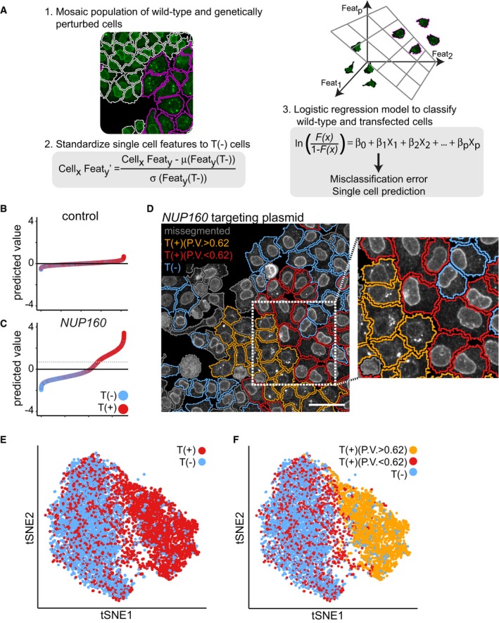
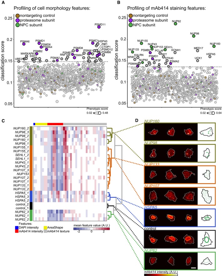
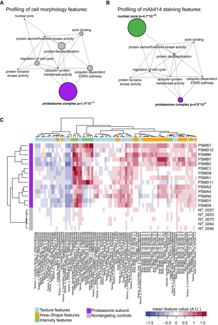
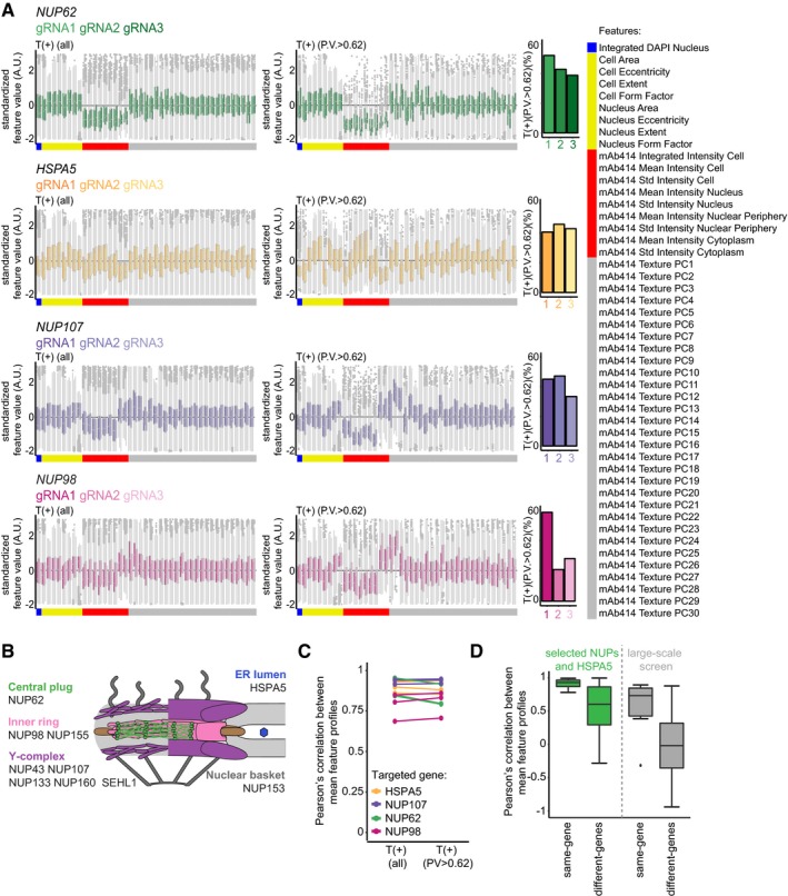
High‐content imaging using automated microscopy and computer vision allows multivariate profiling of single‐cell phenotypes. Here, we present methods for the application of the CISPR‐Cas9 system in large‐scale, image‐based, gene perturbation experiments. We show that CRISPR‐Cas9‐mediated gene perturbation can be achieved in human tissue culture cells in a timeframe that is compatible with image‐based phenotyping. We developed a pipeline to construct a large‐scale arrayed library of 2,281 sequence‐verified CRISPR‐Cas9 targeting plasmids and profiled this library for genes affecting cellular morphology and the subcellular localization of components of the nuclear pore complex (NPC). We conceived a machine‐learning method that harnesses genetic heterogeneity to score gene perturbations and identify phenotypically perturbed cells for in‐depth characterization of gene perturbation effects. This approach enables genome‐scale image‐based multivariate gene perturbation profiling using CRISPR‐Cas9.
Overview
The claims and statistics of this paper haven’t been checked yet.
Free: it reads the open-access text and checks the first 5 claims that cite a source we can check. Create a free account to check every claim.
Integrity signals
Registry
Cites
Statistics
Figures

Verification
Claims the paper makes
The claims and statistics of this paper haven’t been checked yet.
Free: it reads the open-access text and checks the first 5 claims that cite a source we can check. Create a free account to check every claim.
Claims other papers make about this one
Each is checked against this paper’s own text: does it say what the citing paper says it does?
The source backs part of the claim, not all of it — or backs it for a different population, condition or comparison.
In the citing paper
“In recent years, there has been wide use of pooled genome-scale CRISPR screens based on enriching cells via fitness or reporter fluorescence in various model systems, but only one large-scale (2,281 sgRNA) CRISPR imaging screen (Groot et al., 2018).”
From Optical Pooled Screens in Human Cells (2019) · cites this paper as Groot et al., 2018
Experimental — accuracy not yet measured
Self-check
Percentages, sums and reported differences re-computed from the paper’s own numbers.
The self-check needs the paper’s full text.
Citations
Cites
(2008) · Journal of Machine Learning Research · Cited by 33,051
Regularization and Variable Selection Via the Elastic Net
Zou, H., & Hastie, T. (2005) · Journal of the Royal Statistical Society Series B: Statistical Methodology · doi:10.1111/j.1467-9868.2005.00503.x · Cited by 20,957
Regularization Paths for Generalized Linear Models via Coordinate Descent
Friedman, J., Hastie, T., & Tibshirani, R. (2010) · Journal of Statistical Software · doi:10.18637/jss.v033.i01 · Cited by 19,927 · cited 2×
“We randomly selected 500 T(+) and T( ) cells (with replacement) form every targeted cell population and trained a 10- fold cross-validated logistic regression model on the single-cell data using the R software package glmnet (Friedman et al, 2010).”
MethodGenome engineering using the CRISPR-Cas9 system
Ran, F. A., Hsu, P. D., Wright, J., Agarwala, V., Scott, D. A., & Zhang, F. (2013) · Nature Protocols · doi:10.1038/nprot.2013.143 · Cited by 11,133
Genome-scale CRISPR-Cas9 knockout screening in human cells
Shalem, O., Sanjana, N. E., Hartenian, E., Shi, X., Scott, D. A., Mikkelsen, T. S., Heckl, D., Ebert, B. L., Root, D. E., Doench, J. G., et al. (2014) · Science · doi:10.1126/science.1247005 · Cited by 5,234 · cited 1×
“Most screens performed to date employ a pooled screening strategy, which can identify genes that cause differential growth in screening conditions (Koike-Yusa et al, 2013; Shalem et al, 2014; Wang et al, 2014).”
BackgroundGenetic screens in human cells using the CRISPR-Cas9 system
Wang, T., Wei, J. J., Sabatini, D. M., & Lander, E. S. (2014) · Science · doi:10.1126/science.1246981 · Cited by 2,856 · cited 1×
“Most screens performed to date employ a pooled screening strategy, which can identify genes that cause differential growth in screening conditions (Koike-Yusa et al, 2013; Shalem et al, 2014; Wang et al, 2014).”
BackgroundDixit, A., Parnas, O., Li, B., Chen, J., Fulco, C. P., Jerby-Arnon, L., Marjanovic, N. D., Dionne, D., Burks, T., Raychowdhury, R., et al. (2016) · Cell · doi:10.1016/j.cell.2016.11.038 · Cited by 1,863
Rational design of highly active sgRNAs for CRISPR-Cas9-mediated gene inactivation
Doench, J. G., Hartenian, E., Graham, D. B., Tothova, Z., Hegde, M., Smith, I., Sullender, M., Ebert, B. L., Xavier, R. J., & Root, D. E. (2014) · Nature Biotechnology · doi:10.1038/nbt.3026 · Cited by 1,617 · cited 2×
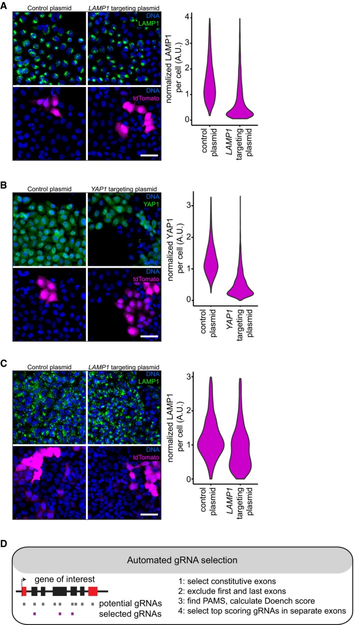
“To systematically test our approach across multiple genes, we automated the selection of gRNA sequences with high predicted ontarget efficacy (Doench et al, 2014).”
MethodGarcía-Alonso, C. R., Pérez-Naranjo, L. M., & Fernández-Caballero, J. C. (2014) · Annals of Operations Research · doi:10.1007/s10479-011-0841-3 · Cited by 1,353
High-throughput functional genomics using CRISPR-Cas9
Shalem, O., Sanjana, N. E., & Zhang, F. (2015) · Nature Reviews Genetics · doi:10.1038/nrg3899 · Cited by 1,233 · cited 2×
“Importantly, CRISPR-Cas9 is not 100% effective in all targeted cells, which can be the result of in-frame repair of the CRISPR-Cas9induced DNA lesions, a failure to target all functional alleles or limited efficacy of the CRISPR-Cas9 system (Shalem et al, 2015).”
BackgroundMethod
Cited by
Optical Pooled Screens in Human Cells
Feldman, D., Singh, A., Schmid-Burgk, J. L., Carlson, R. J., Mezger, A., Garrity, A. J., Zhang, F., & Blainey, P. C. (2019) · Cell · doi:10.1016/j.cell.2019.09.016 · Cited by 321
A new era in functional genomics screens
Przybyla, L., & Gilbert, L. A. (2022) · Nature Reviews Genetics · doi:10.1038/s41576-021-00409-w · Cited by 281
MIGA2 Links Mitochondria, the ER, and Lipid Droplets and Promotes De Novo Lipogenesis in Adipocytes
Freyre, C. A. C., Rauher, P. C., Ejsing, C. S., & Klemm, R. W. (2019) · Molecular Cell · doi:10.1016/j.molcel.2019.09.011 · Cited by 253
Machine learning and image-based profiling in drug discovery
Scheeder, C., Heigwer, F., & Boutros, M. (2018) · Current Opinion in Systems Biology · doi:10.1016/j.coisb.2018.05.004 · Cited by 169
Christopher, J. A., Stadler, C., Martin, C. E., Morgenstern, M., Pan, Y., Betsinger, C. N., Rattray, D. G., Mahdessian, D., Gingras, A.-C., Warscheid, B., et al. (2021) · Nature Reviews Methods Primers · doi:10.1038/s43586-021-00029-y · Cited by 129
Metadata
- Authors
- de Groot, R., Lüthi, J., Lindsay, H., Holtackers, R., & Pelkmans, L.
- Affiliations
- University of Zurich (CH)
- Corresponding author
- Pelkmans, Lucas
- Venue
- Molecular Systems Biology
- Published
- 2018-01-01
- Volume
- 14
- Issue
- 1
- Pages
- e8064-e8064
- ISSN
- 1744-4292 (ISSN-L)
- PMC
- PMC5787707
- OpenAlex
- W2785088317
- Cited by
- 74
- References
- 35
- Citations by year
- 2026: 3 · 2025: 3 · 2024: 8 · 2023: 6 · 2022: 11 · 2021: 9 · 2020: 16 · 2019: 14 · 2018: 4
- FWCI
- 3.82
- Type
- journal article
- Language
- en
- Open access
- gold
- doi.org
- Licence
- other-oa
- creativecommons.org/licenses/by/4.0/ (tdm)
- creativecommons.org/licenses/by/4.0/ (vor, 22 d)
- Locations
- Molecular Systems Biology (publishedVersion, other-oa)
- PubMed Central (publishedVersion, cc-by)
- PubMed (publishedVersion)
- Zurich Open Repository and Archive (University of Zurich) (acceptedVersion, cc-by)
- DOAJ (DOAJ: Directory of Open Access Journals) (submittedVersion)
- Repository for Publications and Research Data (ETH Zurich) (submittedVersion, cc-by)
- Zurich Open Repository and Archive (University of Zurich) (submittedVersion, cc-by)
- ETH Zürich Research Collection (cc-by)
- Zurich Open Repository and Archive (University of Zurich)
- Funders
- SystemsX.ch (TPdF2015/342)
- Topics
- Life Sciences › Biochemistry, Genetics and Molecular Biology › Molecular Biology › CRISPR and Genetic Engineering
- Life Sciences › Biochemistry, Genetics and Molecular Biology › Biophysics › Cell Image Analysis Techniques
- Life Sciences › Biochemistry, Genetics and Molecular Biology › Molecular Biology › Single-cell and spatial transcriptomics
- Keywords
- image-based phenotyping, nuclear pore complex, cell morphology, subcellular localization, genetic heterogeneity, high-content imaging
- MeSH terms
- Machine Learning, HeLa Cells, Humans, Phenotype, Gene Library, Nuclear Pore, Gene Knockout Techniques, Single-Cell Analysis, CRISPR-Cas Systems
About these checks
Registry, retraction, Cites and statistics results are computed, not judged, with one exception: a printed reference that no index lists is read once by a model before it counts as a citation error. Claims are extracted by a model and kept only when their quote appears verbatim in the text; each cited source is then checked, with a model’s help, for whether it says what the claim relies on. Those verdicts can be wrong — read the claim’s quote and the cited source before relying on one.
Statistics and arithmetic are recomputed, not judged.
Confidence bands are shown only for fields calibrated on our annotated test set. None is calibrated yet, so no band is shown.
What counts as a claim ›How we measure confidence ›How we check this ›
Are you an author, or do you see a problem with this page? Request a review
How to cite
Cite CoChat for the checks on this page, and the paper for its own findings.
- This page
- CoChat. (2026). Citation check: Large-scale image-based profiling of single-cell phenotypes in arrayed CRISPR-Cas9 gene perturbation screens. https://app.cochat.ai/papers/10.15252/msb.20178064
- The paper
- de Groot, R., Lüthi, J., Lindsay, H., Holtackers, R., & Pelkmans, L. (2018). Large-scale image-based profiling of single-cell phenotypes in arrayed CRISPR-Cas9 gene perturbation screens. Molecular Systems Biology. https://doi.org/10.15252/msb.20178064
Data sources
Bibliographic data: Semantic Scholar, OpenAlex, Crossref.
Contains information from the Semantic Scholar Academic Graph Datasets, made available under the ODC Attribution License (ODC-BY 1.0).
Semantic Scholar’s data is described in: Kinney, R., et al. (2023). The Semantic Scholar Open Data Platform. arXiv:2301.10140.
