

Large-scale image-based profiling of single-cell phenotypes in arrayed CRISPR-Cas9 gene perturbation screens
Open accessThe DOI matches its Crossref record; 35 of the 35 references it cites were found in a registry or an index.
Abstract
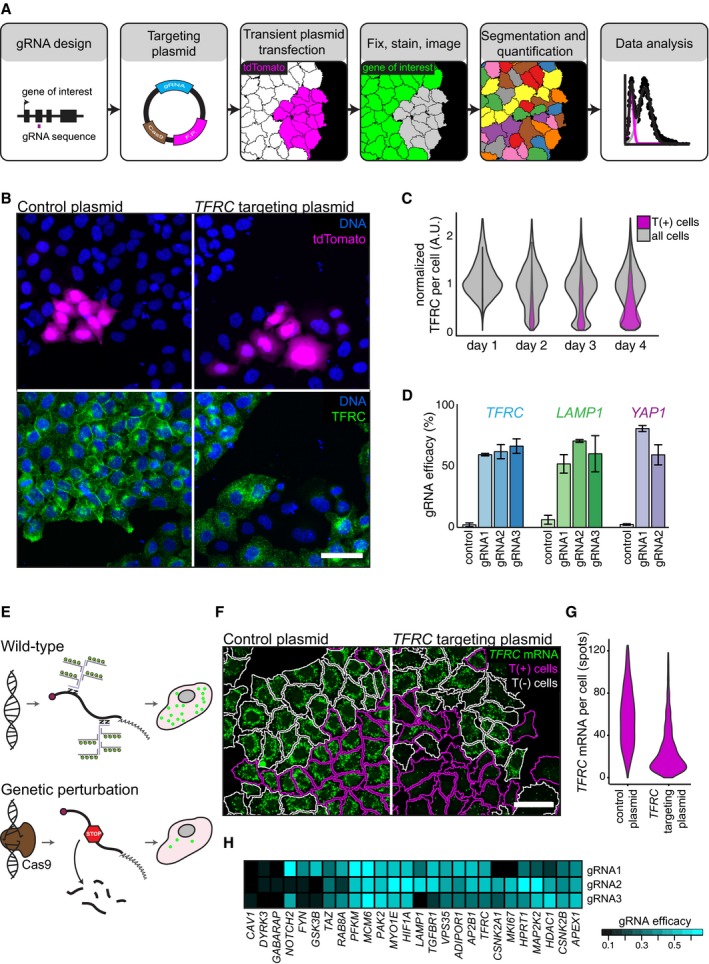
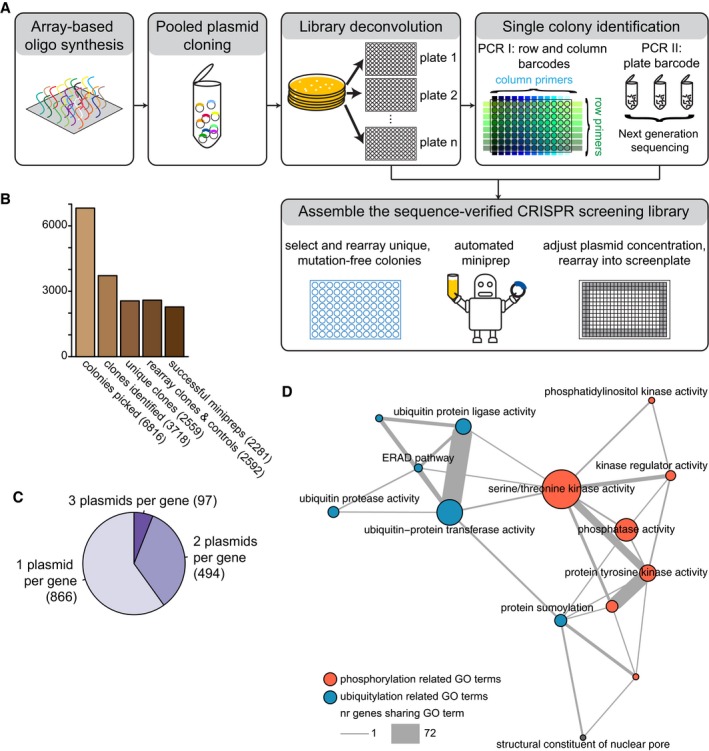
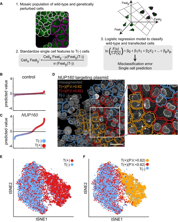
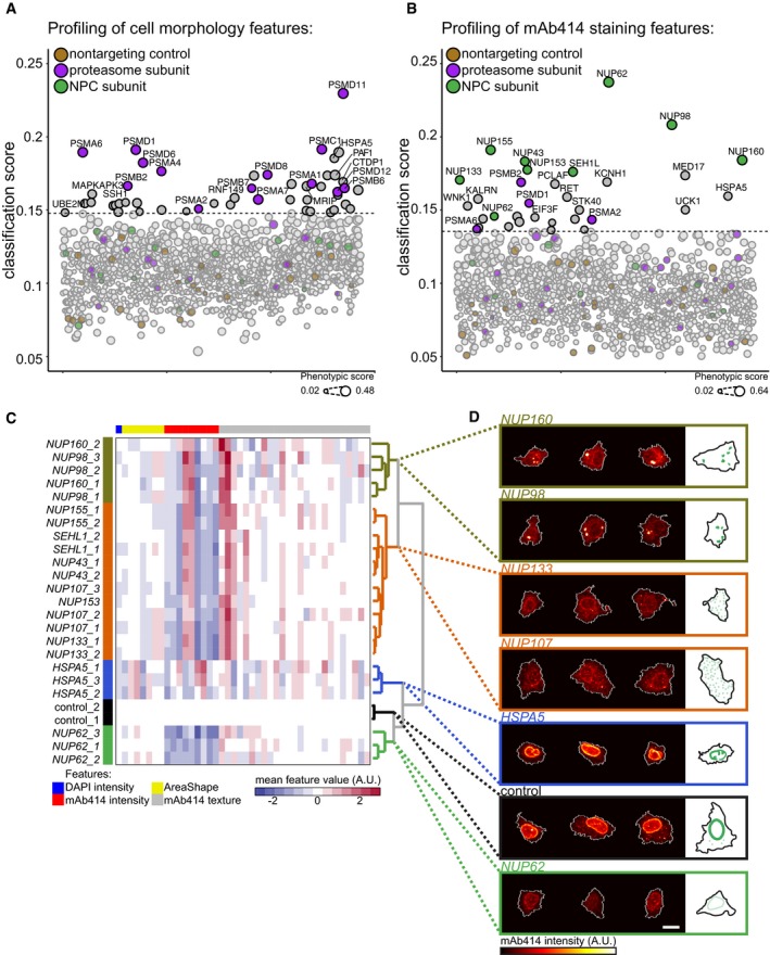
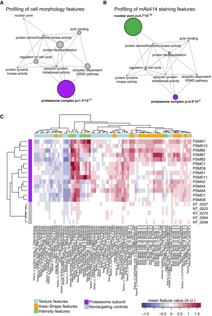
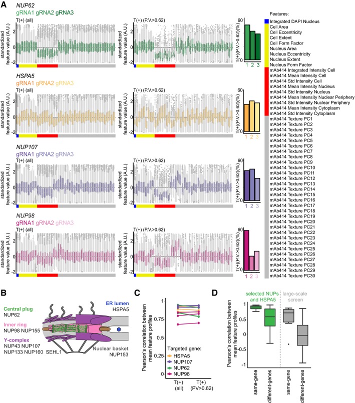
High‐content imaging using automated microscopy and computer vision allows multivariate profiling of single‐cell phenotypes. Here, we present methods for the application of the CISPR‐Cas9 system in large‐scale, image‐based, gene perturbation experiments. We show that CRISPR‐Cas9‐mediated gene perturbation can be achieved in human tissue culture cells in a timeframe that is compatible with image‐based phenotyping. We developed a pipeline to construct a large‐scale arrayed library of 2,281 sequence‐verified CRISPR‐Cas9 targeting plasmids and profiled this library for genes affecting cellular morphology and the subcellular localization of components of the nuclear pore complex (NPC). We conceived a machine‐learning method that harnesses genetic heterogeneity to score gene perturbations and identify phenotypically perturbed cells for in‐depth characterization of gene perturbation effects. This approach enables genome‐scale image‐based multivariate gene perturbation profiling using CRISPR‐Cas9.
Overview
The claims and statistics of this paper haven’t been checked yet.
Free: it reads the open-access text and checks the first 5 claims that cite a source we can check. Create a free account to check every claim.
Integrity signals
Registry
Cites
Statistics
Figures

Verification
Claims the paper makes
The claims and statistics of this paper haven’t been checked yet.
Free: it reads the open-access text and checks the first 5 claims that cite a source we can check. Create a free account to check every claim.
Claims other papers make about this one
Each is checked against this paper’s own text: does it say what the citing paper says it does?
The source backs part of the claim, not all of it — or backs it for a different population, condition or comparison.
In the citing paper
“In recent years, there has been wide use of pooled genome-scale CRISPR screens based on enriching cells via fitness or reporter fluorescence in various model systems, but only one large-scale (2,281 sgRNA) CRISPR imaging screen (Groot et al., 2018).”
From Optical Pooled Screens in Human Cells (2019) · cites this paper as Groot et al., 2018
Experimental — accuracy not yet measured
Self-check
Percentages, sums and reported differences re-computed from the paper’s own numbers.
The self-check needs the paper’s full text.
Citations
Cites
(2008) · Journal of Machine Learning Research · Cited by 33,051
Regularization and Variable Selection Via the Elastic Net
Zou, H., & Hastie, T. (2005) · Journal of the Royal Statistical Society Series B: Statistical Methodology · doi:10.1111/j.1467-9868.2005.00503.x · Cited by 20,957
Regularization Paths for Generalized Linear Models via Coordinate Descent
Friedman, J., Hastie, T., & Tibshirani, R. (2010) · Journal of Statistical Software · doi:10.18637/jss.v033.i01 · Cited by 19,927 · cited 2×
“We randomly selected 500 T(+) and T( ) cells (with replacement) form every targeted cell population and trained a 10- fold cross-validated logistic regression model on the single-cell data using the R software package glmnet (Friedman et al, 2010).”
MethodGenome engineering using the CRISPR-Cas9 system
Ran, F. A., Hsu, P. D., Wright, J., Agarwala, V., Scott, D. A., & Zhang, F. (2013) · Nature Protocols · doi:10.1038/nprot.2013.143 · Cited by 11,133
Genome-scale CRISPR-Cas9 knockout screening in human cells
Shalem, O., Sanjana, N. E., Hartenian, E., Shi, X., Scott, D. A., Mikkelsen, T. S., Heckl, D., Ebert, B. L., Root, D. E., Doench, J. G., et al. (2014) · Science · doi:10.1126/science.1247005 · Cited by 5,234 · cited 1×
“Most screens performed to date employ a pooled screening strategy, which can identify genes that cause differential growth in screening conditions (Koike-Yusa et al, 2013; Shalem et al, 2014; Wang et al, 2014).”
BackgroundGenetic screens in human cells using the CRISPR-Cas9 system
Wang, T., Wei, J. J., Sabatini, D. M., & Lander, E. S. (2014) · Science · doi:10.1126/science.1246981 · Cited by 2,856 · cited 1×
“Most screens performed to date employ a pooled screening strategy, which can identify genes that cause differential growth in screening conditions (Koike-Yusa et al, 2013; Shalem et al, 2014; Wang et al, 2014).”
BackgroundDixit, A., Parnas, O., Li, B., Chen, J., Fulco, C. P., Jerby-Arnon, L., Marjanovic, N. D., Dionne, D., Burks, T., Raychowdhury, R., et al. (2016) · Cell · doi:10.1016/j.cell.2016.11.038 · Cited by 1,863
Rational design of highly active sgRNAs for CRISPR-Cas9-mediated gene inactivation
Doench, J. G., Hartenian, E., Graham, D. B., Tothova, Z., Hegde, M., Smith, I., Sullender, M., Ebert, B. L., Xavier, R. J., & Root, D. E. (2014) · Nature Biotechnology · doi:10.1038/nbt.3026 · Cited by 1,617 · cited 2×
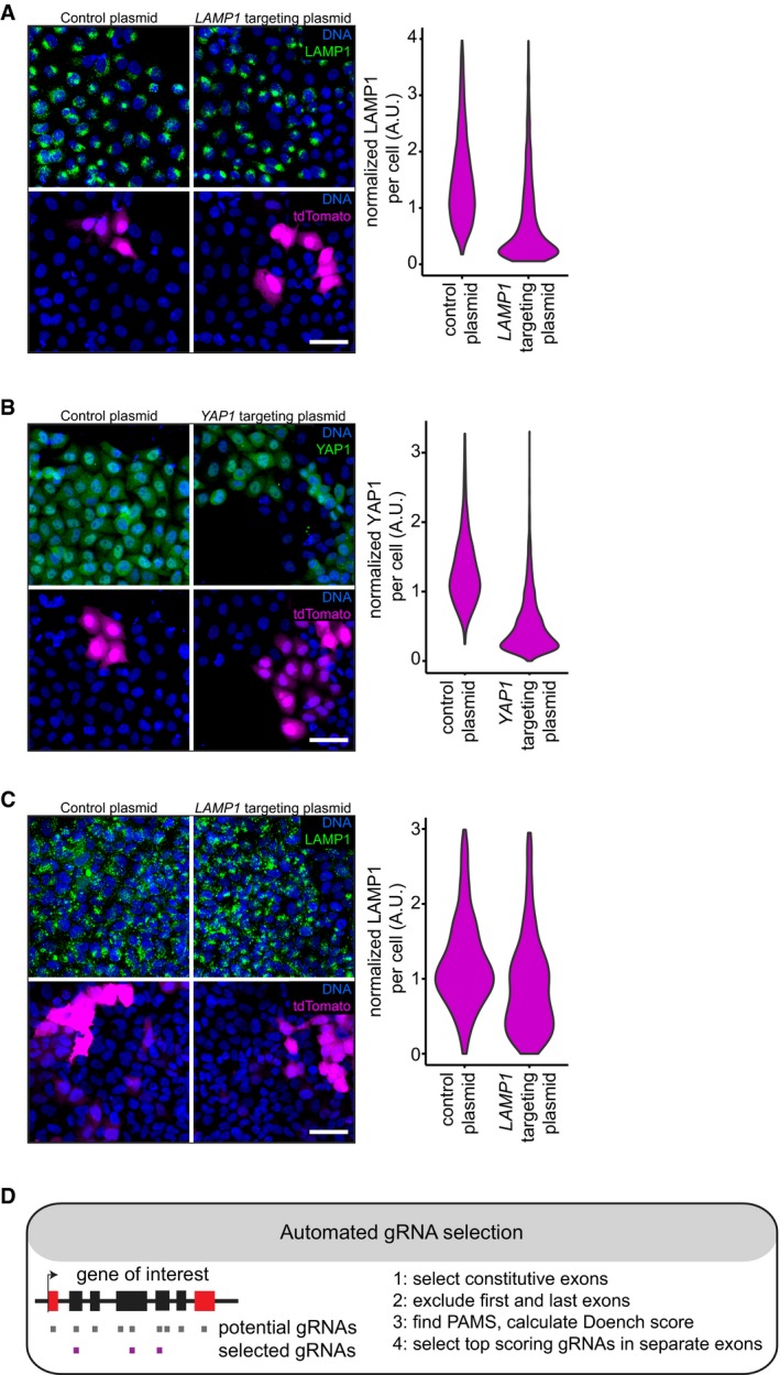
“To systematically test our approach across multiple genes, we automated the selection of gRNA sequences with high predicted ontarget efficacy (Doench et al, 2014).”
MethodGarcía-Alonso, C. R., Pérez-Naranjo, L. M., & Fernández-Caballero, J. C. (2014) · Annals of Operations Research · doi:10.1007/s10479-011-0841-3 · Cited by 1,353
High-throughput functional genomics using CRISPR-Cas9
Shalem, O., Sanjana, N. E., & Zhang, F. (2015) · Nature Reviews Genetics · doi:10.1038/nrg3899 · Cited by 1,233 · cited 2×
“Importantly, CRISPR-Cas9 is not 100% effective in all targeted cells, which can be the result of in-frame repair of the CRISPR-Cas9induced DNA lesions, a failure to target all functional alleles or limited efficacy of the CRISPR-Cas9 system (Shalem et al, 2015).”
BackgroundMethod
Cited by
Optical Pooled Screens in Human Cells
Feldman, D., Singh, A., Schmid-Burgk, J. L., Carlson, R. J., Mezger, A., Garrity, A. J., Zhang, F., & Blainey, P. C. (2019) · Cell · doi:10.1016/j.cell.2019.09.016 · Cited by 321
A new era in functional genomics screens
Przybyla, L., & Gilbert, L. A. (2022) · Nature Reviews Genetics · doi:10.1038/s41576-021-00409-w · Cited by 281
MIGA2 Links Mitochondria, the ER, and Lipid Droplets and Promotes De Novo Lipogenesis in Adipocytes
Freyre, C. A. C., Rauher, P. C., Ejsing, C. S., & Klemm, R. W. (2019) · Molecular Cell · doi:10.1016/j.molcel.2019.09.011 · Cited by 253
Machine learning and image-based profiling in drug discovery
Scheeder, C., Heigwer, F., & Boutros, M. (2018) · Current Opinion in Systems Biology · doi:10.1016/j.coisb.2018.05.004 · Cited by 169
Christopher, J. A., Stadler, C., Martin, C. E., Morgenstern, M., Pan, Y., Betsinger, C. N., Rattray, D. G., Mahdessian, D., Gingras, A.-C., Warscheid, B., et al. (2021) · Nature Reviews Methods Primers · doi:10.1038/s43586-021-00029-y · Cited by 129
Kilchrist, K. V., Dimobi, S. C., Jackson, M. A., Evans, B. C., Werfel, T. A., Dailing, E. A., Bedingfield, S. K., Kelly, I. B., & Duvall, C. L. (2019) · ACS Nano · doi:10.1021/acsnano.8b05482 · Cited by 120
Imaging-based pooled CRISPR screening reveals regulators of lncRNA localization
Wang, C., Lu, T., Emanuel, G., Babcock, H. P., & Zhuang, X. (2019) · Proceedings of the National Academy of Sciences · doi:10.1073/pnas.1903808116 · Cited by 111
Image-based high-content screening in drug discovery
Lin, S., Schorpp, K., Rothenaigner, I., & Hadian, K. (2020) · Drug Discovery Today · doi:10.1016/j.drudis.2020.06.001 · Cited by 124
Pooled CRISPR screens with imaging on microraft arrays reveals stress granule-regulatory factors
Wheeler, E. C., Vu, A. Q., Einstein, J. M., DiSalvo, M., Ahmed, N., Van Nostrand, E. L., Shishkin, A. A., Jin, W., Allbritton, N. L., & Yeo, G. W. (2020) · Nature Methods · doi:10.1038/s41592-020-0826-8 · Cited by 87
“Subcellular phenotypes account for both physiological and pathological changes in cell identity and function, such as transcription factor translocation into the nucleus 9 , protein localization to cellular sub-structures 10 , or mis-localization of proteins into disease-associated aggregates 11 .”
High-content imaging-based pooled CRISPR screens in mammalian cells
Yan, X., Stuurman, N., Ribeiro, S. A., Tanenbaum, M. E., Horlbeck, M. A., Liem, C. R., Jost, M., Weissman, J. S., & Vale, R. D. (2021) · Journal of Cell Biology · doi:10.1083/jcb.202008158 · Cited by 87
High-throughput genome-wide phenotypic screening via immunomagnetic cell sorting
Mair, B., Aldridge, P. M., Atwal, R. S., Philpott, D. N., Zhang, M., Masud, S. N., Labib, M., Tong, A. H. Y., Sargent, E. H., Angers, S., et al. (2019) · Nature Biomedical Engineering · doi:10.1038/s41551-019-0454-8 · Cited by 88
Taemaitree, L., Shivalingam, A., El-Sagheer, A. H., & Brown, T. (2019) · Nature Communications · doi:10.1038/s41467-019-09600-4 · Cited by 68
Ušaj, M. M., Sahin, N., Friesen, H., Pons, C., Ušaj, M., Masinas, M. P. D., Shuteriqi, E., Shkurin, A., Aloy, P., Morris, Q., et al. (2020) · Molecular Systems Biology · doi:10.15252/msb.20199243 · Cited by 60
“…conserved bioprocess that plays a central role in eukaryotic cell biology, mediating the internalization of receptors, nutrients and other molecules, controlling the lipid and protein composition of the plasma membrane and the coupling of different intracellular compartments (Goode et al , 2015).”
Pooled genetic perturbation screens with image-based phenotypes
Feldman, D., Funk, L., Le, A., Carlson, R. J., Leiken, M. D., Tsai, F., Soong, B., Singh, A., & Blainey, P. C. (2022) · Nature Protocols · doi:10.1038/s41596-021-00653-8 · Cited by 59
Single-cell image analysis to explore cell-to-cell heterogeneity in isogenic populations
Ušaj, M. M., Yeung, C. H. L., Friesen, H., Boone, C. M., & Andrews, B. (2021) · Cell Systems · doi:10.1016/j.cels.2021.05.010 · Cited by 46
Application of Machine Learning in Spatial Proteomics
Mou, M., Pan, Z., Lu, M., Sun, H., Wang, Y., Luo, Y., & Zhu, F. (2022) · Journal of Chemical Information and Modeling · doi:10.1021/acs.jcim.2c01161 · Cited by 50
Massively Parallel CRISPR‐Based Genetic Perturbation Screening at Single‐Cell Resolution
Cheng, J., Lin, G., Wang, T., Wang, Y., Guo, W., Liao, J., Yang, P., Chen, J., Shao, X., Lu, X., et al. (2022) · Advanced Science · doi:10.1002/advs.202204484 · Cited by 48
Yeast genetic interaction screens in the age of CRISPR/Cas
Adames, N. R., Gallegos, J. E., & Peccoud, J. (2018) · Current Genetics · doi:10.1007/s00294-018-0887-8 · Cited by 42
Genetic interaction networks in cancer cells
Mair, B., Moffat, J., Boone, C. M., & Andrews, B. (2019) · Current Opinion in Genetics & Development · doi:10.1016/j.gde.2019.03.002 · Cited by 35
Yin, J., Frick, L. D., Scheidmann, M. C., Liu, T., Trevisan, C., Dhingra, A., Spinelli, A., Wu, Y., Yao, L., Vena, D. L., et al. (2024) · Nature Biomedical Engineering · doi:10.1038/s41551-024-01278-4 · Cited by 59
Self-citationFolkerts, J., Gaudenzio, N., Maurer, M., Hendriks, R. W., Stadhouders, R., Tam, S., & Galli, S. J. (2020) · Nature Protocols · doi:10.1038/s41596-019-0288-6 · Cited by 37
Morphological profiling of human T and NK lymphocytes by high-content cell imaging
German, Y., Vulliard, L., Kamnev, A., Pfajfer, L., Huemer, J., Mautner, A.-K., Rubio, A., Kalinichenko, A., Boztuǧ, K., Ferrand, A., et al. (2021) · Cell Reports · doi:10.1016/j.celrep.2021.109318 · Cited by 38
Ross‐Thriepland, D., Bornot, A., Butler, L., Desai, A. S., Jaiswal, H., Peel, S. E., Hunter, M. R., Odunze, U., Isherwood, B. J., & Gianni, D. (2020) · SLAS DISCOVERY · doi:10.1177/2472555220925770 · Cited by 28
Blank spots on the map: some current questions on nuclear organization and genome architecture
Adriaens, C., Serebryannyy, L., Feric, M., Schibler, A., Meaburn, K. J., Kubben, N., Trzaskoma, P., Shachar, S., Vidak, S., Finn, E. H., et al. (2018) · Histochemistry and Cell Biology · doi:10.1007/s00418-018-1726-1 · Cited by 33
Stossi, F., Singh, P. K., Safari, K., Marini, M., Labate, D., & Mancini, M. A. (2023) · Biochemical Pharmacology · doi:10.1016/j.bcp.2023.115770 · Cited by 33
“Some recent examples coupling CRISPR and imaging can be found in [126 – 128].”
BackgroundMethodDiscovering metabolic disease gene interactions by correlated effects on cellular morphology
Jiao, Y., Ahmed, U., Sim, M. F. M., Bejar, A., Zhang, X., Talukder, M., Rice, R., Flannick, J. A., Podgornaia, A., Reilly, D. F., et al. (2019) · Molecular Metabolism · doi:10.1016/j.molmet.2019.03.001 · Cited by 23
“Cellular morphology has been successfully deployed to interrogate subcellular mechanisms [6] and several recent studies have demonstrated the feasibility of combining morphological readout with gain/loss-of-function genetic perturbations to functionally annotate genes into molecular pathways [7,8].”
Backgroundle Sage, C., Lawo, S., & Cross, B. C. S. (2019) · SLAS DISCOVERY · doi:10.1177/2472555219883621 · Cited by 24
Co-translational targeting of transcripts to endosomesPreprint
Popovic, D., Nijenhuis, W., Kapitein, L. C., & Pelkmans, L. (2020) · bioRxiv (Cold Spring Harbor Laboratory) · doi:10.1101/2020.07.17.208652 · Cited by 21
Self-citationYin, J., Frick, L. D., Scheidmann, M. C., Liu, T., Trevisan, C., Dhingra, A., Spinelli, A., Wu, Y., Yao, L., Vena, D. L., et al. (2022) · bioRxiv (Cold Spring Harbor Laboratory) · doi:10.1101/2022.05.25.493370 · Cited by 19
“E, Robust ablation of genes inadequately disrupted by single sgRNAs(de Groot et al., 2018).”
BackgroundMethodSelf-citationProtein research in millets: current status and way forward
Ceasar, S. A., Prabhu, S., & Ebeed, H. T. (2024) · Planta · doi:10.1007/s00425-024-04478-z · Cited by 19
“Imaging has been an increasingly effective method for establishing a genetic connection to phenotype, including subcellular location alterations, thanks to recent developments in genome-wide RNA interference technologies and CRISPR-Cas9-based tools (De Groot et al. 2018).”
Guerriero, M. L., Corrigan, A. M., Bornot, A., Firth, M. A., O'Shea, P. J., Ross‐Thriepland, D., & Peel, S. E. (2020) · SLAS DISCOVERY · doi:10.1177/2472555220921132 · Cited by 13
Applications of Functional Genomics for Drug Discovery
Kabadi, A. M., McDonnell, E., Frank, C. L., & Drowley, L. (2020) · SLAS DISCOVERY · doi:10.1177/2472555220902092 · Cited by 16
Robertson, N. R., Trivedi, V., Lupish, B., Ramesh, A., Aguilar, Y., Carrera, S., Lee, S. C., Arteaga, A., Nguyen, A., Lenert-Mondou, C., et al. (2024) · Metabolic Engineering · doi:10.1016/j.ymben.2024.09.005 · Cited by 16
A solid‐phase transfection platform for arrayed CRISPR screens
Serçin, Ö., Reither, S., Roidos, P., Ballin, N., Palikyras, S., Baginska, A., Rein, K., Prada, M. L., Halavatyi, A. A., Winter, H., et al. (2019) · Molecular Systems Biology · doi:10.15252/msb.20198983 · Cited by 11
Daga, K. R., Priyadarshani, P., Larey, A. M., Rui, K., Mortensen, L. J., & Marklein, R. A. (2021) · Current Opinion in Biomedical Engineering · doi:10.1016/j.cobme.2021.100352 · Cited by 11
Identifying chromatin features that regulate gene expression distribution
Zhang, T., Foreman, R., & Wollman, R. (2020) · Scientific Reports · doi:10.1038/s41598-020-77638-2 · Cited by 8
Wong, C. H., Wingett, S., Chen, Q., Taliaferro, J. M., Ross‐Thriepland, D., & Bullock, S. L. (2023) · bioRxiv (Cold Spring Harbor Laboratory) · doi:10.1101/2023.03.01.530592 · Cited by 10
Yenkin, A. L., Bramley, J. C., Kremitzki, C. L., Waligorski, J. E., Liebeskind, M. J., Xu, X. E., Chandrasekaran, V., Vakaki, M. A., Bachman, G. W., Mitra, R. D., et al. (2022) · Communications Biology · doi:10.1038/s42003-022-04089-y · Cited by 10
The necessity and power of random, under-sampled experiments in biologyPreprint
Cleary, B., & Regev, A. (2020) · arXiv (Cornell University) · doi:10.48550/arxiv.2012.12961 · Cited by 17
“More recently, CRISPR perturbations have been paired with a high-content optical readout 66,67 , enabling even larger scale studies 67 .”
BackgroundO'Shea, P. J., Wildenhain, J., Leveridge, M., Revankar, C. M., Yang, J., Bradley, J., Firth, M. A., Pilling, J. E., Piper, D. R., Chesnut, J. D., et al. (2020) · SLAS DISCOVERY · doi:10.1177/2472555220926160 · Cited by 7
Labitigan, R. L. D., Sanborn, A. L., Hao, C. V., Chan, C. K., Belliveau, N. M., Brown, E. M., Mehrotra, M., & Theriot, J. A. (2024) · eLife · doi:10.7554/elife.94964.1 · Cited by 9
TheCellVision.org: A Database for Visualizing and Mining High-Content Cell Imaging Projects
Masinas, M. P. D., Ušaj, M. M., Ušaj, M., Boone, C. M., & Andrews, B. (2020) · G3 Genes Genomes Genetics · doi:10.1534/g3.120.401570 · Cited by 7
Machine learning approach for discrimination of genotypes based on bright-field cellular images
Suzuki, G., Saitō, Y., Seki, M., Evans-Yamamoto, D., Negishi, M., Kakoi, K., Kawai, H., Landry, C. R., Yachie, N., & Mitsuyama, T. (2021) · npj Systems Biology and Applications · doi:10.1038/s41540-021-00190-w · Cited by 6
Wong, C. H., Wingett, S., Chen, Q., Hunter, M. R., Taliaferro, J. M., Ross‐Thriepland, D., & Bullock, S. L. (2024) · The Journal of Cell Biology · doi:10.1083/jcb.202306048 · Cited by 14
“This strategy has previously been used to assess cell state (Way et al., 2021), predict biological activity of small molecules (Simm et al., 2018), and identify co-functional genes in functional genomics data (Rohban et al., 2017; de Groot et al., 2018; Duan et al., 2021; Funk et al., 2022).”
Labitigan, R. L. D., Sanborn, A. L., Hao, C. V., Chan, C. K., Belliveau, N. M., Brown, E. M., Mehrotra, M., & Theriot, J. A. (2022) · bioRxiv (Cold Spring Harbor Laboratory) · doi:10.1101/2022.12.27.522042 · Cited by 7
Barragán-Álvarez, C. P., Flores-Fernández, J. M., Hernández‐Pérez, O. R., Ávila‐González, D., Díaz, N. F., Padilla‐Camberos, E., Dublán‐García, O., Gómez‐Oliván, L. M., & Diaz-Martinez, N. E. (2022) · Frontiers in Cell and Developmental Biology · doi:10.3389/fcell.2022.947769 · Cited by 6
Taemaitree, L., Shivalingam, A., El-Sagheer, A. H., & Brown, T. (2020) · Methods in molecular biology · doi:10.1007/978-1-0716-0687-2_5 · Cited by 3
Genetic approaches to revealing the principles of nuclear architecture
Askjaer, P., & Harr, J. C. (2020) · Current Opinion in Genetics & Development · doi:10.1016/j.gde.2020.11.001 · Cited by 4
Robertson, N. R., Trivedi, V., Lupish, B., Ramesh, A., Aguilar, Y., Arteaga, A., Nguyen, A., Lee, S. C., Lenert-Mondou, C., Harland‐Dunaway, M., et al. (2024) · bioRxiv (Cold Spring Harbor Laboratory) · doi:10.1101/2024.06.20.599746 · Cited by 4
Bao, F., Li, L., Hammerlindl, H., Shen, S. Q., Hammerlindl, S., Altschuler, S. J., & Wu, L. F. (2025) · Nature Biotechnology · doi:10.1038/s41587-025-02729-2 · Cited by 6
“Since the introduction of cell phenotypic profiling two decades ago 16 , academic and industrial laboratories have applied HCSs to diverse applications, creating a rapidly growing number of datasets 29–40 .”
Kim, C. S., Cairns, J., Quarantotti, V., Kaczkowski, B., Wang, Y., Könings, P., & Zhang, X. (2024) · PLoS ONE · doi:10.1371/journal.pone.0307445 · Cited by 5
Mutagenomics for Functional Analysis of Plant Genome using CRISPR Library Screen
Dongre, N., Kumari, D. D., Mahto, B. K., Arya, S. S., & Lenka, S. K. (2021) · Concepts and strategies in plant sciences · doi:10.1007/978-3-030-64994-4_15 · Cited by 3
Adding a Chemical Biology Twist to CRISPR Screening
Huang, S., & Baskin, J. M. (2022) · Israel Journal of Chemistry · doi:10.1002/ijch.202200056 · Cited by 3
Labitigan, R. L. D., Sanborn, A. L., Hao, C. V., Chan, C. K., Belliveau, N. M., Brown, E. M., Mehrotra, M., & Theriot, J. A. (2024) · eLife · doi:10.7554/elife.94964 · Cited by 10
A Comprehensive Review on CRISPR-Based Screening and Its Applications
Sichani, A. S., Hassani, M., Gila, F., Shafieipour, N., Dabbaghipour, R., Heidari, Z., Sisakht, M., Hassani, M., & Fallahi, J. (2026) · Molecular Biotechnology · doi:10.1007/s12033-026-01562-5 · Cited by 2
“Arrayed gRNA libraries have been developed in various formats, including viral vector-based [78], chemically synthesized [79], and plasmid-based systems [80, 81].”
AI in Genomics: From Variant Calling to Multi‐Omics Integration
Sultana, H., Mohanty, S., Solomon, A. D., Iqbal, M. Y., Wani, A. K., Kumar, V., & Khattri, A. (2026) · BioEssays · doi:10.1002/bies.70160 · Cited by 3
Applying Systems Toxicology Methods to Drug Safety
Talikka, M., Belcastro, V., Boué, S., Marescotti, D., Hoeng, J., & Peitsch, M. C. (2019) · Elsevier eBooks · doi:10.1016/b978-0-12-801238-3.11522-3 · Cited by 2
Ušaj, M. M., Sahin, N., Friesen, H., Pons, C., Ušaj, M., Masinas, M. P. D., Shuteriqi, E., Shkurin, A., Aloy, P., Morris, Q., et al. (2023) · doi:10.32920/22149032.v1 · Cited by 2
Application of Arrayed CRISPR/cas9 Screen and its Data Analysis: a Systematic Review
Sun, S., Chang, C., Huang, C., & Zhang, X. D. (2018) · IEEE International Conference on Bioinformatics and Biomedicine (BIBM) · doi:10.1109/bibm.2018.8621522 · Cited by 2
“Reinoud de Groot and his team published their highthroughput arrayed screen strategy combined with single-cell phenotypes profiling which can enable the screen with more informative readouts [43].”
BackgroundHCS Methodology for Helping in Lab Scale Image-Based Assays
Soriano, J., Mata, G., & Megías, D. (2019) · Methods in molecular biology · doi:10.1007/978-1-4939-9686-5_15 · Cited by 1
Scalable, FACS-Free Genome-Wide Phenotypic ScreeningPreprint
Mair, B., Aldridge, P. M., Atwal, R. S., Masud, S. N., Zhang, M., Philpott, D. N., Tong, A. H. Y., Sargent, E. H., Angers, S., Moffat, J., et al. (2019) · bioRxiv (Cold Spring Harbor Laboratory) · doi:10.1101/612887 · Cited by 1
Mitochondrial Phenotypes Distinguish Pathogenic MFN2 Mutations by Pooled Functional Genomics ScreenPreprint
Yenkin, A. L., Bramley, J. C., Kremitzki, C. L., Waligorski, J. E., Liebeskind, M. J., Xu, X. E., Vakaki, M. A., Chandrasekaran, V. D., Mitra, R. D., Milbrandt, J., et al. (2021) · bioRxiv (Cold Spring Harbor Laboratory) · doi:10.1101/2021.03.12.434746 · Cited by 1
Shaping vessels and microenvironment: adipose stromal cells in retinal-related diseases
Terlizzi, V. (2019) · University of Groningen research database (University of Groningen / Centre for Information Technology)
High-Content Screening in Cell Biology
Ketteler, R., & Kriston-Vizi, J. (2022) · Elsevier eBooks · doi:10.1016/b978-0-12-821618-7.00032-8
CRISPR/Cas-based Functional Genomic Approaches to Phenotypic Screening
Isherwood, B. J., Peel, S. E., Dunn, S., & McDermott, U. (2020) · Royal Society of Chemistry eBooks · doi:10.1039/9781839160721-00058
Commercially Available Reagents and Contract Research Services for CRISPR‐Based Studies
Maratou, K., Cheng, A. T., Behan, F. M., Sun, N., & Lu, Q. (2022) · Wiley eBooks · doi:10.1002/9781119671404.ch4
D’Gama, J. D., Park, J. S., & Waldor, M. K. (2022) · Wiley eBooks · doi:10.1002/9781683673798.ch13
TheCellVision.org: A Database for Visualizing and Mining High-Content Cell Imaging Projects
Masinas, M. P. D., Ušaj, M. M., Ušaj, M., Boone, C. M., & Andrews, B. (2023) · doi:10.32920/22149035.v1
TheCellVision.org: A Database for Visualizing and Mining High-Content Cell Imaging Projects
Masinas, M. P. D., Ušaj, M. M., Ušaj, M., Boone, C. M., & Andrews, B. (2023) · doi:10.32920/22149035
Ušaj, M. M., Sahin, N., Friesen, H., Pons, C., Ušaj, M., Masinas, M. P. D., Shuteriqi, E., Shkurin, A., Aloy, P., Morris, Q., et al. (2023) · doi:10.32920/22149032
Ziegler, M., Böger, C., Alecu, J. E., Kim, H., Saffari, A., Davies, A. K., Şahin, M., & Ebrahimi‐Fakhari, D. (2025) · bioRxiv (Cold Spring Harbor Laboratory) · doi:10.1101/2025.02.22.639634 · Cited by 2
Ziegler, M., Böger, C., Alecu, J. E., Kim, H., Saffari, A., Davies, A. K., Şahin, M., & Ebrahimi‐Fakhari, D. (2025) · SSRN Electronic Journal · doi:10.2139/ssrn.5165225
ZIEGLER, M. L., Guenter, C., Alecu, J. E., Xue, X., Kim, H. M., Saffari, A., Davies, A. K., Şahin, M., & Ebrahimi‐Fakhari, D. (2026) · JCI Insight · doi:10.1172/jci.insight.202204
“However, only a limited number of arrayed CRISPR screens have been conducted to date (23–25).”
Metadata
- Authors
- de Groot, R., Lüthi, J., Lindsay, H., Holtackers, R., & Pelkmans, L.
- Affiliations
- University of Zurich (CH)
- Corresponding author
- Pelkmans, Lucas
- Venue
- Molecular Systems Biology
- Published
- 2018-01-01
- Volume
- 14
- Issue
- 1
- Pages
- e8064-e8064
- ISSN
- 1744-4292 (ISSN-L)
- PMC
- PMC5787707
- OpenAlex
- W2785088317
- Cited by
- 74
- References
- 35
- Citations by year
- 2026: 3 · 2025: 3 · 2024: 8 · 2023: 6 · 2022: 11 · 2021: 9 · 2020: 16 · 2019: 14 · 2018: 4
- FWCI
- 3.82
- Type
- journal article
- Language
- en
- Open access
- gold
- doi.org
- Licence
- other-oa
- creativecommons.org/licenses/by/4.0/ (tdm)
- creativecommons.org/licenses/by/4.0/ (vor, 22 d)
- Locations
- Molecular Systems Biology (publishedVersion, other-oa)
- PubMed Central (publishedVersion, cc-by)
- PubMed (publishedVersion)
- Zurich Open Repository and Archive (University of Zurich) (acceptedVersion, cc-by)
- DOAJ (DOAJ: Directory of Open Access Journals) (submittedVersion)
- Repository for Publications and Research Data (ETH Zurich) (submittedVersion, cc-by)
- Zurich Open Repository and Archive (University of Zurich) (submittedVersion, cc-by)
- ETH Zürich Research Collection (cc-by)
- Zurich Open Repository and Archive (University of Zurich)
- Funders
- SystemsX.ch (TPdF2015/342)
- Topics
- Life Sciences › Biochemistry, Genetics and Molecular Biology › Molecular Biology › CRISPR and Genetic Engineering
- Life Sciences › Biochemistry, Genetics and Molecular Biology › Biophysics › Cell Image Analysis Techniques
- Life Sciences › Biochemistry, Genetics and Molecular Biology › Molecular Biology › Single-cell and spatial transcriptomics
- Keywords
- image-based phenotyping, nuclear pore complex, cell morphology, subcellular localization, genetic heterogeneity, high-content imaging
- MeSH terms
- Machine Learning, HeLa Cells, Humans, Phenotype, Gene Library, Nuclear Pore, Gene Knockout Techniques, Single-Cell Analysis, CRISPR-Cas Systems
About these checks
Registry, retraction, Cites and statistics results are computed, not judged, with one exception: a printed reference that no index lists is read once by a model before it counts as a citation error. Claims are extracted by a model and kept only when their quote appears verbatim in the text; each cited source is then checked, with a model’s help, for whether it says what the claim relies on. Those verdicts can be wrong — read the claim’s quote and the cited source before relying on one.
Statistics and arithmetic are recomputed, not judged.
Confidence bands are shown only for fields calibrated on our annotated test set. None is calibrated yet, so no band is shown.
What counts as a claim ›How we measure confidence ›How we check this ›
Are you an author, or do you see a problem with this page? Request a review
Data sources
Bibliographic data: Semantic Scholar, OpenAlex, Crossref.
Contains information from the Semantic Scholar Academic Graph Datasets, made available under the ODC Attribution License (ODC-BY 1.0).
Semantic Scholar’s data is described in: Kinney, R., et al. (2023). The Semantic Scholar Open Data Platform. arXiv:2301.10140.
