

The DOI matches its Crossref record; 5 of the 5 references it cites were found in a registry or an index.
miR-212-3p attenuates neuroinflammation of rats with Alzheimer's disease via regulating the SP1/BACE1/NLRP3/Caspase-1 signaling pathway
Abstract
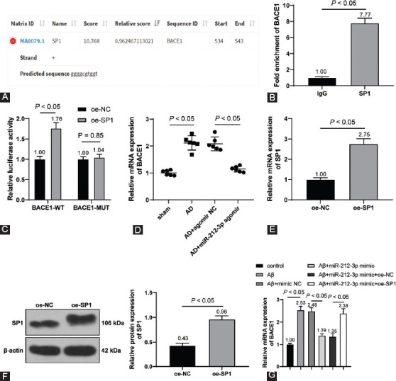
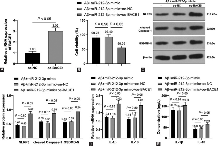
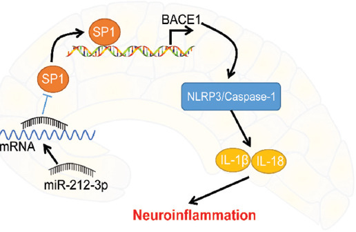
Alzheimer's disease (AD) ranks as the leading cause of dementia. MicroRNA (miR)-212-3p has been identified to exert neuroprotective effects on brain disorders. The current study analyzed the protective role of miR-212-3p in AD rats via regulating the nucleotide-binding oligomerization domain-like receptor family pyrin domain containing 3 (NLRP3)/Caspase-1 signaling pathway. The AD rat model was established via injection of amyloid-β 1-42 (Aβ1-42), followed by the Morris water maze test. The morphology and functions of neurons were observed. Furthermore, miR-212-3p, NLRP3, cleaved Caspase-1, gasdermin D N-terminus, interleukin (IL)-1β and IL-18 expressions were measured. H19-7 cells were treated with Aβ1-42 to establish the AD cell model, followed by an assessment of cell viability and pyroptosis. Downstream targets of miR-212-3p and specificity protein 1 (SP1), as well as beta-site amyloid precursor protein cleaving enzyme 1 (BACE1) were predicted by databases and testified using dual-luciferase and chromatin immunoprecipitation assays. miR-212-3p was weakly expressed in AD rats. miR-212-3p overexpression was linked to improved learning and memory capacities of AD rats and reduced neuronal pyroptosis linked to neuroinflammation attenuation. In vitro, miR-212-3p improved viability and suppressed pyroptosis of neurons via inhibiting NLRP3/Caspase-1. Overall, miR-212-3p inhibited SP1 expression to block BACE1-induced activation of NLRP3/Caspase-1, thereby attenuating neuroinflammation of AD rats.
Overview
The claims and statistics of this paper haven’t been checked yet.
Run the check — free, no account needed. It reads the open-access text and checks the sources it cites.
Integrity signals
Registry
Cites
Statistics
Figures
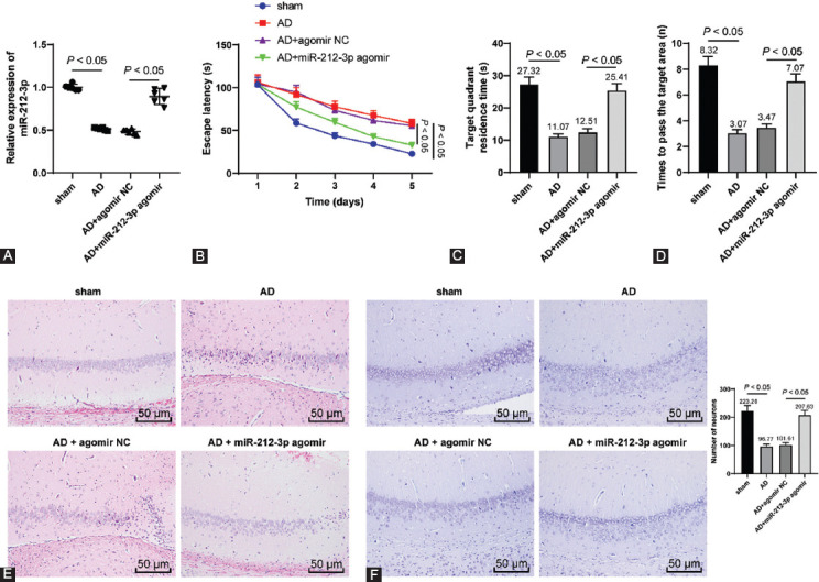
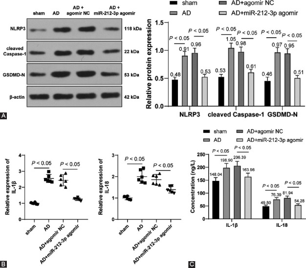
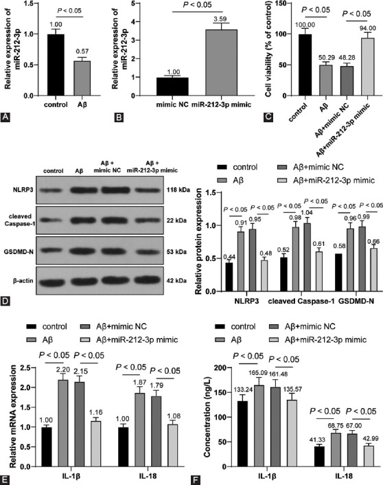
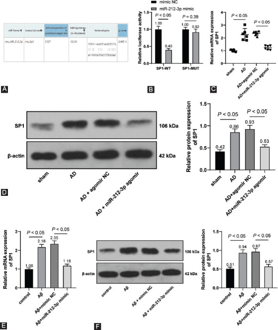
Verification
Claims the paper makes
The claims and statistics of this paper haven’t been checked yet.
Run the check — free, no account needed. It reads the open-access text and checks the sources it cites.
Claims other papers make about this one
Each is checked against this paper’s own text: does it say what the citing paper says it does?
The source backs part of the claim, not all of it — or backs it for a different population, condition or comparison.
In the citing paper
“Additional studies suggest that targeting miR-212-5p expression is neuroprotective in models of PD, AD, and traumatic brain injury [63–65].”
From A role for astrocytic miR-129-5p in frontotemporal dementia (2025) · cites this paper as [65]
Experimental — accuracy not yet measured
Self-check
Percentages, sums and reported differences re-computed from the paper’s own numbers.
The self-check needs the paper’s full text.
Citations
Cites
The NLRP3 Inflammasome: an overview of mechanisms of activation and regulation
Kelley, N., Jeltema, D., Duan, Y., & He, Y. (2019) · International Journal of Molecular Sciences · doi:10.3390/ijms20133328 · Cited by 3,539
Comprehensive Review on Alzheimer’s Disease: Causes and Treatment
Breijyeh, Z., & Karaman, R. (2020) · Molecules · doi:10.3390/molecules25245789 · Cited by 2,789
Brites, D., & Fernandes, A. (2015) · Frontiers in Cellular Neuroscience · doi:10.3389/fncel.2015.00476 · Cited by 599 · cited 1×
“Hard done work of our peers has unveiled that dysregulation of miRNAs is associated with neuroinflammation in AD [39].”
BackgroundManagement of oxidative stress and other pathologies in Alzheimer’s disease
Simunkova, M., Alwasel, S. H., Alhazza, I. M., Jomova, K., Kollar, V., Rusko, M., & Valko, M. (2019) · Archives of Toxicology · doi:10.1007/s00204-019-02538-y · Cited by 303
Neuroinflammation and neurogenesis in Alzheimer's disease and potential therapeutic approaches
Sung, P.-S., Lin, P.-Y., Liu, C.-H., Su, H.-C., & Tsai, K.-J. (2020) · International Journal of Molecular Sciences · doi:10.3390/ijms21030701 · Cited by 193
Research works printed but not linked: 48
- [1]Soria Lopez JA Gonzalez HM Leger GC Alzheimer's disease Handb Clin Neurol 2019 167 231 55 https://doi.org/10.1016/B978-0-12-804766-8.00013-3 31753135 · DOI
- [3]Tanaka M Toldi J Vecsei L Exploring the etiological links behind neurodegenerative diseases:Inflammatory cytokines and bioactive kynurenines Int J Mol Sci 2020 21 7 2431 https://doi.org/10.3390/ijms21072431 PMC7177899 32244523 · DOI
- [4]Brunetti D Catania A Viscomi C Deleidi M Bindoff LA Ghezzi D Role of PITRM1 in mitochondrial dysfunction and neurodegeneration Biomedicines 2021 9 7 833 https://doi.org/10.3390/biomedicines9070833 34356897 PMC8301332 · DOI
- [5]Sanchez-Sarasua S Fernandez-Perez I Espinosa-Fernandez V Sanchez-Perez AM Ledesma JC Can we treat neuroinflammation in Alzheimer's disease? Int J Mol Sci 2020 21 22 8751 https://doi.org/10.3390/ijms2122∯ PMC7699542 33228179 · DOI
- [8]Voet S Srinivasan S Lamkanfi M van Loo G Inflammasomes in neuroinflammatory and neurodegenerative diseases EMBO Mol Med 2019 11 6 e10248 https://doi.org/10.15252/emmm.201810248 31015277 PMC6554670 · DOI · Paper page
- [10]Li J Zhuang L Luo X Liang J Sun E He Y Protection of MCC950 against Alzheimer's disease via inhibiting neuronal pyroptosis in SAMP8 mice Exp Brain Res 2020 238 11 2603 14 https://doi.org/10.1007/s00221-020-05916-6 32892233 · DOI
- [11]Thawkar BS Kaur G Inhibitors of NF-kappaB and P2X7/NLRP3/Caspase 1 pathway in microglia:Novel therapeutic opportunities in neuroinflammation induced early-stage Alzheimer's disease J Neuroimmunol 2019 326 62 74 https://doi.org/10.1016/j.jneuroim.2018.11.010 30502599 · DOI
- [12]Brito LM Ribeiro-Dos-Santos A Vidal AF de Araujo GS Differential expression and miRNA-gene interactions in early and late mild cognitive impairment Biology (Basel) 2020 9 9 251 https://doi.org/10.3390/biology9090251 PMC7565463 32872134 · DOI
- [13]Catanesi M d'Angelo M Tupone MG Benedetti E Giordano A Castelli V MicroRNAs dysregulation and mitochondrial dysfunction in neurodegenerative diseases Int J Mol Sci 2020 21 17 5986 https://doi.org/10.3390/ijms21175986 PMC7504116 32825273 · DOI
- [14]Swarbrick S Wragg N Ghosh S Stolzing A Systematic review of miRNA as biomarkers in Alzheimer's disease Mol Neurobiol 2019 56 9 6156 67 https://doi.org/10.1007/s12035-019-1500-y 30734227 PMC6682547 · DOI
- [15]Aten S Hansen KF Hoyt KR Obrietan K The miR-132/212 locus:A complex regulator of neuronal plasticity, gene expression and cognition RNA Dis 2016 3 2 e1375 27713923 PMC5049877
- [16]Wang Y Chang Q MicroRNA miR-212 regulates PDCD4 to attenuate Abeta25-35-induced neurotoxicity via PI3K/AKT signaling pathway in Alzheimer's disease Biotechnol Lett 2020 42 9 1789 97 https://doi.org/10.1007/s10529-020-02915-z 32474742 · DOI
- [17]Wang Y Veremeyko T Wong AH El Fatimy R Wei Z Cai W Downregulation of miR-132/212 impairs S-nitrosylation balance and induces tau phosphorylation in Alzheimer's disease Neurobiol Aging 2017 51 156 66 https://doi.org/10.1016/j.neurobiolaging.2016.12.015 28089352 · DOI · Paper page
- [18]Li X Zou ZZ Wen M Xie YZ Peng KJ Luo T ZLM-7 inhibits the occurrence and angiogenesis of breast cancer through miR-212-3p/Sp1/VEGFA signal axis Mol Med 2020 26 1 109 33187481 PMC7666510
- [19]Wang R Yang Y Wang H He Y Li C MiR-29c protects against inflammation and apoptosis in Parkinson's disease model in vivo and in vitro by targeting SP1 Clin Exp Pharmacol Physiol 2020 47 3 372 82 https://doi.org/10.1111/1440-1681.13212 31732967 · DOI
- [20]Qin J Zhang X Wang Z Li J Zhang Z Gao L Presenilin 2 deficiency facilitates Abeta-induced neuroinflammation and injury by upregulating P2X7 expression Sci China Life Sci 2017 60 2 189 201 https://doi.org/10.1007/s11427-016-0347-4 28120269 · DOI
- [21]Zhang H Wang X Xu P Ji X Chi T Liu P Tolfenamic acid inhibits GSK-3beta and PP2A mediated tau hyperphosphorylation in Alzheimer's disease models J Physiol Sci 2020 70 1 29 32517647 PMC10717460
- [22]Hou X Adeosun SO Zhang Q Barlow B Brents M Zheng B Differential contributions of ApoE4 and female sex to BACE1 activity and expression mediate Abeta deposition and learning and memory in mouse models of Alzheimer's disease Front Aging Neurosci 2015 7 207 https://doi.org/10.3389/fnagi.2015.00207 26582141 PMC4628114 · DOI
- [23]Lombardo S Chiacchiaretta M Tarr A Kim W Cao T Sigal G BACE1 partial deletion induces synaptic plasticity deficit in adult mice Sci Rep 2019 9 1 19877 https://doi.org/10.1038/s41598-019-56329-7 31882662 PMC6934620 · DOI
- [24]National Research Council (US) Committee for the Update of the Guide for the Care and Use of Laboratory Animals Guide for the Care and Use of Laboratory Animals 2011 Washington, DC National Academies Press US 21595115
- [25]Paxinos G Watson CR Emson PC AChE-stained horizontal sections of the rat brain in stereotaxic coordinates J Neurosci Methods 1980 3 2 129 49 https://doi.org/10.1016/0165-0270(80)90021-7 6110810 · DOI
- [26]Ruan Z Li Y He R Li X Inhibition of microRNA-10b-5p up-regulates HOXD10 to attenuate Alzheimer's disease in rats via the Rho/ROCK signalling pathway J Drug Target 2021 29 5 531 40 https://doi.org/10.1080/1061186X.2020.1⇇39 33307856 · DOI
- [27]Wu GD Li ZH Li X Zheng T Zhang DK microRNA-592 blockade inhibits oxidative stress injury in Alzheimer's disease astrocytes via the KIAA0319-mediated Keap1/Nrf2/ARE signaling pathway Exp Neurol 2020 324 113128 https://doi.org/10.1016/j.expneurol.2019.113128 31759899 · DOI
- [28]Zeng Z Liu Y Zheng W Liu L Yin H Zhang S MicroRNA-129-5p alleviates nerve injury and inflammatory response of Alzheimer's disease via downregulating SOX6 Cell Cycle 2019 18 22 3095 110 https://doi.org/10.1080/15384101.2019.1669388 31564203 PMC6816367 · DOI
- [29]Sirin S Aslim B Characterization of lactic acid bacteria derived exopolysaccharides for use as a defined neuroprotective agent against amyloid beta1-42-induced apoptosis in SH-SY5Y cells Sci Rep 2020 10 1 8124 32415207 PMC7229009
- [30]Xu W Li K Fan Q Zong B Han L Knockdown of long non-coding RNA SOX21-AS1 attenuates amyloid-beta-induced neuronal damage by sponging miR-107 Biosci Rep 2020 40 3 BSR20194295 https://doi.org/10.1042/BSR20194295 32124921 PMC7103586 · DOI
- [31]Yu L Li H Liu W Zhang L Tian Q Li H MiR-485-3p serves as a biomarker and therapeutic target of Alzheimer's disease via regulating neuronal cell viability and neuroinflammation by targeting AKT3 Mol Genet Genomic Med 2021 9 1 e1548 https://doi.org/10.1002/mgg3.1548 33220166 PMC7963426 · DOI
- [32]Herrera-Espejo S Santos-Zorrozua B Alvarez-Gonzalez P Lopez-Lopez E Garcia-Orad A A systematic review of MicroRNA expression as biomarker of late-onset Alzheimer's disease Mol Neurobiol 2019 56 12 8376 91 https://doi.org/10.1007/s12035-019-01676-9 31240600 · DOI · Paper page
- [33]Wu L Du Q Wu C CircLPAR1/miR-212-3p/ZNF217 feedback loop promotes amyloid beta-induced neuronal injury in Alzheimer's disease Brain Res 2021 1770 147622 https://doi.org/10.1016/j.brainres.2021.147622 34403662 · DOI
- [34]Ren C Chen M Mu G Peng S Liu X Ou C NLRP3 inflammasome mediates neurodegeneration in rats with chronic neuropathic pain Shock 2021 56 5 840 9 https://doi.org/10.1097/SHK.0000000000001832 34265833 · DOI
- [35]Wei C Zhang W Zhou Q Zhao C Du Y Yan Q Mithramycin a alleviates cognitive deficits and reduces neuropathology in a transgenic mouse model of Alzheimer's disease Neurochem Res 2016 41 8 1924 38 https://doi.org/10.1007/s11064-016-1903-3 27072684 · DOI
- [36]Gao X Zhang C Zheng P Dan Q Luo H Ma X Arsenic suppresses GDF1 expression via ROS-dependent downregulation of specificity protein 1 Environ Pollut 2021 271 116302 https://doi.org/10.1016/j.envpol.2020.116302 33360347 · DOI
- [37]Du Y Du Y Zhang Y Huang Z Fu M Li J MKP-1 reduces Abeta generation and alleviates cognitive impairments in Alzheimer's disease models Signal Transduct Target Ther 2019 4 58 https://doi.org/10.1038/s41392-019-0091-4 31840000 PMC6895219 · DOI
- [38]Sha S Shen X Cao Y Qu L Mesenchymal stem cells-derived extracellular vesicles ameliorate Alzheimer's disease in rat models via the microRNA-29c-3p/BACE1 axis and the Wnt/beta-catenin pathway Aging (Albany NY) 2021 13 11 15285 306 https://doi.org/10.18632/aging.203088 34086603 PMC8221351 · DOI
- [40]Pichler S Gu W Hartl D Gasparoni G Leidinger P Keller A The miRNome of Alzheimer's disease:Consistent downregulation of the miR-132/212 cluster Neurobiol Aging 2017 50 167.e1 ne110 https://doi.org/10.1016/j.neurobiolaging.2016.09.019 27816213 · DOI · Paper page
- [41]Hernandez-Rapp J Smith PY Filali M Goupil C Planel E Magill ST Memory formation and retention are affected in adult miR-132/212 knockout mice Behav Brain Res 2015 287 15 26 https://doi.org/10.1016/j.bbr.2015.03.032 25813747 · DOI · Paper page
- [42]Smith PY Hernandez-Rapp J Jolivette F Lecours C Bisht K Goupil C miR-132/212 deficiency impairs tau metabolism and promotes pathological aggregation in vivo Hum Mol Genet 2015 24 23 6721 35 https://doi.org/10.1093/hmg/ddv377 26362250 PMC4634376 · DOI · Paper page
- [43]Wong HK Veremeyko T Patel N Lemere CA Walsh DM Esau C De-repression of FOXO3a death axis by microRNA-132 and -212 causes neuronal apoptosis in Alzheimer's disease Hum Mol Genet 2013 22 15 3077 92 23585551 PMC13387022
- [44]He Y Hara H Nunez G Mechanism and regulation of NLRP3 inflammasome activation Trends Biochem Sci 2016 41 12 1012 21 https://doi.org/10.1016/j.tibs.2016.09.002 27669650 PMC5123939 · DOI · Paper page
- [45]Xiao X Jiang Y Liang W Wang Y Cao S Yan H miR-212-5p attenuates ferroptotic neuronal death after traumatic brain injury by targeting Ptgs2 Mol Brain 2019 12 1 78 https://doi.org/10.1186/s13041-019-0501-0 31533781 PMC6749650 · DOI · Paper page
- [46]Chen W Bian H Xie X Yang X Bi B Li C Negative feedback loop of ERK/CREB/miR-212-3p inhibits HBeAg-induced macrophage activation J Cell Mol Med 2020 24 18 10935 45 https://doi.org/10.1111/jcmm.15723 32767729 PMC7521245 · DOI
- [47]Chen W Ma X Zhang P Li Q Liang X Liu J MiR-212-3p inhibits LPS-induced inflammatory response through targeting HMGB1 in murine macrophages Exp Cell Res 2017 350 2 318 26 https://doi.org/10.1016/j.yexcr.2016.12.008 27940320 · DOI
- [48]Rossner S Sastre M Bourne K Lichtenthaler SF Transcriptional and translational regulation of BACE1 expression--implications for Alzheimer's disease Prog Neurobiol 2006 79 2 95 111 https://doi.org/10.1016/j.pneurobio.2006.06.001 16904810 · DOI
- [49]Hampel H Vassar R De Strooper B Hardy J Willem M Singh N The beta-Secretase BACE1 in Alzheimer's disease Biol Psychiatry 2021 89 8 745 56 https://doi.org/10.1016/j.biopsych.2020.02.001 32223911 PMC7533042 · DOI · Paper page
- [50]Moussa-Pacha NM Abdin SM Omar HA Alniss H Al-Tel TH BACE1 inhibitors:Current status and future directions in treating Alzheimer's disease Med Res Rev 2020 40 1 339 84 https://doi.org/10.1002/med.21622 31347728 · DOI · Paper page
- [51]Huang N Li Y Zhou Y Zhou Y Feng F Shi S Neuroprotective effect of tanshinone IIA-incubated mesenchymal stem cells on Abeta25-35-induced neuroinflammation Behav Brain Res 2019 365 48 55 https://doi.org/10.1016/j.bbr.2019.03.001 30831140 · DOI
- [52]Kim N Do J Ju IG Jeon SH Lee JK Oh MS Picrorhiza kurroa prevents memory deficits by inhibiting NLRP3 inflammasome activation and BACE1 expression in 5xFAD mice Neurotherapeutics 2020 17 1 189 99 https://doi.org/10.1007/s13311-019-00792-7 31741224 PMC7007473 · DOI
- [53]Ruan Y Qiu X Lv YD Dong D Wu XJ Zhu J Kainic acid Induces production and aggregation of amyloid beta-protein and memory deficits by activating inflammasomes in NLRP3- and NF-kappaB-stimulated pathways Aging (Albany NY) 2019 11 11 3795 810 https://doi.org/10.18632/aging.102017 31182681 PMC6594814 · DOI
Cited by
Sorted by citations
A role for astrocytic miR-129-5p in frontotemporal dementia
Kaurani, L., Pradhan, R., Schröder, S., Burkhardt, S., Schuetz, A.-L., Krüger, D. M., Pena, T., Heutink, P., Sananbenesi, F., & Fischer, A. (2025) · Translational Psychiatry · doi:10.1038/s41398-025-03338-y · Cited by 10
Metadata
- Authors
- Nong, W., Bao, C., Chen, Y., & Wei, Z.
- ORCID
- Wei, Zhiquan
- Venue
- Bosnian Journal of Basic Medical Sciences
- Published
- 2022-02-12
- ISSN
- 1840-4812
- 1512-8601
- PMC
- PMC9392983
- Cited by
- 14
- References
- 5
About these checks
Registry, retraction, Cites and statistics results are computed, not judged, with one exception: a printed reference that no index lists is read once by a model before it counts as a citation error. Claims are extracted by a model and kept only when their quote appears verbatim in the text; each cited source is then checked, with a model’s help, for whether it says what the claim relies on. Those verdicts can be wrong — read the claim’s quote and the cited source before relying on one.
Statistics and arithmetic are recomputed, not judged.
Confidence bands are shown only for fields calibrated on our annotated test set. None is calibrated yet, so no band is shown.
What counts as a claim ›How we measure confidence ›How we check this ›
Are you an author, or do you see a problem with this page? Request a review
