Cited by 44

The DOI matches its Crossref record; 3 of the 3 references it cites were found in a registry or an index.

HIF-1α/microRNA-128-3p axis protects hippocampal neurons from apoptosis via the Axin1-mediated Wnt/β-catenin signaling pathway in Parkinson’s disease models

Zhang, Guangping, Chen, Luzhu, Liu, Jing, Jin, Yan, Lin, Zaihong, Du, Shu, Fu, Zenghui, Chen, Tuantuan, Qin, Yinghui, Sui, Fenghu, Jiang, Yan·2020·Aging

Overview

The claims and statistics of this paper haven’t been checked yet.

Run the check — free, no account needed. It reads the open-access text and checks the sources it cites.

A few free checks a day · papers already verified open instantly · results are public

Integrity signals

Registry

Crossref · 10.18632/aging.102636

Cites

3 of 3 references verified0 citation errors · 0 not checkable

Statistics

Figures

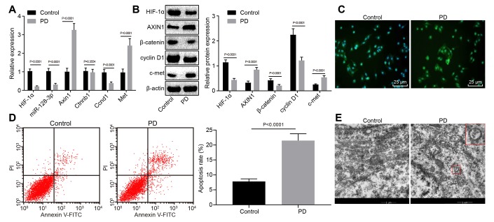
Figure 1 The expression pattern of HIF-1α/miR-128-3p/AXIN1 and the Wnt/β-catenin signaling pathway-related proteins (β-catenin, cyclin D1 and c-met) in hippocampal neurons of normal mice (n = 10) and the MPTP-lesioned mouse model of PD (n = 30). (A) The miR-128-3p expression and mRNA levels of Hif1a, Axin1, Ctnnb1, Ccnd1, and Met determined by RT-qPCR. (B) The protein levels of HIF-1α, AXIN1, β-catenin, cyclin D1, and c-met normalized to β-actin as determined by western blot analysis. (C) The localization of β-catenin protein in hippocampal tissues detected by the immunofluorescence assay (scale bar = 25 μm). (D) The hippocampal neuron apoptosis identified by flow cytometry. (E) The ultrastructure of hippocampal neurons through electron microscopy. * p < 0.05 vs. the control group (primary hippocampal neurons of normal mice).
Figure 1 The expression pattern of HIF-1α/miR-128-3p/AXIN1 and the Wnt/β-catenin signaling pathway-related proteins (β-catenin, cyclin D1 and c-met) in hippocampal neurons of normal mice (n = 10) and the MPTP-lesioned mouse model of PD (n = 30). (A) The miR-128-3p expression and mRNA levels of Hif1a, Axin1, Ctnnb1, Ccnd1, and Met determined by RT-qPCR. (B) The protein levels of HIF-1α, AXIN1, β-catenin, cyclin D1, and c-met normalized to β-actin as determined by western blot analysis. (C) The localization of β-catenin protein in hippocampal tissues detected by the immunofluorescence assay (scale bar = 25 μm). (D) The hippocampal neuron apoptosis identified by flow cytometry. (E) The ultrastructure of hippocampal neurons through electron microscopy. * p < 0.05 vs. the control group (primary hippocampal neurons of normal mice).CC BY · PubMed Central
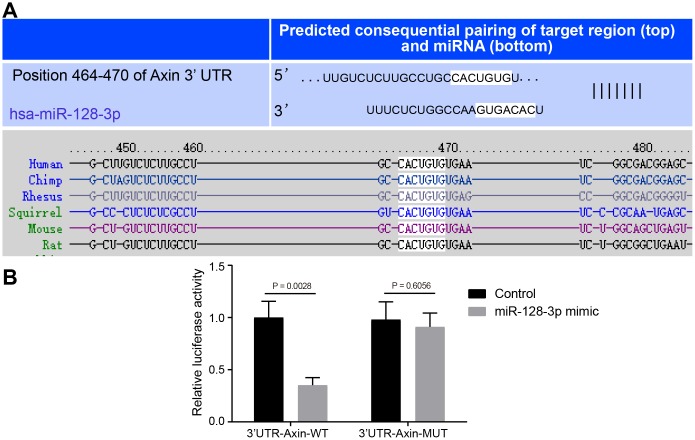
Figure 2 The targeting relationship between Axin1 and miR-128-3p. (A) The binding sites between miR-128-3p and Axin1 3'UTR predicted by Targetscan (http://www.targetscan.org/vert_71/). (B) The targeting relationship between miR-128-3p and Axin1 verified using dual-luciferase reporter gene assay. In the X-axis, 3'UTR-Axin1-WT refers to Axin1 wild type 3’UTR and 3'UTR-Axin1-MUT refers to Axin1 mutant 3’UTR. * p < 0.05 vs. the control group (primary hippocampal neurons of normal mice). The experiment was repeated three times independently.
Figure 2 The targeting relationship between Axin1 and miR-128-3p. (A) The binding sites between miR-128-3p and Axin1 3'UTR predicted by Targetscan (http://www.targetscan.org/vert_71/). (B) The targeting relationship between miR-128-3p and Axin1 verified using dual-luciferase reporter gene assay. In the X-axis, 3'UTR-Axin1-WT refers to Axin1 wild type 3’UTR and 3'UTR-Axin1-MUT refers to Axin1 mutant 3’UTR. * p < 0.05 vs. the control group (primary hippocampal neurons of normal mice). The experiment was repeated three times independently.CC BY · PubMed Central
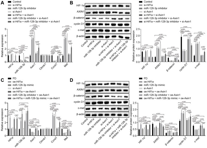
Figure 3 The expression pattern of HIF-1α/miR-128-3p/AXIN1 and the Wnt/β-catenin signaling pathway-related proteins (β-catenin, cyclin D1 and c-met) in hippocampal neurons of normal mice (n = 10) and in the MPTP-lesioned mouse model of PD (n = 30) after transduction. (A) The expression levels of Hif1a, miR-128-3p, Axin1, Ctnnb1, Ccnd1, and Met in primary hippocampal neurons of normal mice after treated with si-Hif1a, miR-128-3p inhibitor, or si-Axin1, determined by RT-qPCR. (B) The protein band patterns and the protein levels of HIF-1α, AXIN1, β-catenin, cyclin D1, and c-met normalized to β-actin in primary hippocampal neurons of normal mice after treatment with si-Hif1a, miR-128-3p inhibitor, or si-Axin1, all as determined by western blot analysis. * p < 0.05 vs. the control group (primary hippocampal neurons of normal mice); # p < 0.05 vs. the si-Hif1a group (hippocampal neurons of normal mice treated with si-Hif1a); & p < 0.05 vs. the miR-128-3p inhibitor group (hippocampal neurons of normal mice treated with miR-128-3p inhibitor); @ p < 0.05 vs. the si-Hif1a + si-Axin1 group (hippocampal neurons of normal mice treated with si-Hif1a + si-Axin1); $ p < 0.05 vs. the miR-128-3p inhibitor + si-Axin1 group (hippocampal neurons of normal mice treated with miR-128-3p inhibitor + si-Axin1). (C) The expression levels of Hif1a, miR-128-3p, Axin1, Ctnnb1, Ccnd1, and Met in primary hippocampal neurons cultured from the MPTP-lesioned mouse model of PD after treatment with oe-Hif1a, miR-128-3p mimic, or oe-Axin1, as determined by RT-qPCR. (D) The protein band patterns and the protein levels of HIF-1α, AXIN1, β-catenin, cyclin D1, and c-met normalized to β-actin in primary hippocampal neurons of the MPTP-lesioned mouse model of PD after treatment with oe-Hif1a, miR-128-3p mimic, or oe-Axin1, as determined by western blot analysis. * p < 0.05 vs. the PD group (primary hippocampal neurons of MPTP-lesioned mouse model of PD); # p < 0.05 vs. the oe-Hif1a group (hippocampal neurons of MPTP-lesioned mouse model of PD treated with oe-Hif1a); & p < 0.05 vs. the miR-128-3p mimic group (hippocampal neurons of MPTP-lesioned mouse model of PD treated with miR-128-3p mimic); @ p < 0.05 vs. the oe-Hif1a + oe-Axin1 group (hippocampal neurons of MPTP-lesioned mouse model of PD treated with oe-Hif1a + oe-Axin1); $ p < 0.05 vs. the miR-128-3p mimic + oe-Axin1 group (hippocampal neurons of MPTP-lesioned mouse model of PD treated with miR-128-3p mimic + oe-Axin1). The experiment was repeated tree times independently.
Figure 3 The expression pattern of HIF-1α/miR-128-3p/AXIN1 and the Wnt/β-catenin signaling pathway-related proteins (β-catenin, cyclin D1 and c-met) in hippocampal neurons of normal mice (n = 10) and in the MPTP-lesioned mouse model of PD (n = 30) after transduction. (A) The expression levels of Hif1a, miR-128-3p, Axin1, Ctnnb1, Ccnd1, and Met in primary hippocampal neurons of normal mice after treated with si-Hif1a, miR-128-3p inhibitor, or si-Axin1, determined by RT-qPCR. (B) The protein band patterns and the protein levels of HIF-1α, AXIN1, β-catenin, cyclin D1, and c-met normalized to β-actin in primary hippocampal neurons of normal mice after treatment with si-Hif1a, miR-128-3p inhibitor, or si-Axin1, all as determined by western blot analysis. * p < 0.05 vs. the control group (primary hippocampal neurons of normal mice); # p < 0.05 vs. the si-Hif1a group (hippocampal neurons of normal mice treated with si-Hif1a); & p < 0.05 vs. the miR-128-3p inhibitor group (hippocampal neurons of normal mice treated with miR-128-3p inhibitor); @ p < 0.05 vs. the si-Hif1a + si-Axin1 group (hippocampal neurons of normal mice treated with si-Hif1a + si-Axin1); $ p < 0.05 vs. the miR-128-3p inhibitor + si-Axin1 group (hippocampal neurons of normal mice treated with miR-128-3p inhibitor + si-Axin1). (C) The expression levels of Hif1a, miR-128-3p, Axin1, Ctnnb1, Ccnd1, and Met in primary hippocampal neurons cultured from the MPTP-lesioned mouse model of PD after treatment with oe-Hif1a, miR-128-3p mimic, or oe-Axin1, as determined by RT-qPCR. (D) The protein band patterns and the protein levels of HIF-1α, AXIN1, β-catenin, cyclin D1, and c-met normalized to β-actin in primary hippocampal neurons of the MPTP-lesioned mouse model of PD after treatment with oe-Hif1a, miR-128-3p mimic, or oe-Axin1, as determined by western blot analysis. * p < 0.05 vs. the PD group (primary hippocampal neurons of MPTP-lesioned mouse model of PD); # p < 0.05 vs. the oe-Hif1a group (hippocampal neurons of MPTP-lesioned mouse model of PD treated with oe-Hif1a); & p < 0.05 vs. the miR-128-3p mimic group (hippocampal neurons of MPTP-lesioned mouse model of PD treated with miR-128-3p mimic); @ p < 0.05 vs. the oe-Hif1a + oe-Axin1 group (hippocampal neurons of MPTP-lesioned mouse model of PD treated with oe-Hif1a + oe-Axin1); $ p < 0.05 vs. the miR-128-3p mimic + oe-Axin1 group (hippocampal neurons of MPTP-lesioned mouse model of PD treated with miR-128-3p mimic + oe-Axin1). The experiment was repeated tree times independently.CC BY · PubMed Central
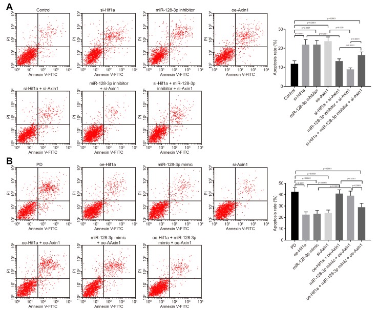
Figure 4 The apoptosis of hippocampal neurons cultured from normal mice and an MPTP-lesioned mouse model of PD mediated by the HIF-1α/miR-128-3p/AXIN1 axis. (A) The apoptosis of hippocampal neurons of normal mice after treatment with si-Hif1a, miR-128-3p inhibitor, or si-Axin1 as detected by flow cytometry. * p < 0.05 vs. the control group (primary hippocampal neurons of normal mice); # p < 0.05 vs. the si-Hif1a group (hippocampal neurons of normal mice treated with si-Hif1a); & p < 0.05 vs. the miR-128-3p inhibitor group (hippocampal neurons of normal mice treated with miR-128-3p inhibitor); @ p < 0.05 vs. the si-Hif1a + si-Axin1 group (hippocampal neurons of normal mice treated with si-Hif1a + si-Axin1); $ p < 0.05 vs. the miR-128-3p inhibitor + si-Axin1 group (hippocampal neurons of normal mice treated with miR-128-3p inhibitor + si-Axin1). (B) The apoptosis of hippocampal neurons in the MPTP-lesioned mouse model of PD after treatment with oe-Hif1a, miR-128-3p mimic, or oe-Axin1 detected by flow cytometry. * p < 0.05 vs. the PD group (primary hippocampal neurons of the MPTP-lesioned mouse model of PD); # p < 0.05 vs. the oe-Hif1a group (hippocampal neurons of the MPTP-lesioned mouse model of PD treated with oe-Hif1a); & p < 0.05 vs. the miR-128-3p mimic group (hippocampal neurons of MPTP-lesioned mouse model of PD treated with miR-128-3p mimic); @ p < 0.05 vs. the oe-Hif1a + oe-Axin1 group (hippocampal neurons of MPTP-lesioned mouse model of PD treated with oe-Hif1a + oe-Axin1); $ p < 0.05 vs. the miR-128-3p mimic + oe-Axin1 group (hippocampal neurons of MPTP-lesioned mouse model of PD treated with miR-128-3p mimic + oe-Axin1). The experiment was repeated three times independently.
Figure 4 The apoptosis of hippocampal neurons cultured from normal mice and an MPTP-lesioned mouse model of PD mediated by the HIF-1α/miR-128-3p/AXIN1 axis. (A) The apoptosis of hippocampal neurons of normal mice after treatment with si-Hif1a, miR-128-3p inhibitor, or si-Axin1 as detected by flow cytometry. * p < 0.05 vs. the control group (primary hippocampal neurons of normal mice); # p < 0.05 vs. the si-Hif1a group (hippocampal neurons of normal mice treated with si-Hif1a); & p < 0.05 vs. the miR-128-3p inhibitor group (hippocampal neurons of normal mice treated with miR-128-3p inhibitor); @ p < 0.05 vs. the si-Hif1a + si-Axin1 group (hippocampal neurons of normal mice treated with si-Hif1a + si-Axin1); $ p < 0.05 vs. the miR-128-3p inhibitor + si-Axin1 group (hippocampal neurons of normal mice treated with miR-128-3p inhibitor + si-Axin1). (B) The apoptosis of hippocampal neurons in the MPTP-lesioned mouse model of PD after treatment with oe-Hif1a, miR-128-3p mimic, or oe-Axin1 detected by flow cytometry. * p < 0.05 vs. the PD group (primary hippocampal neurons of the MPTP-lesioned mouse model of PD); # p < 0.05 vs. the oe-Hif1a group (hippocampal neurons of the MPTP-lesioned mouse model of PD treated with oe-Hif1a); & p < 0.05 vs. the miR-128-3p mimic group (hippocampal neurons of MPTP-lesioned mouse model of PD treated with miR-128-3p mimic); @ p < 0.05 vs. the oe-Hif1a + oe-Axin1 group (hippocampal neurons of MPTP-lesioned mouse model of PD treated with oe-Hif1a + oe-Axin1); $ p < 0.05 vs. the miR-128-3p mimic + oe-Axin1 group (hippocampal neurons of MPTP-lesioned mouse model of PD treated with miR-128-3p mimic + oe-Axin1). The experiment was repeated three times independently.CC BY · PubMed Central
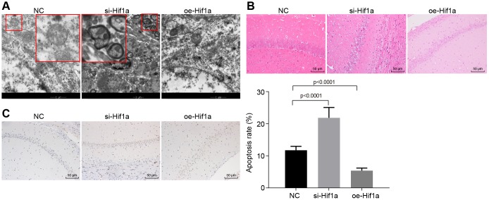
Figure 5 The pathological changes of hippocampal neurons in the MPTP-lesioned mouse model of PD. (A) The ultrastructure of hippocampal neurons treated with si-Hif1a or oe-Hif1a to electron microscopy. (B) The pathological changes of hippocampal neurons treated with si-Hif1a or oe-Hif1a revealed by HE staining (scale bar = 50 μm). (C) The apoptosis of hippocampal neurons treated with si-Hif1a or oe-Hif1a detected by TUNEL staining (scale bar = 50 μm). * p < 0.05 vs. the NC group (MPTP-lesioned mice treated with NC plasmids). N = 10.
Figure 5 The pathological changes of hippocampal neurons in the MPTP-lesioned mouse model of PD. (A) The ultrastructure of hippocampal neurons treated with si-Hif1a or oe-Hif1a to electron microscopy. (B) The pathological changes of hippocampal neurons treated with si-Hif1a or oe-Hif1a revealed by HE staining (scale bar = 50 μm). (C) The apoptosis of hippocampal neurons treated with si-Hif1a or oe-Hif1a detected by TUNEL staining (scale bar = 50 μm). * p < 0.05 vs. the NC group (MPTP-lesioned mice treated with NC plasmids). N = 10.CC BY · PubMed Central
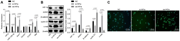
Figure 6 The expression pattern of HIF-1α/miR-128-3p/Axin1 and the Wnt/β-catenin signaling pathway-related proteins (β-catenin, cyclin D1 and c-met) in hippocampal neurons of the MPTP-lesioned mouse model of PD after transfection with si-Hif1a or oe-Hif1a. (A) The miR-128-3p expression and mRNA levels of Hif1a, Axin1, Ctnnb1, Ccnd1, and Met in hippocampal neurons of the MPTP-lesioned mouse model of PD determined by RT-qPCR. (B) The protein band patterns and the protein levels of HIF-1α, AXIN1, β-catenin, cyclin D1, and c-met normalized to β-actin in hippocampal neurons of the MPTP-lesioned mouse model of PD determined by western blot analysis. (C) The localization of β-catenin protein in hippocampal tissues of the MPTP-lesioned mouse model of PD detected by the immunofluorescence assay (scale bar = 25 μm). * p < 0.05 vs. the NC group (hippocampal neurons of the MPTP-lesioned mouse model of PD treated with NC plasmids). N = 10.
Figure 6 The expression pattern of HIF-1α/miR-128-3p/Axin1 and the Wnt/β-catenin signaling pathway-related proteins (β-catenin, cyclin D1 and c-met) in hippocampal neurons of the MPTP-lesioned mouse model of PD after transfection with si-Hif1a or oe-Hif1a. (A) The miR-128-3p expression and mRNA levels of Hif1a, Axin1, Ctnnb1, Ccnd1, and Met in hippocampal neurons of the MPTP-lesioned mouse model of PD determined by RT-qPCR. (B) The protein band patterns and the protein levels of HIF-1α, AXIN1, β-catenin, cyclin D1, and c-met normalized to β-actin in hippocampal neurons of the MPTP-lesioned mouse model of PD determined by western blot analysis. (C) The localization of β-catenin protein in hippocampal tissues of the MPTP-lesioned mouse model of PD detected by the immunofluorescence assay (scale bar = 25 μm). * p < 0.05 vs. the NC group (hippocampal neurons of the MPTP-lesioned mouse model of PD treated with NC plasmids). N = 10.CC BY · PubMed Central

Verification

Claims the paper makes

The claims and statistics of this paper haven’t been checked yet.

Run the check — free, no account needed. It reads the open-access text and checks the sources it cites.

A few free checks a day · papers already verified open instantly · results are public

Claims other papers make about this one

Each is checked against this paper’s own text: does it say what the citing paper says it does?

Self-check

Percentages, sums and reported differences re-computed from the paper’s own numbers.

The self-check needs the paper’s full text.

Citations

Cites

Research works printed but not linked: 42

  1. Lang, 2015, Parkinson’s disease., Lancet, 38, 896, 10.1016/S0140-6736(14)61393-3 · DOI · Paper page
  2. Storstein, 2017, Epidemiology of Parkinson’s disease., J Neural Transm (Vienna), 12, 901, 10.1007/s00702-017-1686-y · DOI
  3. Martí, 2011, MicroRNA profiling of Parkinson’s disease brains identifies early downregulation of miR-34b/c which modulate mitochondrial function., Hum Mol Genet, 2, 3067, 10.1093/hmg/ddr210 · DOI
  4. Schwarzschild, 2016, The epidemiology of Parkinson’s disease: risk factors and prevention., Lancet Neurol, 1, 1257, 10.1016/S1474-4422(16)30230-7 · DOI
  5. Dawson, 2011, MicroRNAs in Parkinson’s disease., J Chem Neuroanat, 4, 127, 10.1016/j.jchemneu.2011.01.005 · DOI
  6. Jimenez-Mateos, 2017, MicroRNAs in Neurodegenerative Diseases., Int Rev Cell Mol Biol, 33, 309, 10.1016/bs.ircmb.2017.04.002 · DOI · Paper page
  7. Xu, 2011, MicroRNA in central nervous system trauma and degenerative disorders., Physiol Genomics, 4, 571, 10.1152/physiolgenomics.00168.2010 · DOI
  8. Schaefer, 2013, MicroRNA-128 governs neuronal excitability and motor behavior in mice., Science, 34, 1254, 10.1126/science.1244193 · DOI · Paper page
  9. Qiu, 2015, IDH1R(1)(3)(2)H decreases the proliferation of U87 glioma cells through upregulation of microRNA-128a., Mol Med Rep, 1, 6695, 10.3892/mmr.2015.4241 · DOI
  10. Wu, 2018, Therapeutic potential of a prolyl hydroxylase inhibitor FG-4592 for Parkinson’s diseasesand: regulation of redox biology and mitochondrial function., Front Aging Neurosci, 1, 121, 10.3389/fnagi.2018.00121 · DOI
  11. Park, 2013, HIF-1α-induced β-catenin activation prevents prion-mediated neurotoxicity., Int J Mol Med, 3, 931, 10.3892/ijmm.2013.1457 · DOI
  12. Pluchino, 2013, Uncovering novel actors in astrocyte-neuron crosstalk in Parkinson’s disease: the Wnt/β-catenin signaling cascade as the common final pathway for neuroprotection and self-repair., Eur J Neurosci, 3, 1550, 10.1111/ejn.12166 · DOI
  13. Arenas, 2010, Emerging roles of Wnts in the adult nervous system., Nat Rev Neurosci, 1, 77, 10.1038/nrn2755 · DOI
  14. Zhao, 2016, Neuroprotective effects of ginsenoside Rg1 through the Wnt/β-catenin signaling pathway in both in vivo and in vitro models of Parkinson’s disease., Neuropharmacology, 10, 480, 10.1016/j.neuropharm.2015.10.024 · DOI
  15. Chen, 2011, Generation of Axin1 conditional mutant mice., Genesis, 4, 98, 10.1002/dvg.20703 · DOI
  16. Chien, 2012, Wnt/β-catenin signaling and AXIN1 regulate apoptosis triggered by inhibition of the mutant kinase BRAFV600E in human melanoma., Sci Signal, 5, ra3, 10.1126/scisignal.2002274 · DOI
  17. Qi, 2013, Alterations of axis inhibition protein 1 (AXIN1) in hepatitis B virus-related hepatocellular carcinoma and overexpression of AXIN1 induces apoptosis in hepatocellular cancer cells., Oncol Res, 2, 281, 10.3727/096504013X13639794277608 · DOI
  18. Cong, 2017, Axin1 up-regulated 1 accelerates stress-induced cardiomyocytes apoptosis through activating Wnt/β-catenin signaling., Exp Cell Res, 35, 441, 10.1016/j.yexcr.2017.08.027 · DOI
  19. Saeed, 2018, Genomic convergence of locus-based GWAS meta-analysis identifies AXIN1 as a novel Parkinson’s gene., Immunogenetics, 7, 563, 10.1007/s00251-018-1068-0 · DOI
  20. Wang, 2018, MicroRNA-128 protects dopamine neurons from apoptosis and upregulates the expression of excitatory amino acid transporter 4 in Parkinson’s disease by binding to AXIN1., Cell Physiol Biochem, 5, 2275, 10.1159/000495872 · DOI · Paper page
  21. Jho, 2011, Downregulation of Wnt/β-catenin signaling causes degeneration of hippocampal neurons in vivo., Neurobiol Aging, 3, 10.1016/j.neurobiolaging.2010.03.013 · DOI
  22. Pan, 2010, Neuroprotection of deferoxamine on rotenone-induced injury via accumulation of HIF-1 alpha and induction of autophagy in SH-SY5Y cells., Neurochem Int, 5, 198, 10.1016/j.neuint.2010.05.008 · DOI
  23. Karabay, 2013, Protein kinase C activation causes neurite retraction via cyclinD1 and p60-katanin increase in rat hippocampal neurons., Eur J Neurosci, 3, 1610, 10.1111/ejn.12185 · DOI
  24. Walikonis, 2008, Hepatocyte growth factor and c-Met promote dendritic maturation during hippocampal neuron differentiation via the Akt pathway., Cell Signal, 2, 825, 10.1016/j.cellsig.2007.12.013 · DOI · Paper page
  25. Andersen, 2016, Regulation of ATP13A2 via PHD2-HIF1α signaling is critical for cellular iron homeostasis: implications for Parkinson’s disease., J Neurosci, 3, 1086, 10.1523/JNEUROSCI.3117-15.2016 · DOI
  26. Espinosa, 2016, The TIP60 complex is a conserved coactivator of HIF1A., Cell Rep, 1, 37, 10.1016/j.celrep.2016.05.082 · DOI
  27. Dastych, 2013, Epigenetic regulation of CD34 and HIF1A expression during the differentiation of human mast cells., Immunogenetics, 6, 429, 10.1007/s00251-013-0695-8 · DOI
  28. Orlacchio, 2014, miR128 up-regulation correlates with impaired amyloid β(1-42) degradation in monocytes from patients with sporadic Alzheimer’s disease., Neurobiol Aging, 3, 345, 10.1016/j.neurobiolaging.2013.08.003 · DOI · Paper page
  29. Sears, 2009, The Axin1 scaffold protein promotes formation of a degradation complex for c-Myc., EMBO J, 2, 500, 10.1038/emboj.2008.279 · DOI
  30. Sun, 2017, Vitamin D receptor is a novel transcriptional regulator for Axin1., J Steroid Biochem Mol Biol, 16, 430, 10.1016/j.jsbmb.2016.09.002 · DOI
  31. Schwienbacher, 2014, Identification of a set of endogenous reference genes for miRNA expression studies in Parkinson’s disease blood samples., BMC Res Notes, 7, 715, 10.1186/1756-0500-7-715 · DOI
  32. Wu, 2014, Neuroprotection by Orexin-A via HIF-1α induction in a cellular model of Parkinson’s disease., Neurosci Lett, 57, 35, 10.1016/j.neulet.2014.07.014 · DOI
  33. Kim, 2013, Differential expression of MicroRNAs in patients with glioblastoma after concomitant chemoradiotherapy., OMICS, 1, 259, 10.1089/omi.2012.0065 · DOI
  34. Thompson, 2014, Modulating Wnt signaling to improve cell replacement therapy for Parkinson’s disease., J Mol Cell Biol, 6, 54, 10.1093/jmcb/mjt045 · DOI
  35. Li, 2017, Simultaneous overactivation of Wnt/β-catenin and TGFβ signalling by miR-128-3p confers chemoresistance-associated metastasis in NSCLC., Nat Commun, 8, 15870, 10.1038/ncomms15870 · DOI
  36. Serafino, 2018, Atrial natriuretic peptide acts as a neuroprotective agent inmodels of Parkinson’s disease via up-regulation of the Wnt/β-Catenin pathway., Front Aging Neurosci, 1, 20, 10.3389/fnagi.2018.00020 · DOI
  37. Shi, 2011, Hypoxia inducible factor-1 as a target for neurodegenerative diseases., Curr Med Chem, 1, 4335, 10.2174/092986711797200426 · DOI
  38. Przedborski, 2007, Protocol for the MPTP mouse model of Parkinson’s disease., Nat Protoc, 2, 141, 10.1038/nprot.2006.342 · DOI
  39. Rowe, 1987, Motor function in the 1-methyl-4-phenyl-1,2,3,6-tetrahydropyridine-treated mouse., J Neurol Sci, 7, 185, 10.1016/0022-510X(87)90121-3 · DOI
  40. Watanabe, 1985, A simple quantitative bradykinesia test in MPTP-treated mice., Res Commun Chem Pathol Pharmacol, 5, 435
  41. Ohta, 1998, Novel endogenous 1,2,3,4-tetrahydroisoquinoline derivatives: uptake by dopamine transporter and activity to induce parkinsonism., J Neurochem, 7, 745, 10.1046/j.1471-4159.1998.70020745.x · DOI
  42. Schmittgen, 2001, Analysis of relative gene expression data using real-time quantitative PCR and the 2(-Delta Delta C(T)) Method., Methods, 2, 402, 10.1006/meth.2001.1262 · DOI · Paper page

Cited by

Sorted by citations

See all 44

Metadata

Authors
Zhang, G., Chen, L., Liu, J., Jin, Y., Lin, Z., Du, S., Fu, Z., Chen, T., Qin, Y., Sui, F., & Jiang, Y.
Affiliations
  • The Fourth Ward, Department of Neurology, The Third Affiliated Hospital of Qiqihar Medical University, Qiqihar 161000, P.R. China
  • Department of Health Care, The Third Affiliated Hospital of Qiqihar Medical University, Qiqihar 161000, P.R. China
Venue
Aging
Published
2020-03-12
Volume
12
Issue
5
Pages
4067-4081
ISSN
1945-4589
Cited by
44
References
3
Language
en

About these checks

Registry, retraction, Cites and statistics results are computed, not judged, with one exception: a printed reference that no index lists is read once by a model before it counts as a citation error. Claims are extracted by a model and kept only when their quote appears verbatim in the text; each cited source is then checked, with a model’s help, for whether it says what the claim relies on. Those verdicts can be wrong — read the claim’s quote and the cited source before relying on one.

Statistics and arithmetic are recomputed, not judged.

Confidence bands are shown only for fields calibrated on our annotated test set. None is calibrated yet, so no band is shown.

What counts as a claim ›How we measure confidence ›How we check this ›

Are you an author, or do you see a problem with this page? Request a review