

The DOI matches its Crossref record; 3 of the 3 references it cites were found in a registry or an index.
Engineered exosome-mediated delivery of functionally active miR-26a and its enhanced suppression effect in HepG2 cells
Overview
The claims and statistics of this paper haven’t been checked yet.
Run the check — free, no account needed. It reads the open-access text and checks the sources it cites.
Integrity signals
Registry
Cites
Statistics
Figures
Verification
Claims the paper makes
The claims and statistics of this paper haven’t been checked yet.
Run the check — free, no account needed. It reads the open-access text and checks the sources it cites.
Claims other papers make about this one
Each is checked against this paper’s own text: does it say what the citing paper says it does?
In the citing paper
“This, in turn, induced cell cycle arrest in HCC cells, inhibiting their proliferation and metastasis [121].”
From Emerging role of exosomes in cancer therapy: progress and challenges (2025) · cites this paper as [121]
In the citing paper
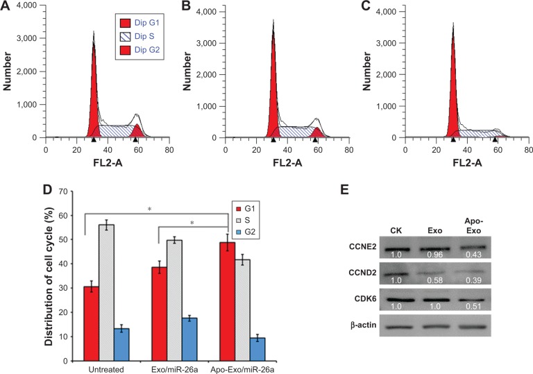
“The exosomes were efficiently absorbed by HepG2 cells, resulting in the downregulation of Cyclin D2, Cyclin E2, and CDK6.”
From Emerging role of exosomes in cancer therapy: progress and challenges (2025) · cites this paper as [121]
Self-check
Percentages, sums and reported differences re-computed from the paper’s own numbers.
The self-check needs the paper’s full text.
Citations
Cites
Tian, T., Zhu, Y.-L., Zhou, Y.-Y., Liang, G.-F., Wang, Y.-Y., Hu, F.-H., & Xiao, Z.-D. (2014) · Journal of Biological Chemistry · doi:10.1074/jbc.m114.588046 · Cited by 726
Self-citationYim, N., Ryu, S.-W., Choi, K., Lee, K. R., Lee, S., Choi, H., Kim, J., Shaker, M. R., Sun, W., Park, J.-H., et al. (2016) · Nature Communications · doi:10.1038/ncomms12277 · Cited by 578
Malhotra, H., Sheokand, N., Kumar, S., Chauhan, A. S., Kumar, M., Jakhar, P., Boradia, V. M., Raje, C. I., & Raje, M. (2016) · Journal of Biomedical Nanotechnology · doi:10.1166/jbn.2016.2229 · Cited by 114
Research works printed but not linked: 46
- [1]Niidome T Huang L Gene therapy progress and prospects: nonviral vectors Gene Ther 2002 9 24 1647 1652 12457277
- [2]Lee SJ Kim MJ Kwon IC Roberts TM Delivery strategies and potential targets for siRNA in major cancer types Adv Drug Deliv Rev 2016 104 2 15 27259398 PMC4958528
- [3]Liang G Li Y Feng W Polyethyleneimine-coated quantum dots for miRNA delivery and its enhanced suppression in HepG2 cells Int J Nanomedicine 2016 11 6079 6088 27895481 PMC5117883
- [4]Alvarez-Erviti L Seow Y Yin H Betts C Lakhal S Wood MJ Delivery of siRNA to the mouse brain by systemic injection of targeted exosomes Nat Biotechnol 2011 29 4 341 345 21423189
- [5]Dai L Gu N Chen BA Marriott G Human platelets repurposed as vehicles for in vivo imaging of myeloma xenotransplants Oncotarget 2016 7 16 21076 21090 27049725 PMC5008270
- [6]Marriott G Engineering platelets for tumour targeting Aging (Albany NY) 2016 8 8 1572 1573 27508429 PMC5032680
- [7]Li J Sharkey CC Wun B Liesveld JL King MR Genetic engineering of platelets to neutralize circulating tumor cells J Control Release 2016 228 38 47 26921521 PMC4828270
- [8]Shi J Kundrat L Pishesha N Engineered red blood cells as carriers for systemic delivery of a wide array of functional probes Proc Natl Acad Sci U S A 2014 111 28 10131 10136 24982154 PMC4104923
- [9]Denzer K Kleijmeer MJ Heijnen HF Stoorvogel W Geuze HJ Exosome: from internal vesicle of the multivesicular body to intercellular signaling device J Cell Sci 2000 113 3365 3374 10984428
- [10]Fuhrmann G Herrmann IK Stevens MM Cell-derived vesicles for drug therapy and diagnostics: opportunities and challenges Nano Today 2015 10 3 397 409 28458718 PMC5409525
- [11]Eckard SC Rice GI Fabre A The SKIV2L RNA exosome limits activation of the RIG-I-like receptors Nat Immunol 2014 15 9 839 845 25064072 PMC4139417
- [12]Gidlof O van der Brug M Ohman J Platelets activated during myocardial infarction release functional miRNA, which can be taken up by endothelial cells and regulate ICAM1 expression Blood 2013 121 9 3908 3917 23493781
- [13]Karantalis V Hare JM Use of mesenchymal stem cells for therapy of cardiac disease Circ Res 2015 116 8 1413 1430 25858066 PMC4429294
- [14]Powers S Pollack RE Inducing stable reversion to achieve cancer control Nat Rev Cancer 2016 16 4 266 270 27458638
- [16]Ghai V Wang K Recent progress toward the use of circulating micro RNAs as clinical biomarkers Arch Toxicol 2016 90 12 2959 2978 27585665
- [17]Lee C Carney RP Hazari S 3D plasmonic nanobowl platform for the study of exosomes in solution Nanoscale 2015 7 20 9290 9297 25939587 PMC11781986
- [18]Tian Y Li S Song J A doxorubicin delivery platform using engineered natural membrane vesicle exosomes for targeted tumor therapy Biomaterials 2014 35 7 2383 2390 24345736
- [19]Kalra H Adda CG Liem M Comparative proteomics evaluation of plasma exosome isolation techniques and assessment of the stability of exosomes in normal human blood plasma Proteomics 2013 13 22 3354 3364 24115447
- [20]Rana S Malinowska K Zoeller M Exosomal tumor microRNA modulates premetastatic organ cells Neoplasia 2013 15 3 281 295 23479506 PMC3593151
- [21]Didiot MC Hall LM Coles AH Exosome-mediated delivery of hydrophobically modified siRNA for huntingtin mRNA silencing Mol Ther 2016 24 10 1836 1847 27506293 PMC5112038
- [22]Morishita M Takahashi Y Matsumoto A Nishikawa M Takakura Y Exosome-based tumor antigens-adjuvant co-delivery utilizing genetically engineered tumor cell-derived exosomes with immunostimulatory CpG DNA Biomaterials 2016 111 55 65 27723556
- [23]O’Connor RM Gururajan A Dinan TG Kenny PJ Cryan JF All roads lead to the miRNome: miRNAs Have a central role in the molecular pathophysiology of psychiatric disorders Trends Pharmacol Sci 2016 37 12 1029 1044 27832923
- [24]Kortylewski M Nechaev S How to train your dragon: targeted delivery of MicroRNA to cancer cells in vivo Mol Ther 2014 22 6 1070 1071 24881764 PMC4048907
- [25]Rincon MY VandenDriessche T Chuah MK Gene therapy for cardiovascular disease: advances in vector development, targeting, and delivery for clinical translation Cardiovasc Res 2015 108 1 4 20 26239654 PMC4571836
- [26]Hatakeyama H Murata M Sato Y The systemic administration of an anti-miRNA oligonucleotide encapsulated pH-sensitive liposome results in reduced level of hepatic microRNA-122 in mice J Control Release 2014 173 1 43 50 24511611
- [27]Scomparin A Polyak D Krivitsky A Satchi-Fainaro R Achieving successful delivery of oligonucleotides – From physico-chemical characterization to in vivo evaluation Biotechnol Adv 2015 33 6 Pt 3 1294 1309 25916823
- [28]Kheirolomoom A Kim CW Seo JW Multifunctional nanoparticles facilitate molecular targeting and miRNA delivery to inhibit atherosclerosis in ApoE(−/−) mice Acs Nano 2015 9 9 8885 8897 26308181 PMC4581466
- [29]Chen F Huang P Zhu YJ Wu J Zhang CL Cui DX The photoluminescence, drug delivery and imaging properties of multifunctional Eu3+/Gd3+ dual-doped hydroxyapatite nanorods Biomaterials 2011 32 34 9031 9039 21875748
- [30]Kota J Chivukula RR O’Donnell KA Therapeutic microRNA delivery suppresses tumorigenesis in a murine liver cancer model Cell 2009 137 6 1005 1017 19524505 PMC2722880
- [31]Liang G Zhu Y Jing A Cationic microRNA-delivering nanocarriers for efficient treatment of colon carcinoma in xenograft model Gene Ther 2016 23 12 829 838 27482839
- [32]Urban S Zieseniss S Werder M Hauser H Budzinski R Engelmann B Scavenger receptor BI transfers major lipoprotein-associated phospholipids into the cells J Biol Chem 2000 275 43 33409 33415 10938082
- [33]Yamamoto S Fukuhara T Ono C Lipoprotein receptors redundantly participate in entry of hepatitis C virus PLoS Pathog 2016 12 5 e1005610 27152966 PMC4859476
- [34]Kharaziha P Chioureas D Rutishauser D Molecular profiling of prostate cancer derived exosomes may reveal a predictive signature for response to docetaxel Oncotarget 2015 6 25 21740 21754 25844599 PMC4673300
- [35]Lazar I Clement E Ducoux-Petit M Proteome characterization of melanoma exosomes reveals a specific signature for metastatic cell lines Pigment Cell Melanoma Res 2015 28 4 464 475 25950383
- [37]Hung ME Leonard JN Stabilization of exosome-targeting peptides via engineered glycosylation J Biol Chem 2015 290 13 8166 8172 25657008 PMC4375473
- [38]Mo ZC Ren K Liu X Tang ZL Yi GH A high-density lipoprotein-mediated drug delivery system Adv Drug Deliv Rev 2016 106 Pt A 132 147 27208399
- [39]Kanada M Bachmann MH Hardy JW Differential fates of biomolecules delivered to target cells via extracellular vesicles Proc Natl Acad Sci U S A 2015 112 12 E1433 E1442 25713383 PMC4378439
- [40]Lunavat TR Jang SC Nilsson L RNAi delivery by exosome-mimetic nanovesicles - Implications for targeting c-Myc in cancer Biomaterials 2016 102 231 238 27344366
- [41]Chen L Zheng J Zhang Y Tumor-specific expression of MicroRNA-26a suppresses human hepatocellular carcinoma growth via cyclin-dependent and -independent pathways Mol Ther 2011 19 8 1521 1528 21610700 PMC3149175
- [42]Gandhi NS Tekade RK Chougule MB Nanocarrier mediated delivery of siRNA/miRNA in combination with chemotherapeutic agents for cancer therapy: current progress and advances J Control Release 2014 194 238 256 25204288 PMC4254052
- [43]Tkach M Thery C Communication by extracellular vesicles: where we are and where we need to go Cell 2016 164 6 1226 1232 26967288
- [45]Choi JS Yoon HI Lee KS Exosomes from differentiating human skeletal muscle cells trigger myogenesis of stem cells and provide biochemical cues for skeletal muscle regeneration J Control Release 2016 222 107 115 26699421
- [46]Tian Y Li S Song J A doxorubicin delivery platform using engineered natural membrane vesicle exosomes for targeted tumor therapy Biomaterials 2014 35 7 2383 2390 24345736
- [47]Martinez LO Jacquet S Esteve JP Ectopic beta-chain of ATP synthase is an apolipoprotein A-I receptor in hepatic HDL endocytosis Nature 2003 421 6918 75 79 12511957
- [48]Tominaga N Yoshioka Y Ochiya T A novel platform for cancer therapy using extracellular vesicles Adv Drug Deliv Rev 2015 95 50 55 26482189
- [49]Jo W Kim J Yoon J Large-scale generation of cell-derived nanovesicles Nanoscale 2014 6 20 12056 12064 25189198
Cited by
Sorted by citations
Engineering exosomes for targeted drug delivery
Liang, Y., Duan, L., Lu, J., & Xia, J. (2021) · Theranostics · doi:10.7150/thno.52570 · Cited by 1,547
Engineered Extracellular Vesicles for Cancer Therapy
Zhang, X., Zhang, H., Gu, J., Zhang, J., Shi, H., Qian, H., Wang, D., Xu, W., Pan, J., & Santos, H. A. (2021) · Advanced Materials · doi:10.1002/adma.202005709 · Cited by 428
Recent advances in exosome-mediated nucleic acid delivery for cancer therapy
Zhang, Y., Liu, Q., Zhang, X., Huang, H., Tang, S., Chai, Y., Xu, Z., Li, M., Chen, X., Liu, J., et al. (2022) · Journal of Nanobiotechnology · doi:10.1186/s12951-022-01472-z · Cited by 306
Emerging role of exosomes in cancer therapy: progress and challenges
Li, J., Wang, J., & Chen, Z. (2025) · Molecular Cancer · doi:10.1186/s12943-024-02215-4 · Cited by 297
Improved Loading of Plasma-Derived Extracellular Vesicles to Encapsulate Antitumor miRNAs
Pomatto, M. A. C., Bussolati, B., D’Antico, S., Ghiotto, S., Tetta, C., Brizzi, M. F., & Camussi, G. (2019) · Molecular Therapy - Methods & Clinical Development · doi:10.1016/j.omtm.2019.01.001 · Cited by 243
Metadata
- Authors
- Liang, G., Kan, S., Zhu, Y., Feng, S., Feng, W., & Gao, S.
- Venue
- International Journal of Nanomedicine
- Publisher
- Informa UK Limited
- Published
- 2018-01
- Volume
- Volume 13
- Pages
- 585-599
- ISSN
- 1178-2013
- PMC
- PMC5796471
- Cited by
- 276
- References
- 3
- Language
- en
About these checks
Registry, retraction, Cites and statistics results are computed, not judged, with one exception: a printed reference that no index lists is read once by a model before it counts as a citation error. Claims are extracted by a model and kept only when their quote appears verbatim in the text; each cited source is then checked, with a model’s help, for whether it says what the claim relies on. Those verdicts can be wrong — read the claim’s quote and the cited source before relying on one.
Statistics and arithmetic are recomputed, not judged.
Confidence bands are shown only for fields calibrated on our annotated test set. None is calibrated yet, so no band is shown.
What counts as a claim ›How we measure confidence ›How we check this ›
Are you an author, or do you see a problem with this page? Request a review
How to cite
Cite CoChat for the checks on this page, and the paper for its own findings.
- This page
- CoChat. (2026). Citation check: Engineered exosome-mediated delivery of functionally active miR-26a and its enhanced suppression effect in HepG2 cells. https://app.cochat.ai/papers/10.2147/ijn.s154458
- The paper
- Liang, G., Kan, S., Zhu, Y., Feng, S., Feng, W., & Gao, S. (2018). Engineered exosome-mediated delivery of functionally active miR-26a and its enhanced suppression effect in HepG2 cells. International Journal of Nanomedicine. https://doi.org/10.2147/ijn.s154458
