

The DOI matches its Crossref record; 75 of the 75 references it cites were found in a registry or an index.
HTRF: A Technology Tailored for Drug Discovery - A Review of Theoretical Aspects and Recent Applications
Open accessAbstract
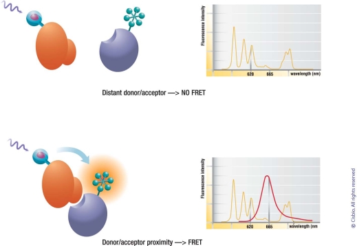
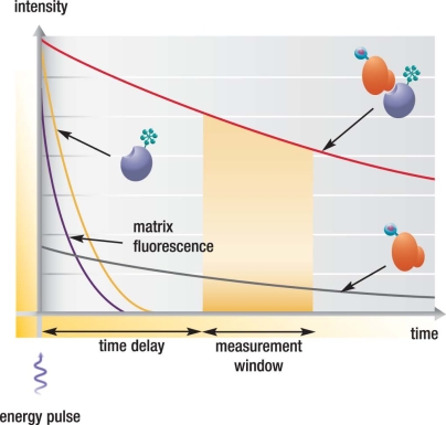
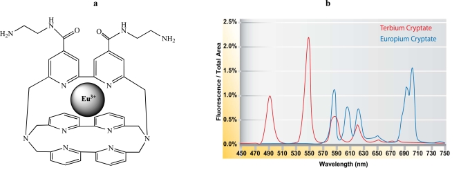
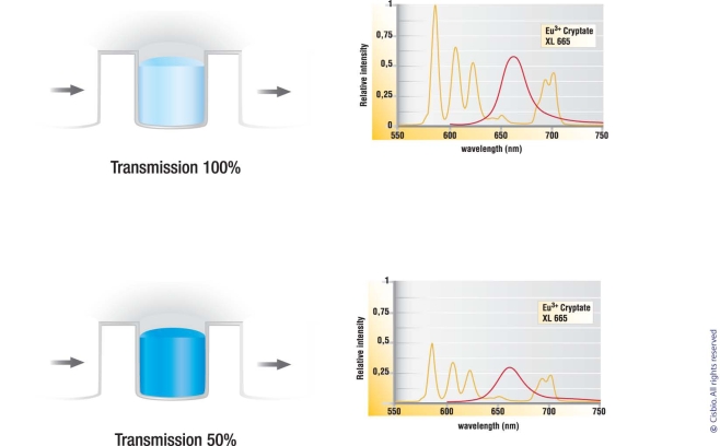
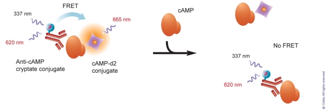
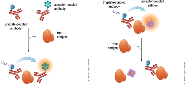
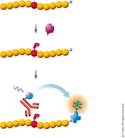
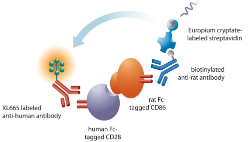
HTRF (Homogeneous Time Resolved Fluorescence) is the most frequently used generic assay technology to measure analytes in a homogenous format, which is the ideal platform used for drug target studies in high-throughput screening (HTS). This technology combines fluorescence resonance energy transfer technology (FRET) with time-resolved measurement (TR). In TR-FRET assays, a signal is generated through fluorescent resonance energy transfer between a donor and an acceptor molecule when in close proximity to each other. Buffer and media interference is dramatically reduced by dual-wavelength detection, and the final signal is proportional to the extent of product formation. The HTRF assay is usually sensitive and robust that can be miniaturized into the 384 and 1536-well plate formats. This assay technology has been applied to many antibody-based assays including GPCR signaling (cAMP and IP-One), kinases, cytokines and biomarkers, bioprocess (antibody and protein production), as well as the assays for protein-protein, proteinpeptide, and protein-DNA/RNA interactions.Since its introduction to the drug-screening world over ten years ago, researchers have used HTRF to expedite the study of GPCRs, kinases, new biomarkers, protein-protein interactions, and other targets of interest. HTRF has also been utilized as an alternative method for bioprocess monitoring. The first-generation HTRF technology, which uses Europium cryptate as a fluorescence donor to monitor reactions between biomolecules, was extended in 2008 through the introduction of a second-generation donor, Terbium cryptate (Tb), enhancing screening performance. Terbium cryptate possesses different photophysical properties compared to Europium, including increased quantum yield and a higher molar extinction coefficient. In addition to being compatible with the same acceptor fluorophors used with Europium, it can serve as a donor fluorophore to green-emitting fluors because it has multiple emission peaks including one at 490 nm. Moreover, all Terbium HTRF assays can be read on the same HTRF-compatible instruments as Europium HTRF assays.Overall, HTRF is a highly sensitive, robust technology for the detection of molecular interactions in vitro and is widely used for primary and secondary screening phases of drug development. This review addresses the general principles of HTRF and its current applications in drug discovery.
Overview
The claims and statistics of this paper haven’t been checked yet.
Run the check — free, no account needed. It reads the open-access text and checks the sources it cites.
Integrity signals
Registry
Cites
Statistics
Figures
Verification
Claims the paper makes
The claims and statistics of this paper haven’t been checked yet.
Run the check — free, no account needed. It reads the open-access text and checks the sources it cites.
Self-check
Percentages, sums and reported differences re-computed from the paper’s own numbers.
The self-check needs the paper’s full text.
Citations
Cites
Blume-Jensen, P., & Hunter, T. (2001) · Nature · doi:10.1038/35077225 · Cited by 3,738
PROTEIN SERINE/THREONINE KINASES
Edelman, A. M., Blumenthal, D., & Krebs, E. G. (1987) · Annual Review of Biochemistry · doi:10.1146/annurev.bi.56.070187.003031 · Cited by 1,435
SIGNALLING BY RECEPTOR TYROSINE KINASES
Fantl, W. J., Johnson, D. E., & Williams, L. T. (1993) · Annual Review of Biochemistry · doi:10.1146/annurev.biochem.62.1.453 · Cited by 1,118
Ciruela, F., Casadó, V., Rodrigues, R. J., Luján, R., Burgueño, J., Canals, M., Borycz, J., Rebola, N., Goldberg, S. R., Mallol, J., et al. (2006) · Journal of Neuroscience · doi:10.1523/jneurosci.3574-05.2006 · Cited by 591
Principles and Biophysical Applications of Lanthanide-Based Probes
Selvin, P. R. (2002) · Annual Review of Biophysics and Biomolecular Structure · doi:10.1146/annurev.biophys.31.101101.140927 · Cited by 549
Maurel, D., Comps‐Agrar, L., Brock, C., Rives, M., Bourrier, E., Ayoub, M. A., Bazin, H., Tinel, N., Durroux, T., Prézeau, L., et al. (2008) · Nature Methods · doi:10.1038/nmeth.1213 · Cited by 500
Rare earth cryptates and homogeneous fluoroimmunoassays with human sera
Mathis, G. (1993) · Clinical Chemistry · doi:10.1093/clinchem/39.9.1953 · Cited by 393
Mathis, G. (1995) · Clinical Chemistry · doi:10.1093/clinchem/41.9.1391 · Cited by 339
Levoye, A., Dam, J., Ayoub, M. A., Guillaume, J., Couturier, C., Delagrange, P., & Jockers, R. (2006) · The EMBO Journal · doi:10.1038/sj.emboj.7601193 · Cited by 335
Dopamine D2 receptors form higher order oligomers at physiological expression levels
Guo, W. X., Urizar, E., Králíková, M., Mobarec, J. C., Shi, L., Filizola, M., & Javitch, J. A. (2008) · The EMBO Journal · doi:10.1038/emboj.2008.153 · Cited by 334
Research works printed but not linked: 1
- [23]Amoravain M Lyotard S Jaga D Lebreton ML Servent F Bomer U Introduction of a new HTRF acceptor, d2: Development of a complete GPCR platform for a Gs, Gi and Gq screening SBS 2005 11th Annual Conference Geneva
Cited by
Sorted by citations
Lanthanide Luminescence for Biomedical Analyses and Imaging
Bünzli, J. G. (2010) · Chemical Reviews · doi:10.1021/cr900362e · Cited by 2,699
Lanthanide NIR luminescence for telecommunications, bioanalyses and solar energy conversion
Bünzli, J. G., & Eliseeva, S. V. (2010) · Journal of Rare Earths · doi:10.1016/s1002-0721(09)60208-8 · Cited by 642
A stable trimeric influenza hemagglutinin stem as a broadly protective immunogen
Impagliazzo, A., Milder, F. J., Kuipers, H., Wagner, M. V., Zhu, X., Hoffman, R. M. B., van Meersbergen, R., Huizingh, J., Wanningen, P., Verspuij, J. W. A., et al. (2015) · Science · doi:10.1126/science.aac7263 · Cited by 591
SMN2 splicing modifiers improve motor function and longevity in mice with spinal muscular atrophy
Naryshkin, N. A., Weetall, M., Dakka, A., Narasimhan, J., Zhao, X., Feng, Z., Ling, K. K. Y., Karp, G. M., Qi, H., Woll, M. G., et al. (2014) · Science · doi:10.1126/science.1250127 · Cited by 524
Modulators of Protein–Protein Interactions
Milroy, L.-G., Grossmann, T. N., Hennig, S., Brunsveld, L., & Ottmann, C. (2014) · Chemical Reviews · doi:10.1021/cr400698c · Cited by 497
Mukhopadhyay, S., Wen, X., Ratti, N., Loktev, A., Rangell, L., Scales, S. J., & Jackson, P. K. (2013) · Cell · doi:10.1016/j.cell.2012.12.026 · Cited by 486
Förster resonance energy transfer – A spectroscopic nanoruler: Principle and applications
Sahoo, H. (2011) · Journal of Photochemistry and Photobiology C Photochemistry Reviews · doi:10.1016/j.jphotochemrev.2011.05.001 · Cited by 430
High-Throughput Screening: today’s biochemical and cell-based approaches
Blay, V., Tolani, B., Ho, S. P., & Arkin, M. R. (2020) · Drug Discovery Today · doi:10.1016/j.drudis.2020.07.024 · Cited by 419
Hang, Y., Boryczka, J., & Wu, N. (2021) · Chemical Society Reviews · doi:10.1039/c9cs00621d · Cited by 386
Doumazane, E., Scholler, P., Zwier, J. M., Trinquet, E., Rondard, P., & Pin, J. (2011) · The FASEB Journal · doi:10.1096/fj.10-163147 · Cited by 324
Zhang, R., & Xie, X. (2012) · Acta Pharmacologica Sinica · doi:10.1038/aps.2011.173 · Cited by 337
Lohse, M. J., Nuber, S., & Hoffmann, C. (2012) · Pharmacological Reviews · doi:10.1124/pr.110.004309 · Cited by 318
Van der Perren, A., Gelders, G., Fenyi, A., Bousset, L., Brito, F., Peelaerts, W., Van den Haute, C., Gentleman, S., Melki, R., & Baekelandt, V. (2020) · Acta Neuropathologica · doi:10.1007/s00401-020-02157-3 · Cited by 277
High density lipoproteins: Measurement techniques and potential biomarkers of cardiovascular risk
Hafiane, A., & Genest, J. J. (2015) · BBA Clinical · doi:10.1016/j.bbacli.2015.01.005 · Cited by 204
Age-dependent SMN expression in disease-relevant tissue and implications for SMA treatment
Ramos, D. M., d’Ydewalle, C. V. O., Gabbeta, V., Dakka, A., Klein, S., Norris, D. A., Matson, J. E., Taylor, S. J., Zaworski, P. G., Prior, T. W., et al. (2019) · Journal of Clinical Investigation · doi:10.1172/jci124120 · Cited by 198
Chemoselective Electrosynthesis Using Rapid Alternating Polarity
Kawamata, Y., Hayashi, K., Carlson, E., Shaji, S., Waldmann, D., Simmons, B. J., Edwards, J. T., Zapf, C. W., Saito, M., & Baran, P. S. (2021) · Journal of the American Chemical Society · doi:10.1021/jacs.1c06572 · Cited by 185
Advances in anion binding and sensing using luminescent lanthanide complexes
Bodman, S. E., & Butler, S. J. (2021) · Chemical Science · doi:10.1039/d0sc05419d · Cited by 179
CA-170 – A Potent Small-Molecule PD-L1 Inhibitor or Not?
Musielak, B., Kocik-Krol, J., Skalniak, Ł., Magiera‐Mularz, K., Sala, D., Czub, M., Stec, M., Siedlar, M., Holak, T. A., & Plewka, J. (2019) · Molecules · doi:10.3390/molecules24152804 · Cited by 173
Emerging degrader technologies engaging lysosomal pathways
Ding, Y., Xing, D., Fei, Y., & Lu, B. (2022) · Chemical Society Reviews · doi:10.1039/d2cs00624c · Cited by 165
Screening Technologies for Small Molecule Discovery: The State of the Art
Janzen, W. P. (2014) · Chemistry & Biology · doi:10.1016/j.chembiol.2014.07.015 · Cited by 156
Cunningham, L., Finckbeiner, S., Hyde, R. K., Southall, N. T., Marugán, J., Yedavalli, V. R. K., Dehdashti, S., Reinhold, W. C., Alemu, L., Zhao, L., et al. (2012) · Proceedings of the National Academy of Sciences · doi:10.1073/pnas.1200037109 · Cited by 136
Sensing with photoluminescent semiconductor quantum dots
Chern, M., Kays, J. C., Bhuckory, S., & Dennis, A. M. (2018) · Methods and Applications in Fluorescence · doi:10.1088/2050-6120/aaf6f8 · Cited by 132
A Selective Luminescent Probe for the Direct Time-Gated Detection of Adenosine Triphosphate
Weitz, E. A., Chang, J. Y., Rosenfield, A. H., & Pierre, V. C. (2012) · Journal of the American Chemical Society · doi:10.1021/ja304373u · Cited by 118
Luminescent Lanthanide Cryptates: from the Bench to the Bedside
Zwier, J. M., Bazin, H., Lamarque, L., & Mathis, G. (2014) · Inorganic Chemistry · doi:10.1021/ic402234k · Cited by 115
Feng, M., Jin, J. Q., Xia, L., Xiao, T., Mei, S., Wang, X., Huang, X., Chen, J., Liu, M., Chen, C., et al. (2019) · Science Advances · doi:10.1126/sciadv.aau5240 · Cited by 111
Kulenkampff, K., Perez, A.-M. W., Sormanni, P., Habchi, J., & Vendruscolo, M. (2021) · Nature Reviews Chemistry · doi:10.1038/s41570-021-00254-9 · Cited by 113
Soulié, M., Latzko, F., Bourrier, E., Placide, V., Butler, S. J., Pál, R., Walton, J. W., Baldeck, P. L., Guennic, B. L., Andraud, C., et al. (2014) · Chemistry - A European Journal · doi:10.1002/chem.201402415 · Cited by 110
Basu, S., Yang, J., Xu, B., Magiera‐Mularz, K., Skalniak, Ł., Musielak, B., Kholodovych, V. V., Holak, T. A., & Hu, L. (2019) · Journal of Medicinal Chemistry · doi:10.1021/acs.jmedchem.9b00795 · Cited by 110
Insulin: a review of analytical methods
Shen, Y., Prinyawiwatkul, W., & Xu, Z. (2019) · The Analyst · doi:10.1039/c9an00112c · Cited by 102
Application of lanthanide luminescence in probing enzyme activity
Hewitt, S. H., & Butler, S. J. (2018) · Chemical Communications · doi:10.1039/c8cc02824a · Cited by 98
Grichine, A., Haefelé, A., Pascal, S., Alain Duperray, B., Michel, R., Andraud, C., & Maury, O. (2014) · Chemical Science · doi:10.1039/c4sc00473f · Cited by 97
Crystal structure of the human oxytocin receptor
Waltenspühl, Y., Schöppe, J., Ehrenmann, J., Kummer, L., & Plückthun, A. (2020) · Science Advances · doi:10.1126/sciadv.abb5419 · Cited by 95
Scholler, P., Névoltris, D., Bundel, D. D., Bossi, S., Moreno‐Delgado, D., Rovira, X., Møller, T. C., Moustaine, D. E., Mathieu, M., Blanc, É., et al. (2017) · Nature Communications · doi:10.1038/s41467-017-01489-1 · Cited by 96
Detailed resume of RNA m6A demethylases
Shen, D., Wang, B., Gao, Y., Zhao, L., Bi, Y., Zhang, J., Wang, N., Kang, H., Pang, J., Liu, Y., et al. (2022) · Acta Pharmaceutica Sinica B · doi:10.1016/j.apsb.2022.01.003 · Cited by 94
Guo, J. L., Braun, D., Fitzgerald, G. A., Hsieh, Y.-T., Rougé, L., Litvinchuk, A., Steffek, M., Propson, N. E., Heffner, C. M., Discenza, C., et al. (2025) · Cell · doi:10.1016/j.cell.2024.10.027 · Cited by 91
Discovering cellular protein‐protein interactions: Technological strategies and opportunities
Titeca, K., Lemmens, I. H., Tavernier, J. H., & Eyckerman, S. (2018) · Mass Spectrometry Reviews · doi:10.1002/mas.21574 · Cited by 95
Prolonged Calcitonin Receptor Signaling by Salmon, but Not Human Calcitonin, Reveals Ligand Bias
Andreassen, K. V., Hjuler, S. T., Furness, S. G., Sexton, P. M., Christopoulos, A., Nosjean, O., Karsdal, M. A., & Henriksen, K. (2014) · PLoS ONE · doi:10.1371/journal.pone.0092042 · Cited by 89
Leyris, J.-P., Roux, T., Trinquet, E., Verdié, P., Fehrentz, J., Oueslati, N., Douzon, S., Bourrier, E., Lamarque, L., Gagne, D., et al. (2010) · Analytical Biochemistry · doi:10.1016/j.ab.2010.09.030 · Cited by 87
Medicinal Chemistry Strategies to Disrupt the p53–MDM2/MDMX Interaction
Lemos, A., Leão, M., Soares, J., Palmeira, A., Pinto, M. M. M., Saraiva, L., & Sousa, M. E. (2016) · Medicinal Research Reviews · doi:10.1002/med.21393 · Cited by 84
Lanthanide-Based Imaging of Protein–Protein Interactions in Live Cells
Rajendran, M., Yapici, E., & Miller, L. W. (2013) · Inorganic Chemistry · doi:10.1021/ic4018739 · Cited by 82
Stabilization of G protein-coupled receptors by point mutations
Heydenreich, F. M., Vuckovic, Z., Matkovic, M., & Veprintsev, D. B. (2015) · Frontiers in Pharmacology · doi:10.3389/fphar.2015.00082 · Cited by 80
McCauliff, L. A., Langan, A., Li, R., Ilnytska, O., Bose, D., Waghalter, M., Lai, K., Kahn, P. C., & Storch, J. (2019) · eLife · doi:10.7554/elife.50832 · Cited by 82
A Homogeneous Chemiluminescent Immunoassay Method
Akhavan-Tafti, H., Binger, D. G., Blackwood, J., Chen, Y., Creager, R. S., de Silva, R., Eickholt, R. A., Gaibor, J. E., Handley, R. S., Kapsner, K. P., et al. (2013) · Journal of the American Chemical Society · doi:10.1021/ja312039k · Cited by 78
Anticancer drug discovery by targeting cullin neddylation
Yu, Q., Jiang, Y., & Sun, Y. (2019) · Acta Pharmaceutica Sinica B · doi:10.1016/j.apsb.2019.09.005 · Cited by 77
Cellular Models and Assays to Study NLRP3 Inflammasome Biology
Zito, G., Buscetta, M., Cimino, M., Dino, P., Bucchieri, F., & Cipollina, C. (2020) · International Journal of Molecular Sciences · doi:10.3390/ijms21124294 · Cited by 80
Functional analysis ofTCF4missense mutations that cause Pitt-Hopkins syndrome
Forrest, M. P., Chapman, R. M., Doyle, A. M., Tinsley, C. L., Waite, A. J., & Blake, D. J. (2012) · Human Mutation · doi:10.1002/humu.22160 · Cited by 73
Bright and stable luminescent probes for target engagement profiling in live cells
Payne, N. C., Kalyakina, A. S., Singh, K., Tye, M. A., & Mazitschek, R. (2021) · Nature Chemical Biology · doi:10.1038/s41589-021-00877-5 · Cited by 74
Zhang, Q., Wu, X., Zhang, H., Wu, Q., Fu, M., Hua, L., Zhu, X., Guo, Y., Zhang, L., You, Q., et al. (2022) · Journal of the American Chemical Society · doi:10.1021/jacs.2c10759 · Cited by 72
Levoye, A., Zwier, J. M., Jaracz‐Ros, A., Klipfel, L., Cottet, M., Maurel, D., Bdioui, S., Balabanian, K., Prézeau, L., Trinquet, E., et al. (2015) · Frontiers in Endocrinology · doi:10.3389/fendo.2015.00167 · Cited by 72
Time-resolved fluorescence ligand binding for G protein–coupled receptors
Emami-Nemini, A., Roux, T., Leblay, M., Bourrier, E., Lamarque, L., Trinquet, E., & Lohse, M. J. (2013) · Nature Protocols · doi:10.1038/nprot.2013.073 · Cited by 71
Time-gated FRET nanoassemblies for rapid and sensitive intra- and extracellular fluorescence imaging
Afsari, H. S., Santos, M. C. D., Lindén, S., Chen, T., Qiu, X., Henegouwen, P. M. P. V. B. E., Jennings, T. L., Susumu, K., Medintz, I. L., Hildebrandt, N., et al. (2016) · Science Advances · doi:10.1126/sciadv.1600265 · Cited by 71
Discovery of a Selective Aurora A Kinase Inhibitor by Virtual Screening
Kilchmann, F., Marcaida, M. J., Kotak, S., Schick, T., Boss, S. D., Awale, M., Gönczy, P., & Reymond, J. (2016) · Journal of Medicinal Chemistry · doi:10.1021/acs.jmedchem.6b00709 · Cited by 71
Comparative Analysis of Multiple Immunoassays for Cytokine Profiling in Drug Discovery
Platchek, M., Lu, Q., Tran, H. T., & Xie, W. (2020) · SLAS DISCOVERY · doi:10.1177/2472555220954389 · Cited by 69
Hoffer, L., Voitovich, Y. V., Raux, B., Carrasco, K., Muller, C., Fedorov, A. Y., Derviaux, C., Amouric, A., Betzi, S., Horvath, D., et al. (2018) · Journal of Medicinal Chemistry · doi:10.1021/acs.jmedchem.8b00653 · Cited by 69
Arezes, J., Foy, N., McHugh, K., Quinkert, D., Benard, S., Sawant, A., Frost, J. N., Armitage, A. E., Pasricha, S., Lim, P. J., et al. (2020) · Blood · doi:10.1182/blood.2019003140 · Cited by 73
Jacobsen, S. E., Nørskov-Lauritsen, L., Thomsen, A. R. B., Smajilovic, S., Wellendorph, P., Larsson, N., Lehmann, A., Bhatia, V. K., & Bräuner‐Osborne, H. (2013) · Journal of Pharmacology and Experimental Therapeutics · doi:10.1124/jpet.113.206276 · Cited by 67
Ma, W., Yang, L., Lv, Y., Fu, J., Zhang, Y., & He, L. (2017) · Journal of Chromatography A · doi:10.1016/j.chroma.2017.04.053 · Cited by 64
Cardiac cAMP: production, hydrolysis, modulation and detection
Boularan, C., & Galès, C. (2015) · Frontiers in Pharmacology · doi:10.3389/fphar.2015.00203 · Cited by 62
In vitro methods for testing antiviral drugs
Rumlová, M., & Ruml, T. (2017) · Biotechnology Advances · doi:10.1016/j.biotechadv.2017.12.016 · Cited by 67
Wildey, M. J., Haunsø, A., Tudor, M., Webb, M. L., & Connick, J. H. (2017) · Annual reports in medicinal chemistry · doi:10.1016/bs.armc.2017.08.004 · Cited by 70
A Novel Bispecific Antibody Targeting PD-L1 and VEGF With Combined Anti-Tumor Activities
Cui, X., Jia, H., Xin, H., Zhang, L. J., Chen, S., Xia, S., Li, X., Xu, W., Chen, X., Feng, Y., et al. (2021) · Frontiers in Immunology · doi:10.3389/fimmu.2021.778978 · Cited by 59
Substrate-selective inhibitors that reprogram the activity of insulin-degrading enzyme
Maianti, J. P., Tan, G. A., Vetere, A., Welsh, A. J., Wagner, B. K., Seeliger, M. A., & Liu, D. R. (2019) · Nature Chemical Biology · doi:10.1038/s41589-019-0271-0 · Cited by 61
Bashore, F. M., Foley, C. A., Ong, H. W., Rectenwald, J. M., Hanley, R. P., Norris-Drouin, J. L., Cholensky, S. H., Mills, C. A., Pearce, K. H., Herring, L. E., et al. (2023) · ACS Chemical Biology · doi:10.1021/acschembio.2c00804 · Cited by 58
Allosteric modulation and functional selectivity of G protein-coupled receptors
Gao, Z., & Jacobson, K. A. (2012) · Drug Discovery Today Technologies · doi:10.1016/j.ddtec.2012.08.004 · Cited by 55
Even-Desrumeaux, K., Névoltris, D., Lavaut, M. N., Alim, K., Borg, J., Audebert, S., Kerfélec, B., Baty, D., & Chames, P. (2013) · Molecular & Cellular Proteomics · doi:10.1074/mcp.o112.025486 · Cited by 55
Loison, S., Cottet, M., Orcel, H., Adihou, H., Rahmeh, R., Lamarque, L., Trinquet, E., Kellenberger, E., Hibert, M., Durroux, T., et al. (2012) · Journal of Medicinal Chemistry · doi:10.1021/jm3006146 · Cited by 55
Lebek, S., Plößl, A., Baier, M. J., Mustroph, J., Tarnowski, D., Lücht, C. M., Schopka, S., Flörchinger, B., Schmid, C., Zausig, Y. A., et al. (2018) · Journal of Molecular and Cellular Cardiology · doi:10.1016/j.yjmcc.2018.03.020 · Cited by 55
Imamura, T., Yanagihara, Y. T., Ohyagi, Y., Nakamura, N., Iinuma, K., Yamasaki, R., Asai, H., Maeda, M., Murakami, K., Irie, K., et al. (2020) · Neurobiology of Disease · doi:10.1016/j.nbd.2020.104739 · Cited by 55
A Reader-Based Assay for m6A Writers and Erasers
Wiedmer, L., Eberle, S. A., Bedi, R. K., Sledz, P., & Caflisch, A. (2019) · Analytical Chemistry · doi:10.1021/acs.analchem.8b05500 · Cited by 53
Toward the identification of viral cap-methyltransferase inhibitors by fluorescence screening assay
Aouadi, W., Eydoux, C., Coutard, B., Martin, B., Debart, F., Vasseur, J., Contreras, J. M., Morice, C., Quérat, G., Jung, M., et al. (2017) · Antiviral Research · doi:10.1016/j.antiviral.2017.06.021 · Cited by 54
Hewitt, S. H., Parris, J., Mailhot, R., & Butler, S. J. (2017) · Chemical Communications · doi:10.1039/c7cc05887j · Cited by 53
Couturier, C., & Deprez, B. (2012) · Frontiers in Endocrinology · doi:10.3389/fendo.2012.00100 · Cited by 52
Target Engagement Assays in Early Drug Discovery
John‐Campbell, S. S., & Bhalay, G. (2025) · Journal of Medicinal Chemistry · doi:10.1021/acs.jmedchem.4c03115 · Cited by 54
Nørskov-Lauritsen, L., Thomsen, A. R. B., & Bräuner‐Osborne, H. (2014) · International Journal of Molecular Sciences · doi:10.3390/ijms15022554 · Cited by 52
In Vitro Expressed GPCR Inserted in Polymersome Membranes for Ligand‐Binding Studies
May, S., Ochsner, M., Fu, Z., Low, Y. X., Tan, D., de Hoog, H. M., Ritz, S., Nallani, M., & Sinner, E. (2012) · Angewandte Chemie International Edition · doi:10.1002/anie.201204645 · Cited by 52
Kopra, K., Vuorinen, E., Blanco, M. A., Wang, Q., Eskonen, V., Gillette, W., Pulliainen, A. T., Holderfield, M. T., & Härmä, H. (2020) · Analytical Chemistry · doi:10.1021/acs.analchem.9b05126 · Cited by 52
Effects of pharmacological modulators of α-synuclein and tau aggregation and internalization
Dominguez‐Meijide, A., Vasili, E., König, A., Cima‐Omori, M., de Opakua, A. I., Leonov, A., Ryazanov, S., Zweckstetter, M., Griesinger, C., & Outeiro, T. F. (2020) · Scientific Reports · doi:10.1038/s41598-020-69744-y · Cited by 52
Time-Gated FRET Detection for Multiplexed Biosensing
Zwier, J. M., & Hildebrandt, N. (2017) · Reviews in fluorescence · doi:10.1007/978-3-319-48260-6_3 · Cited by 50
Recent Advances on Luminescent Enhancement-Based Porous Silicon Biosensors
Jenie, S. N. A., Plush, S. E., & Voelcker, N. H. (2016) · Pharmaceutical Research · doi:10.1007/s11095-016-1889-1 · Cited by 52
Barrile, F., M’Kadmi, C., Francesco, P. N. D., Cabral, A., Romero, G. G., Mustafá, E. R., Cantel, S., Damian, M., Mary, S., Denoyelle, S., et al. (2019) · Molecular and Cellular Endocrinology · doi:10.1016/j.mce.2019.110573 · Cited by 50
HIV-1 integrase tetramers are the antiviral target of pyridine-based allosteric integrase inhibitors
Koneru, P. C., Francis, A. C., Deng, N., Rebensburg, S. V., Hoyte, A. C., Lindenberger, J., Adu-Ampratwum, D., Larue, R. C., Wempe, M. F., Engelman, A. N., et al. (2019) · eLife · doi:10.7554/elife.46344 · Cited by 49
PD-L1 + neutrophils contribute to injury-induced infection susceptibility
Thanabalasuriar, A., Chiang, A. J., Morehouse, C. A., Camara, M., Hawkins, S., Keller, A. E., Koksal, A. C., Caceres, C. S., Berlin, A. A., Holoweckyj, N., et al. (2021) · Science Advances · doi:10.1126/sciadv.abd9436 · Cited by 49
Heeres, J. T., & Hergenrother, P. J. (2010) · Chemical Society Reviews · doi:10.1039/b923660k · Cited by 49
Inability of Some Commercial Assays to Measure Suppression of Glucagon Secretion
Albrechtsen, N. J. W., Veedfald, S., Plamboeck, A., Deacon, C. F., Hartmann, B., Knop, F. K., Vilsbøll, T., & Holst, J. J. (2015) · Journal of Diabetes Research · doi:10.1155/2016/8352957 · Cited by 49
Quantifying autophagy using novel LC3B and p62 TR-FRET assays
Bresciani, A., Spiezia, M. C., Boggio, R., Cariulo, C., Nordheim, A., Altobelli, R., Kuhlbrodt, K., Dominguez, C., Muñoz-Sanjuán, I., Wityak, J., et al. (2018) · PLoS ONE · doi:10.1371/journal.pone.0194423 · Cited by 47
Haas, E., Eltemur, R. D. I., Hentrich, T., Huridou, C., Schmidt, T., Casadei, N., Maringer, Y., Bahl, C., Zimmermann, F., Mills, J. D., et al. (2021) · Molecular Neurobiology · doi:10.1007/s12035-021-02610-8 · Cited by 50
Gul, S., & Hadian, K. (2014) · Expert Opinion on Drug Discovery · doi:10.1517/17460441.2014.954544 · Cited by 49
Fluorescence lifetime plate reader: Resolution and precision meet high-throughput
Petersen, K. J., Peterson, K. C., Muretta, J. M., Higgins, S. E., Gillispie, G. D., & Thomas, D. D. (2014) · Review of Scientific Instruments · doi:10.1063/1.4900727 · Cited by 46
Key role of the CCR2-CCL2 axis in disease modification in a mouse model of tauopathy
Ben-Yehuda, H., Arad, M., Ramos, J. M. P., Sharon, E., Castellani, G., Ferrera, S., Cahalon, L., Colaiuta, S. P., Salame, T., & Schwartz, M. (2021) · Molecular Neurodegeneration · doi:10.1186/s13024-021-00458-z · Cited by 45
Lad, L., Clancy, S., Kovalenko, M. A., Liu, C., Hui, T., Smith, V., & Pagratis, N. C. (2014) · SLAS DISCOVERY · doi:10.1177/1087057114560123 · Cited by 46
Grütter, C., Sreeramulu, S., Sessa, G., & Rauh, D. (2013) · Journal of Molecular Biology · doi:10.1016/j.jmb.2013.07.034 · Cited by 45
The power of combining phenotypic and target-focused drug discovery
Heilker, R., Lessel, U. F., & Bischoff, D. (2018) · Drug Discovery Today · doi:10.1016/j.drudis.2018.10.009 · Cited by 45
A cell-free approach to accelerate the study of protein–protein interactions in vitro
Sierecki, E., Giles, N., Polinkovsky, M. E., Moustaqil, M. D., Alexandrov, K. A., & Gambin, Y. (2013) · Interface Focus · doi:10.1098/rsfs.2013.0018 · Cited by 44
Conley, J. M., Brand, C. S., Bogard, A. S., Pratt, E. P. S., Xu, R., Hockerman, G. H., Ostrom, R. S., Dessauer, C. W., & Watts, V. J. (2013) · The Journal of Pharmacology and Experimental Therapeutics · doi:10.1124/jpet.113.207449 · Cited by 41
Fodale, V., Kegulian, N. C., Verani, M., Cariulo, C., Azzollini, L., Petricca, L., Daldin, M., Boggio, R., Padova, A., Kuhn, R. R., et al. (2014) · PLoS ONE · doi:10.1371/journal.pone.0112262 · Cited by 41
Protein–Protein Interaction Inhibition (2P2I)-Oriented Chemical Library Accelerates Hit Discovery
Milhas, S., Raux, B., Betzi, S., Derviaux, C., Roche, P., Restouin, A., Basse, M.-J., Rebuffet, E., Lugari, A., Badol, M., et al. (2016) · ACS Chemical Biology · doi:10.1021/acschembio.6b00286 · Cited by 42
Studies Pertaining to the Emerging Cannabinoid Hexahydrocannabinol (HHC)
Nasrallah, D. J., & Garg, N. K. (2023) · ACS Chemical Biology · doi:10.1021/acschembio.3c00254 · Cited by 40
Characterization of 12 GnRH peptide agonists – a kinetic perspective
Nederpelt, I., Georgi, V., Schiele, F., Nowak‐Reppel, K., Fernández‐Montalván, A. E., IJzerman, A. P., & Heitman, L. H. (2015) · British Journal of Pharmacology · doi:10.1111/bph.13342 · Cited by 41
Continuous Activity Assay for HDAC11 Enabling Reevaluation of HDAC Inhibitors
Kutil, Z., Mikešová, J., Zessin, M., Meleshin, M., Nováková, Z., Alquicer, G., Kozikowski, A. P., Sippl, W., Bařinka, C., & Schutkowski, M. (2019) · ACS Omega · doi:10.1021/acsomega.9b02808 · Cited by 40
Europium-Complex-Grafted Polymer Dots for Amplified Quenching and Cellular Imaging Applications
Li, Q., Zhang, J., Sun, W., Yu, J., Wu, C., Qin, W., & Chiu, D. T. (2014) · Langmuir · doi:10.1021/la501876m · Cited by 40
Metadata
- Authors
- Degorce, F.
- Venue
- Current Chemical Genomics
- Publisher
- Bentham Science Publishers Ltd.
- Published
- 2009-03-17
- Volume
- 3
- Issue
- 1
- Pages
- 22-32
- ISSN
- 1875-3973 (ISSN-L)
- PMC
- PMC2802762
- OpenAlex
- W2073113196
- Cited by
- 501
- References
- 74
- Citations by year
- 2026: 29 · 2025: 31 · 2024: 35 · 2023: 37 · 2022: 31 · 2021: 53 · 2020: 35 · 2019: 29 · 2018: 34 · 2017: 28 · 2016: 23 · 2015: 25 · 2014: 39 · 2013: 28 · 2012: 26
- FWCI
- 5.90
- Type
- journal article
- Language
- en
- Open access
- diamond
- doi.org
- Licence
- cc-by
- Locations
- Current Chemical Genomics (publishedVersion, cc-by)
- PubMed Central (publishedVersion, cc-by-nc)
- PubMed (publishedVersion)
- Topics
- Life Sciences › Biochemistry, Genetics and Molecular Biology › Molecular Biology › Advanced Biosensing Techniques and Applications
- Life Sciences › Biochemistry, Genetics and Molecular Biology › Biophysics › Advanced Fluorescence Microscopy Techniques
- Life Sciences › Biochemistry, Genetics and Molecular Biology › Molecular Biology › Receptor Mechanisms and Signaling
- Keywords
- homogeneous time-resolved fluorescence, time-resolved fluorescence resonance energy transfer, drug discovery, high-throughput screening, fluorescence resonance energy transfer, europium cryptate, G protein-coupled receptor signaling, kinase activity assay, protein-protein interaction, bioprocess monitoring, antibody-based assays, biomarkers
About these checks
Registry, retraction, Cites and statistics results are computed, not judged, with one exception: a printed reference that no index lists is read once by a model before it counts as a citation error. Claims are extracted by a model and kept only when their quote appears verbatim in the text; each cited source is then checked, with a model’s help, for whether it says what the claim relies on. Those verdicts can be wrong — read the claim’s quote and the cited source before relying on one.
Statistics and arithmetic are recomputed, not judged.
Confidence bands are shown only for fields calibrated on our annotated test set. None is calibrated yet, so no band is shown.
What counts as a claim ›How we measure confidence ›How we check this ›
Are you an author, or do you see a problem with this page? Request a review
