Cited by 9Ubiquitin and proteasome pathwaysEnzyme Structure and FunctionProtein Degradation and Inhibitors

The DOI matches its Crossref record; 4 of the 4 references it cites were found in a registry or an index.

Structural insights into ubiquitin chain cleavage by Legionella ovarian tumor deubiquitinases

Open access
Kang, Sangwoo, Kim, Gyuhee, Choi, Minhyeong, Jeong, Minwoo, van der Heden van Noort, Gerbrand J, Roh, Soung-Hun, Shin, Donghyuk·2023·Life Science Alliance

Published version of a preprint

Abstract

Although ubiquitin is found only in eukaryotes, several pathogenic bacteria and viruses possess proteins that hinder the host ubiquitin system. Legionella , a gram-negative intracellular bacterium, possesses an ovarian tumor (OTU) family of deubiquitinases (Lot DUBs). Herein, we describe the molecular characteristics of Lot DUBs. We elucidated the structure of the LotA OTU1 domain and revealed that entire Lot DUBs possess a characteristic extended helical lobe that is not found in other OTU-DUBs. The structural topology of an extended helical lobe is the same throughout the Lot family, and it provides an S1′ ubiquitin-binding site. Moreover, the catalytic triads of Lot DUBs resemble those of the A20-type OTU-DUBs. Furthermore, we revealed a unique mechanism by which LotA OTU domains cooperate together to distinguish the length of the chain and preferentially cleave longer K48-linked polyubiquitin chains. The LotA OTU1 domain itself cleaves K6-linked ubiquitin chains, whereas it is also essential for assisting the cleavage of longer K48-linked polyubiquitin chains by the OTU2 domain. Thus, this study provides novel insights into the structure and mechanism of action of Lot DUBs.

Overview

The claims and statistics of this paper haven’t been checked yet.

Run the check — free, no account needed. It reads the open-access text and checks the sources it cites.

A few free checks a day · papers already verified open instantly · results are public

Integrity signals

Registry

Crossref · 10.26508/lsa.202201876

Cites

4 of 4 references verified0 citation errors · 0 not checkable

Statistics

Figures

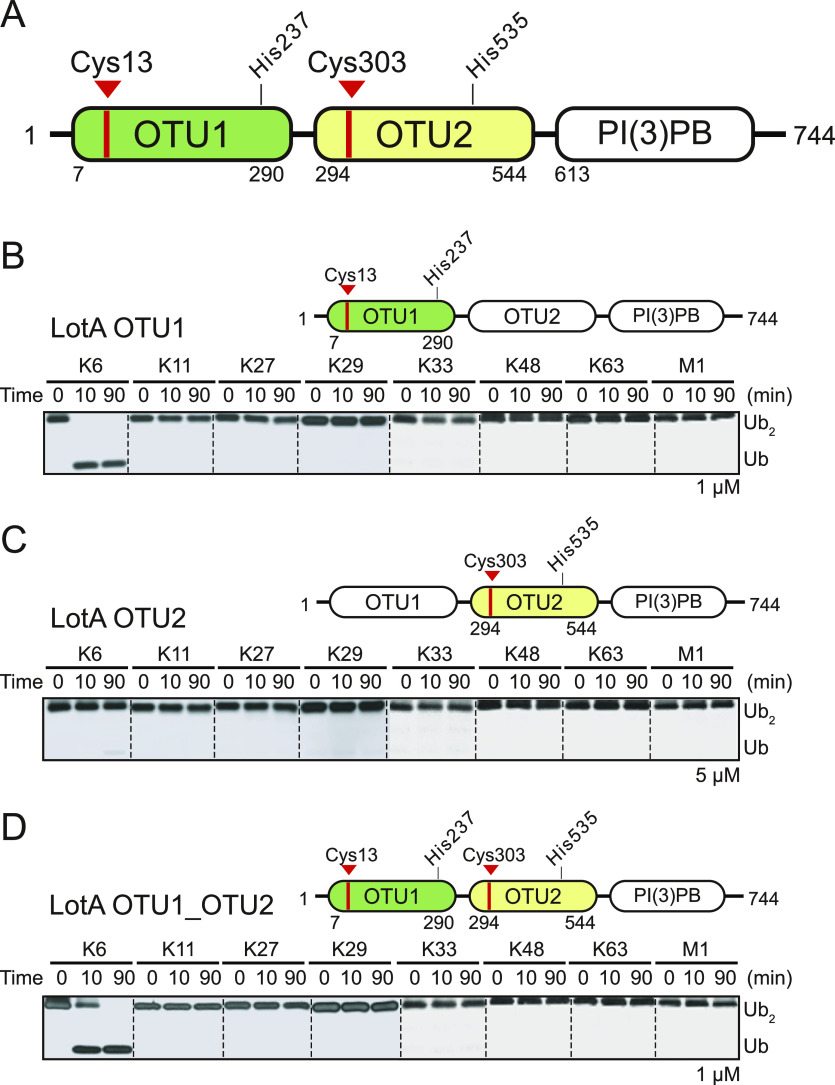
Figure 1. Deubiquitinase activities of both OTU domains of LotA. (A) Schematics of L. pneumophila LotA domain architecture. LotA comprises two OTU domains (LotA OTU1 and LotA OTU2). Catalytic cysteines of each OTU domain are labeled. (B) Diubiquitin cleavage assay by LotA OTU1 (LotA7–290) against eight different diubiquitin linkages. Reactions were quenched at the indicated time-points and analyzed on SDS–PAGE with silver staining. (C) Diubiquitin cleavage assay by LotA OTU2 (LotA294–544) against eight different diubiquitin linkages. Reactions were quenched at the indicated time-points and analyzed on SDS–PAGE with silver staining. (D) Diubiquitin cleavage assay by LotA OTU1_OTU2 (LotA7–544) against eight different diubiquitin linkages. Reactions were quenched at the indicated time-points and analyzed on SDS–PAGE with silver staining. Source data are available for this figure.
Figure 1. Deubiquitinase activities of both OTU domains of LotA. (A) Schematics of L. pneumophila LotA domain architecture. LotA comprises two OTU domains (LotA OTU1 and LotA OTU2). Catalytic cysteines of each OTU domain are labeled. (B) Diubiquitin cleavage assay by LotA OTU1 (LotA7–290) against eight different diubiquitin linkages. Reactions were quenched at the indicated time-points and analyzed on SDS–PAGE with silver staining. (C) Diubiquitin cleavage assay by LotA OTU2 (LotA294–544) against eight different diubiquitin linkages. Reactions were quenched at the indicated time-points and analyzed on SDS–PAGE with silver staining. (D) Diubiquitin cleavage assay by LotA OTU1_OTU2 (LotA7–544) against eight different diubiquitin linkages. Reactions were quenched at the indicated time-points and analyzed on SDS–PAGE with silver staining. Source data are available for this figure.CC BY · PubMed Central
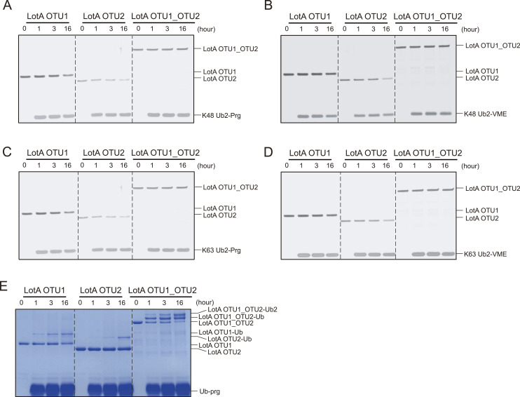
Figure S1. Deubiquitinase activities of LotA constructs. (A) K48-linked diubiquitin propargyl (Prg) activity-based probes (ABPs) were incubated at indicated time-points with LotA OTU constructs (OTU1, OTU2, and OTU1_OTU2) and analyzed at indicated time-points on SDS–PAGE with silver staining. (B) K48-linked diubiquitin vinyl methyl ester ABPs were incubated with LotA OTU constructs (OTU1, OTU2, and OTU1_OTU2) and analyzed at indicated time-points on SDS–PAGE with silver staining. (C) K63-linked diubiquitin propargyl (Prg) ABPs were incubated at indicated time-points with LotA OTU constructs (OTU1, OTU2, and OTU1_OTU2) and analyzed at indicated time-points on SDS–PAGE with silver staining. (D) K63-linked di-ubiquitin vinyl methyl ester ABPs were incubated at indicated time-points with LotA OTU constructs (OTU1, OTU2, and OTU1_OTU2) and analyzed at indicated time-points on SDS–PAGE with silver staining. (E) Ubiquitin propargyl ABPs were incubated at indicated time-points with LotA OTU constructs (OTU1, OTU2, and OTU1_OTU2) and analyzed at indicated time-points on SDS–PAGE with silver staining. Source data are available for this figure.
Figure S1. Deubiquitinase activities of LotA constructs. (A) K48-linked diubiquitin propargyl (Prg) activity-based probes (ABPs) were incubated at indicated time-points with LotA OTU constructs (OTU1, OTU2, and OTU1_OTU2) and analyzed at indicated time-points on SDS–PAGE with silver staining. (B) K48-linked diubiquitin vinyl methyl ester ABPs were incubated with LotA OTU constructs (OTU1, OTU2, and OTU1_OTU2) and analyzed at indicated time-points on SDS–PAGE with silver staining. (C) K63-linked diubiquitin propargyl (Prg) ABPs were incubated at indicated time-points with LotA OTU constructs (OTU1, OTU2, and OTU1_OTU2) and analyzed at indicated time-points on SDS–PAGE with silver staining. (D) K63-linked di-ubiquitin vinyl methyl ester ABPs were incubated at indicated time-points with LotA OTU constructs (OTU1, OTU2, and OTU1_OTU2) and analyzed at indicated time-points on SDS–PAGE with silver staining. (E) Ubiquitin propargyl ABPs were incubated at indicated time-points with LotA OTU constructs (OTU1, OTU2, and OTU1_OTU2) and analyzed at indicated time-points on SDS–PAGE with silver staining. Source data are available for this figure.CC BY · PubMed Central
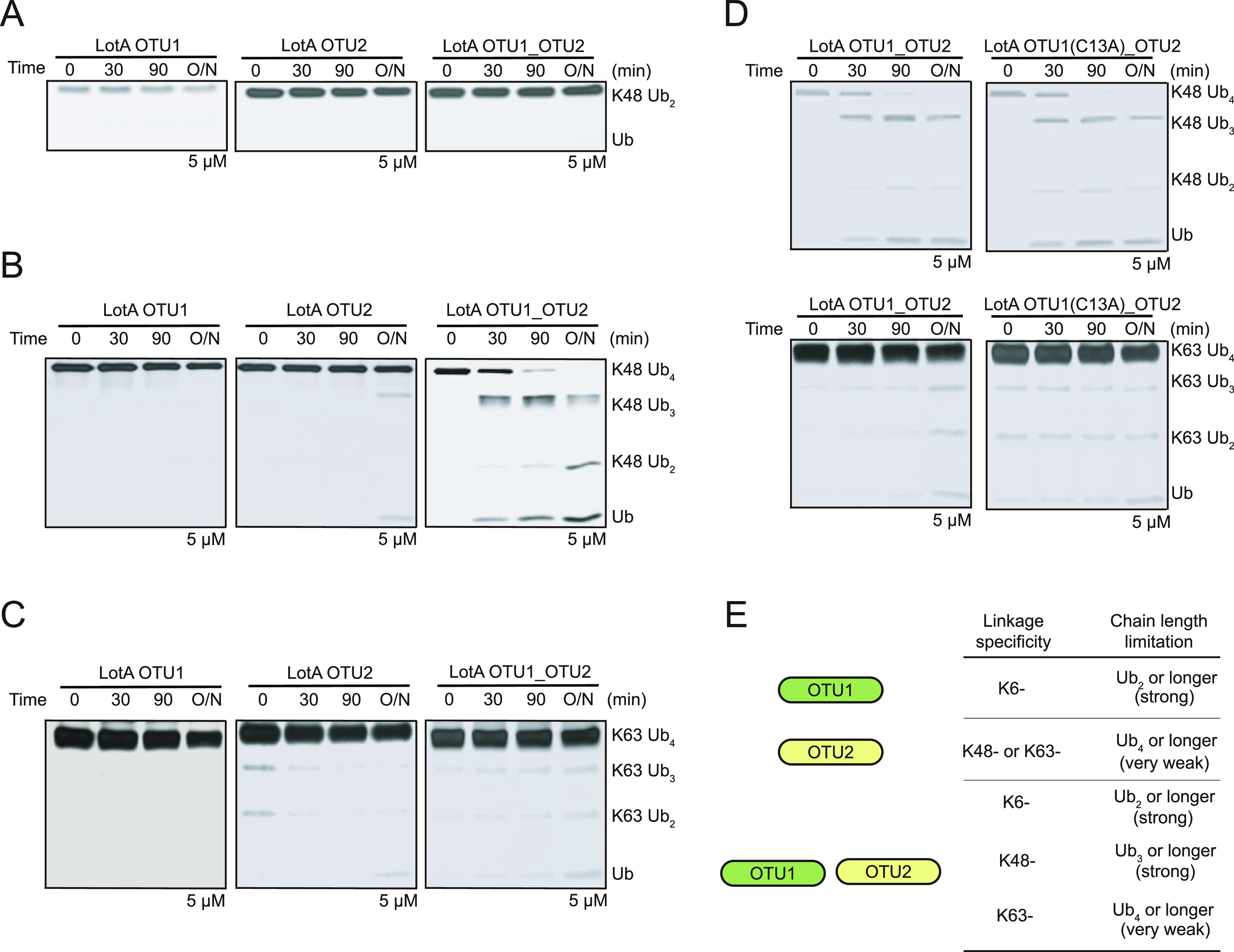
Figure 2. Cooperative catalytic activity of LotA OTU1 and OTU2 domains. (A) K48-linked diubiquitin cleavage assay by LotA OTU constructs (OTU1, OTU2, and OTU1_OTU2). (B) K48-linked tetraubiquitin chain cleavage assay by LotA OTU constructs (OTU1, OTU2, and OTU1_OTU2). Catalytic activity against K48-linked tetraubiquitin chains was remarkably enhanced when intact OTU1 and OTU2 domains (LotA7–544) were used. (C) K63-linked tetraubiquitin chain cleavage assay by LotA OTU constructs (OTU1, OTU2, and OTU1_OTU2). Catalytic activity against K63-linked tetraubiquitin chains was enhanced when intact OTU1 and OTU2 domains (LotA7–544) were used. (D) K48-linked tetraubiquitin chain cleavage assay by WT LotA OTU1_OTU2 and LotA OTU1*_OTU2 (C13A) mutant. (E) Ubiquitin linkage and length preference of LotA OTU constructs (OTU1, OTU2, and OTU1_OTU2). Source data are available for this figure.
Figure 2. Cooperative catalytic activity of LotA OTU1 and OTU2 domains. (A) K48-linked diubiquitin cleavage assay by LotA OTU constructs (OTU1, OTU2, and OTU1_OTU2). (B) K48-linked tetraubiquitin chain cleavage assay by LotA OTU constructs (OTU1, OTU2, and OTU1_OTU2). Catalytic activity against K48-linked tetraubiquitin chains was remarkably enhanced when intact OTU1 and OTU2 domains (LotA7–544) were used. (C) K63-linked tetraubiquitin chain cleavage assay by LotA OTU constructs (OTU1, OTU2, and OTU1_OTU2). Catalytic activity against K63-linked tetraubiquitin chains was enhanced when intact OTU1 and OTU2 domains (LotA7–544) were used. (D) K48-linked tetraubiquitin chain cleavage assay by WT LotA OTU1_OTU2 and LotA OTU1*_OTU2 (C13A) mutant. (E) Ubiquitin linkage and length preference of LotA OTU constructs (OTU1, OTU2, and OTU1_OTU2). Source data are available for this figure.CC BY · PubMed Central
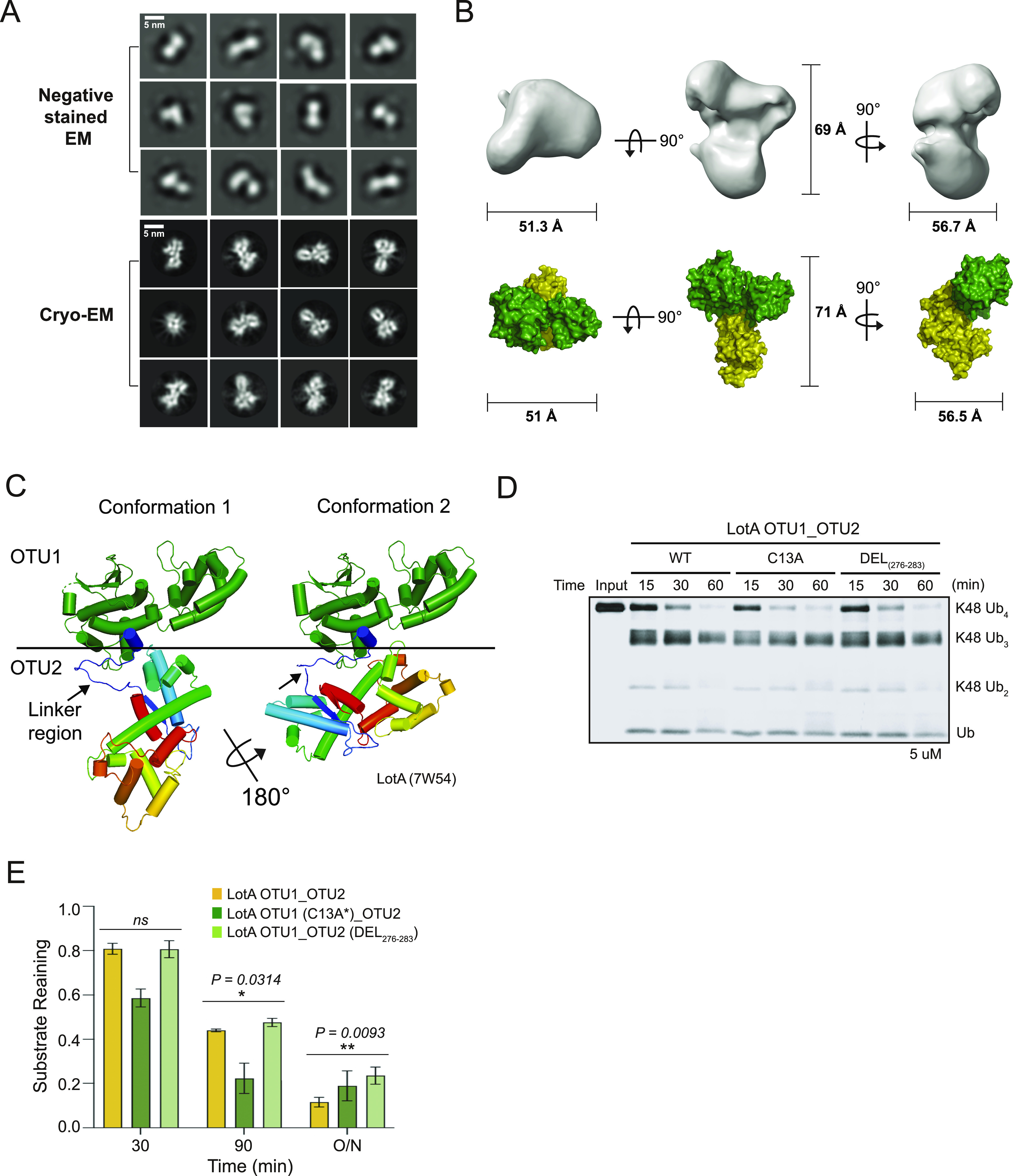
Figure 3. Structure of LotA OTU1_OTU2. (A) Selected 2D classes of negative-stain (upper) and cryo-EM (down) LotA OTU1_OTU2. Scale bar represented 5 nm. (B) LotA7–544 cryo-EM model orientated in X, Y, and Z axis. Each distance is described as angstrom (Å) (upper). Proposed OTU1 and OTU2 orientation of LotA7–544 (down) using crystal structure (PDB: 7W54). (C) Crystal structure of LotA OTU1_OTU2 (7W54). Two completely different conformations of LotA molecules are observed in the asymmetric unit. (D) DUB assay comparing cleavage of K48-linked tetraubiquitin chains by wild-type LotA OTU1_OTU2, LotA OTU1*_OTU2 (C13A) mutant, and linker deletion (residues 276–283) mutant of LotA OTU1_OTU2. (D, E) Quantification of remaining K48-linked tetraubiquitin chains shown in (D). Data are presented as the mean ± S.D (n = 3, * 0.01 < P < 0.05 and ** 0.001 < P < 0.01). Source data are available for this figure.
Figure 3. Structure of LotA OTU1_OTU2. (A) Selected 2D classes of negative-stain (upper) and cryo-EM (down) LotA OTU1_OTU2. Scale bar represented 5 nm. (B) LotA7–544 cryo-EM model orientated in X, Y, and Z axis. Each distance is described as angstrom (Å) (upper). Proposed OTU1 and OTU2 orientation of LotA7–544 (down) using crystal structure (PDB: 7W54). (C) Crystal structure of LotA OTU1_OTU2 (7W54). Two completely different conformations of LotA molecules are observed in the asymmetric unit. (D) DUB assay comparing cleavage of K48-linked tetraubiquitin chains by wild-type LotA OTU1_OTU2, LotA OTU1*_OTU2 (C13A) mutant, and linker deletion (residues 276–283) mutant of LotA OTU1_OTU2. (D, E) Quantification of remaining K48-linked tetraubiquitin chains shown in (D). Data are presented as the mean ± S.D (n = 3, * 0.01 < P < 0.05 and ** 0.001 < P < 0.01). Source data are available for this figure.CC BY · PubMed Central
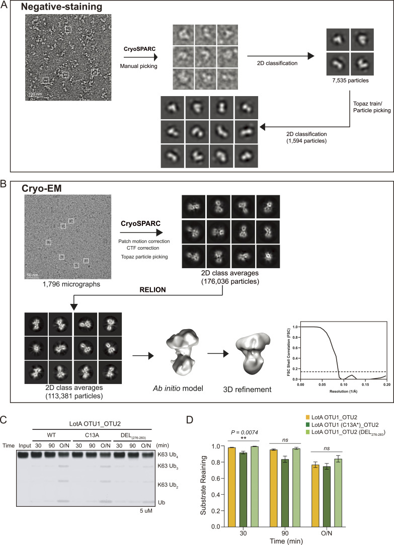
Figure S2. Negative-staining and cryo-EM data processing workflow. (A, B) Negative-staining (A) and single-particle cryo-EM (B) data processing flowchart of LotA OTU1_OTU2. (C) DUB assay comparing cleavage of K63-linked tetraubiquitin chains by wild-type LotA OTU1_OTU2, LotA OTU1*_OTU2 (C13A) mutant, and linker deletion (residues 276–283) mutant of LotA OTU1_OTU2. (D) Quantification of remaining K63-linked tetraubiquitin chains shown in (D). Data are presented as the mean ± S.D (n = 3, ns: not significant, * 0.01 < P < 0.05 and ** 0.001 < P < 0.01). Source data are available for this figure.
Figure S2. Negative-staining and cryo-EM data processing workflow. (A, B) Negative-staining (A) and single-particle cryo-EM (B) data processing flowchart of LotA OTU1_OTU2. (C) DUB assay comparing cleavage of K63-linked tetraubiquitin chains by wild-type LotA OTU1_OTU2, LotA OTU1*_OTU2 (C13A) mutant, and linker deletion (residues 276–283) mutant of LotA OTU1_OTU2. (D) Quantification of remaining K63-linked tetraubiquitin chains shown in (D). Data are presented as the mean ± S.D (n = 3, ns: not significant, * 0.01 < P < 0.05 and ** 0.001 < P < 0.01). Source data are available for this figure.CC BY · PubMed Central
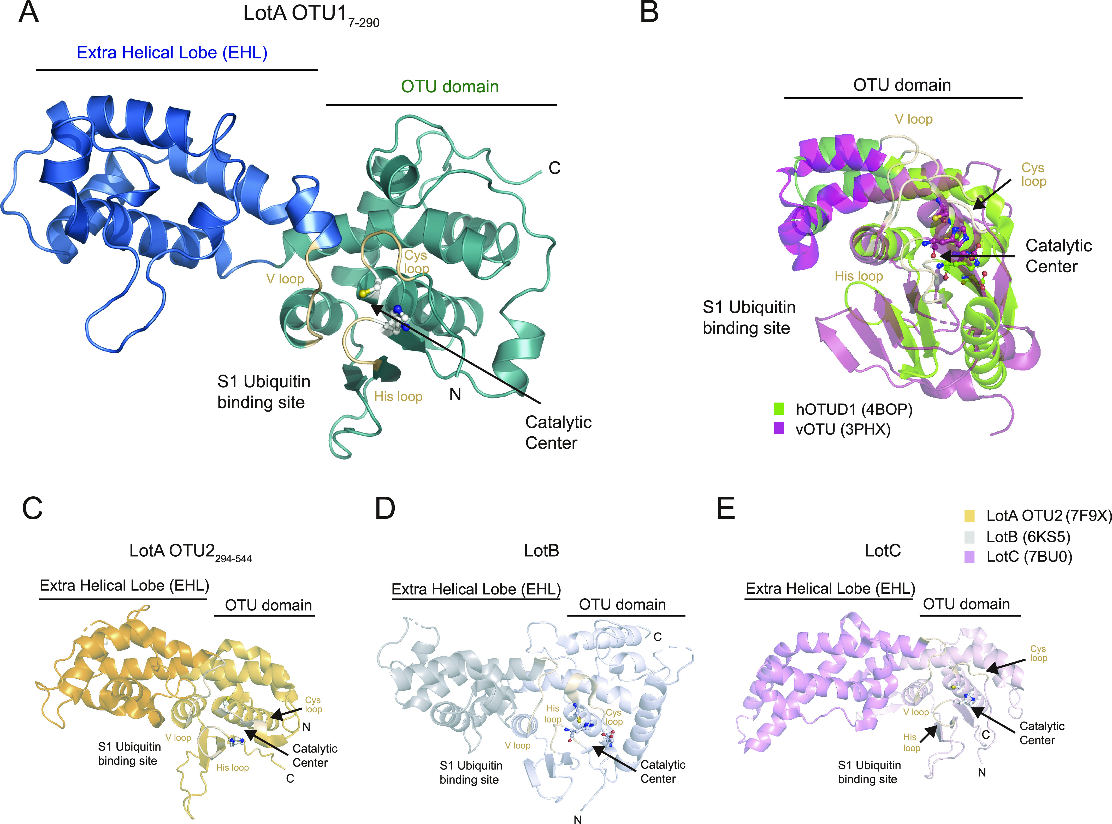
Figure 4. Structural comparison of Legionella OTU deubiquitinases. (A) X-ray crystal structure of LotA OTU1 (LotA7–290) diffracted to 1.54 Å determined by molecular replacement (deposited PDB ID: 8GOK). Catalytic cysteine and histidine of LotA OTU1 are shown as a ball-and-stick model, and Cys loop, His loop, and V loop are highlighted in wheat color. The EHL domain and the catalytic OTU domain are labeled. (B) Structural comparison of hOTUD1 (4BOP, green) and vOTU (3PHX, pink). Structures were aligned by their core OTU domain. (C) Crystal structure of LotA OTU2 (7F9X). Catalytic cysteine and histidine of LotA OTU2 are shown as a ball-and-stick model. The EHL domain and the catalytic OTU domain are labeled. (D) Crystal structure of LotB (6KS5). Catalytic triad residues of LotB are shown as a ball-and-stick model. The EHL domain and the catalytic OTU domain are labeled. (E) Crystal structure of LotC (7BU0). Catalytic triad residues of LotC are shown as a ball-and-stick model. The EHL domain and the catalytic OTU domain are labeled.
Figure 4. Structural comparison of Legionella OTU deubiquitinases. (A) X-ray crystal structure of LotA OTU1 (LotA7–290) diffracted to 1.54 Å determined by molecular replacement (deposited PDB ID: 8GOK). Catalytic cysteine and histidine of LotA OTU1 are shown as a ball-and-stick model, and Cys loop, His loop, and V loop are highlighted in wheat color. The EHL domain and the catalytic OTU domain are labeled. (B) Structural comparison of hOTUD1 (4BOP, green) and vOTU (3PHX, pink). Structures were aligned by their core OTU domain. (C) Crystal structure of LotA OTU2 (7F9X). Catalytic cysteine and histidine of LotA OTU2 are shown as a ball-and-stick model. The EHL domain and the catalytic OTU domain are labeled. (D) Crystal structure of LotB (6KS5). Catalytic triad residues of LotB are shown as a ball-and-stick model. The EHL domain and the catalytic OTU domain are labeled. (E) Crystal structure of LotC (7BU0). Catalytic triad residues of LotC are shown as a ball-and-stick model. The EHL domain and the catalytic OTU domain are labeled.CC BY · PubMed Central
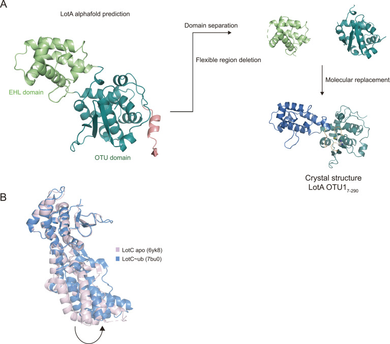
Figure S3. Structure determination process of LotA OTU1. (A) Structure determination process of LotA7–290 using molecular replacement with the AlphaFold-predicted structure as a template model. (B) Structural comparison of LotC apo (6YK8, pink) and LotC bound with ubiquitin (7BU0, blue). Structures were aligned by the core OTU domain.
Figure S3. Structure determination process of LotA OTU1. (A) Structure determination process of LotA7–290 using molecular replacement with the AlphaFold-predicted structure as a template model. (B) Structural comparison of LotC apo (6YK8, pink) and LotC bound with ubiquitin (7BU0, blue). Structures were aligned by the core OTU domain.CC BY · PubMed Central
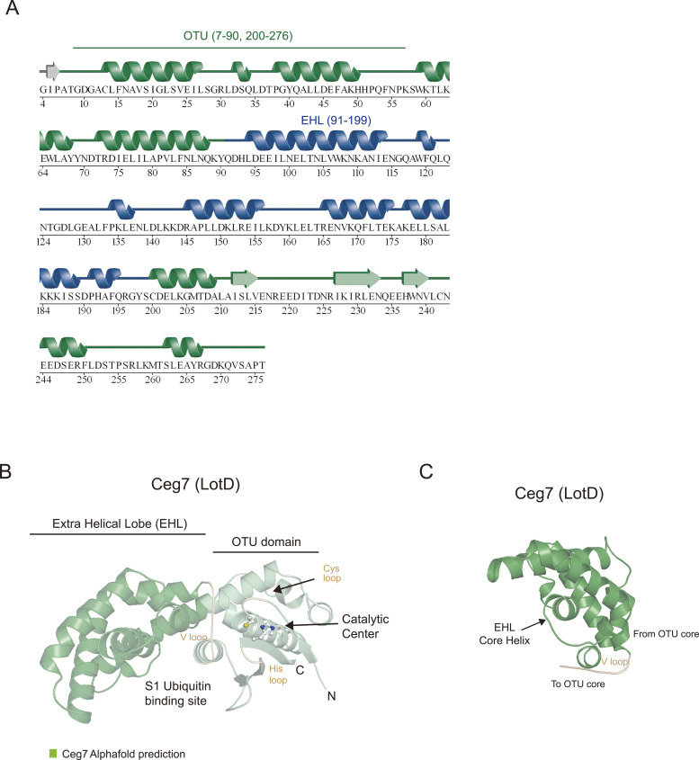
Figure S4. Structural comparison of Legionella OTU deubiquitinases. (A) Secondary structure alignment of LotA OTU1 based on the crystal structure. The OTU domain of LotA is colored green, and the EHL is colored blue. (B) AlphaFold prediction structure of LotD. Catalytic cysteine and histidine of LotD are shown as a ball-and-stick model. Additional EHL compared with hOTUD1 and vOTU is colored dark green. (C) Structure of the EHL domain of LotD. The EHL domain of the LOT DUB family shared roof-like folding.
Figure S4. Structural comparison of Legionella OTU deubiquitinases. (A) Secondary structure alignment of LotA OTU1 based on the crystal structure. The OTU domain of LotA is colored green, and the EHL is colored blue. (B) AlphaFold prediction structure of LotD. Catalytic cysteine and histidine of LotD are shown as a ball-and-stick model. Additional EHL compared with hOTUD1 and vOTU is colored dark green. (C) Structure of the EHL domain of LotD. The EHL domain of the LOT DUB family shared roof-like folding.CC BY · PubMed Central
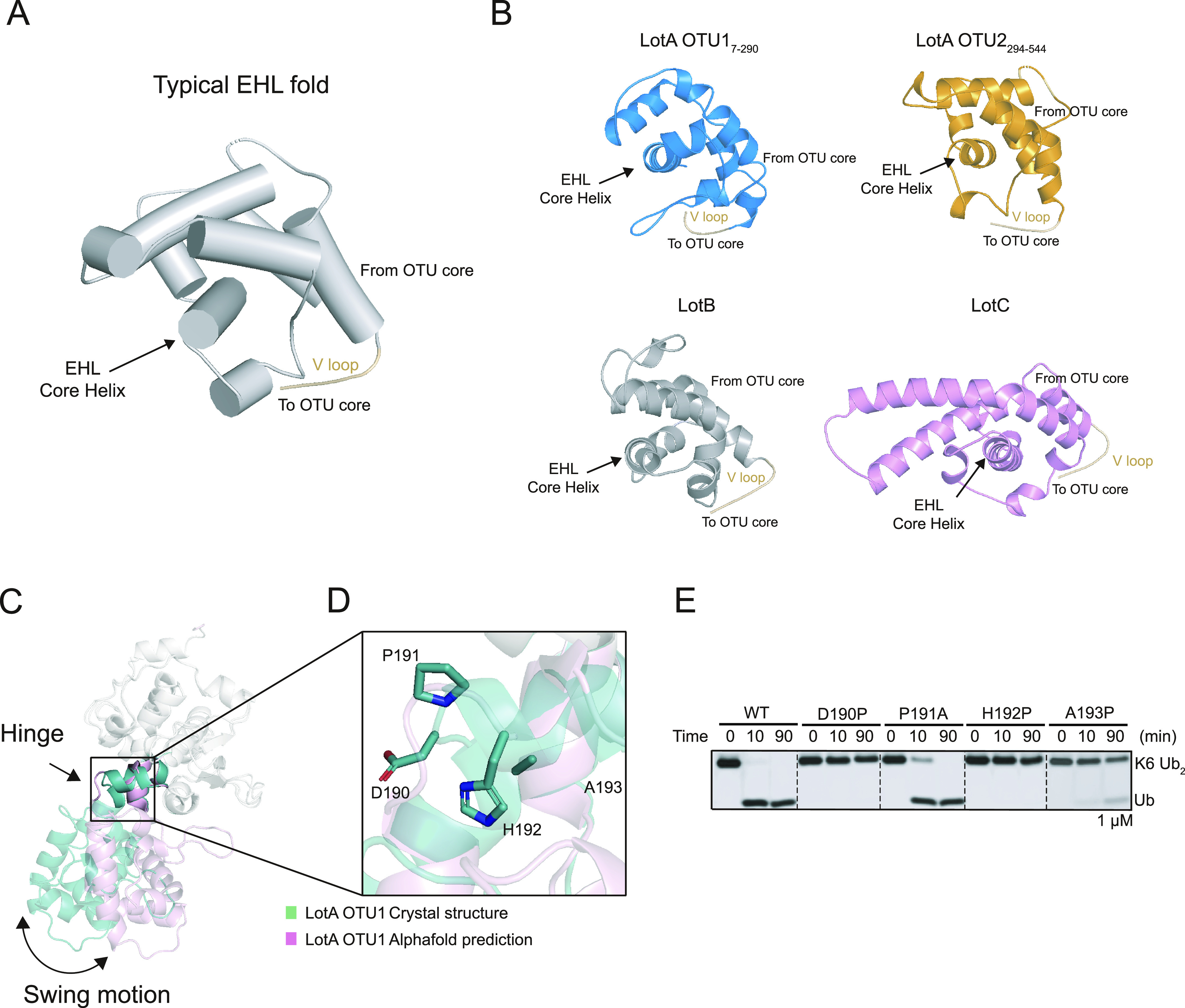
Figure 5. Structural analysis of the EHL domain of Lots. (A) Typical EHL fold architecture of the LOT DUB family. (B) Structure of the EHL domain of the LOT DUB family. The EHL domain of the LOT DUB family shared roof-like folding. (C) Structural comparison of LotA OTU1 crystal structure (cyan) and LotA OTU1 AlphaFold prediction (pink). The OTU domain of two structures is superimposed, and relative orientation of the EHL domain to the OTU domain is presented. (D) Close-up view of hinge region of LotA OTU1. Four amino acids (residues 190–193) in the hinge region between EHL and OTU domains are shown as a stick model. (E) DUB assay against K6-linked diubiquitin with wild-type and the hinge mutants of LotA OTU. Source data are available for this figure.
Figure 5. Structural analysis of the EHL domain of Lots. (A) Typical EHL fold architecture of the LOT DUB family. (B) Structure of the EHL domain of the LOT DUB family. The EHL domain of the LOT DUB family shared roof-like folding. (C) Structural comparison of LotA OTU1 crystal structure (cyan) and LotA OTU1 AlphaFold prediction (pink). The OTU domain of two structures is superimposed, and relative orientation of the EHL domain to the OTU domain is presented. (D) Close-up view of hinge region of LotA OTU1. Four amino acids (residues 190–193) in the hinge region between EHL and OTU domains are shown as a stick model. (E) DUB assay against K6-linked diubiquitin with wild-type and the hinge mutants of LotA OTU. Source data are available for this figure.CC BY · PubMed Central
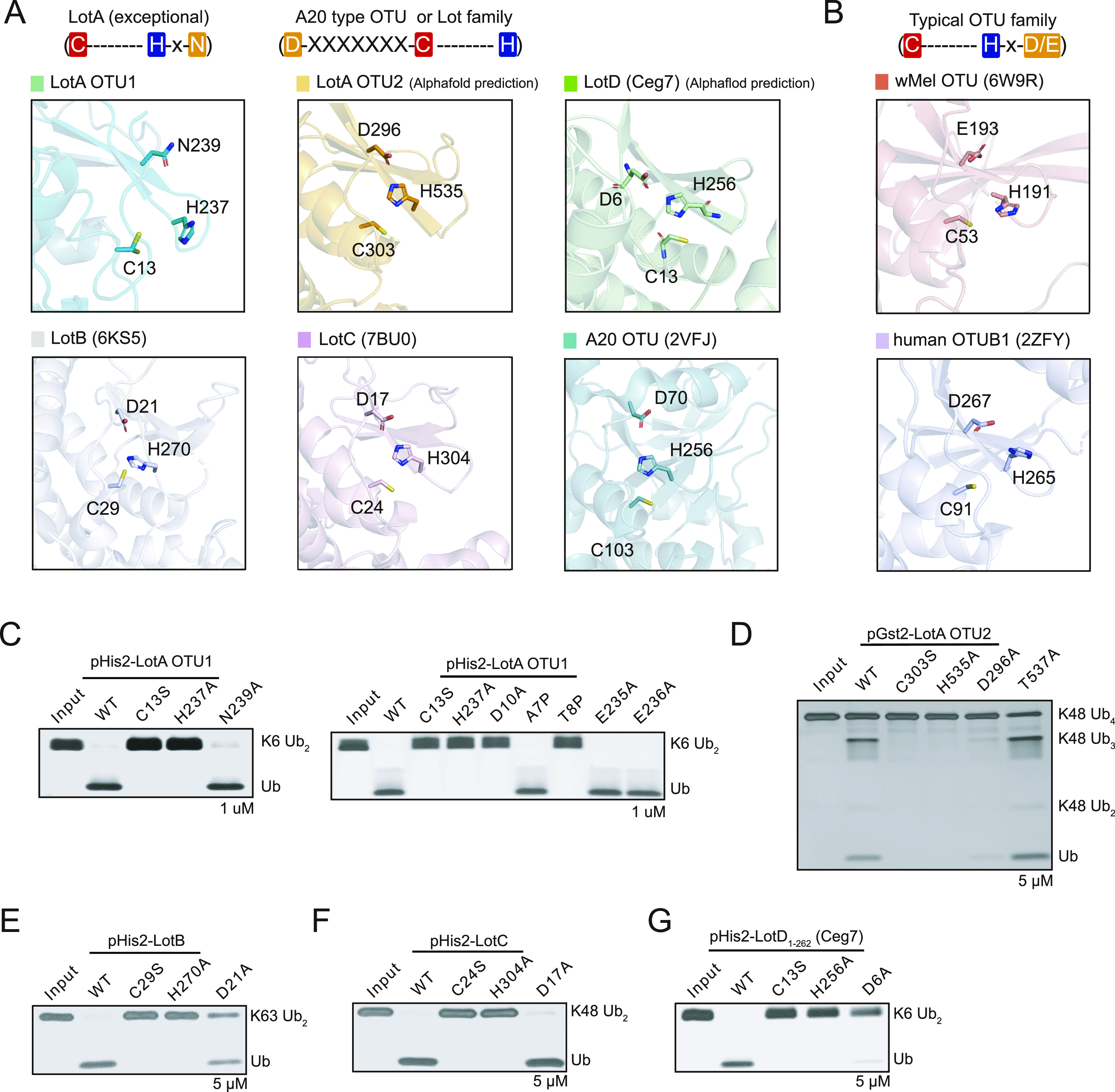
Figure 6. Catalytic triad of the LOT DUB family. (A) Close-up views of the LotA OTU1, LotA OTU2, LotB (6KS5), LotC (7BU0), LotD, and A20 OTU (2VFJ) catalytic sites. Catalytic triads are highlighted as a stick model. Schematic representation of the order of catalytic triads is also presented. (B) Close-up views of the wMel OTU (6W9R) and hOTUB1 (2ZFY) catalytic sites. Catalytic triads are highlighted as a stick model. Schematic representation of the order of catalytic triads is also presented. (C, D, E, F, G) Diubiquitin or polyubiquitin cleavage assay by the LOT DUB family to define catalytic triad residues. (C, D, E, F, G) LotA-OTU1, (D) LotA-OTU2, (E) LotB, (F) LotC, and (G) LotD. The catalytic activity of the LOT DUB family WT and their mutants was tested. Reactions were quenched at the indicated time-points and resolved by SDS–PAGE with silver staining, respectively. Source data are available for this figure.
Figure 6. Catalytic triad of the LOT DUB family. (A) Close-up views of the LotA OTU1, LotA OTU2, LotB (6KS5), LotC (7BU0), LotD, and A20 OTU (2VFJ) catalytic sites. Catalytic triads are highlighted as a stick model. Schematic representation of the order of catalytic triads is also presented. (B) Close-up views of the wMel OTU (6W9R) and hOTUB1 (2ZFY) catalytic sites. Catalytic triads are highlighted as a stick model. Schematic representation of the order of catalytic triads is also presented. (C, D, E, F, G) Diubiquitin or polyubiquitin cleavage assay by the LOT DUB family to define catalytic triad residues. (C, D, E, F, G) LotA-OTU1, (D) LotA-OTU2, (E) LotB, (F) LotC, and (G) LotD. The catalytic activity of the LOT DUB family WT and their mutants was tested. Reactions were quenched at the indicated time-points and resolved by SDS–PAGE with silver staining, respectively. Source data are available for this figure.CC BY · PubMed Central
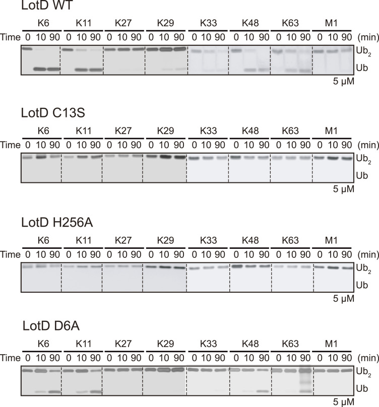
Figure S5. Deubiquitinase activities of LotD. Diubiquitin panel cleavage assay by WT LotD and catalytic triad residues (C13S, H256A, and D6A) of LotD against eight diubiquitin linkages. Reactions were quenched at the indicated time-points and analyzed on SDS–PAGE with silver staining. Source data are available for this figure.
Figure S5. Deubiquitinase activities of LotD. Diubiquitin panel cleavage assay by WT LotD and catalytic triad residues (C13S, H256A, and D6A) of LotD against eight diubiquitin linkages. Reactions were quenched at the indicated time-points and analyzed on SDS–PAGE with silver staining. Source data are available for this figure.CC BY · PubMed Central
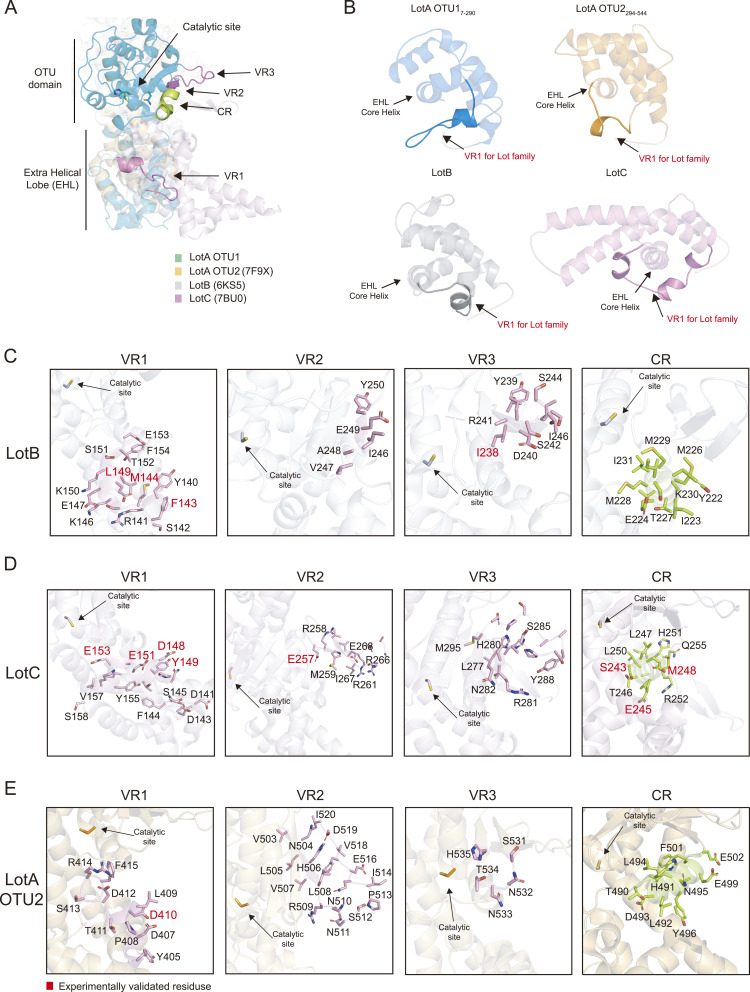
Figure S6. S1 ubiquitin-binding site of the LOT DUB family. (A) Structural overlay of LotA OTU1 (cyan), LotA OTU2 (7F9X, orange), LotB (6KS5, gray), and LotC (7BU0, pink). Structures were aligned by their core OTU domains. Three VRs are highlighted in pink, and CR is colored in green. (B) The EHL domain of LotA OTU1 (blue), LotA OTU2 (7F9X, orange), LotB (6KS5, gray), and LotC (7BU0, pink). VR1 for the LOT DUB family is concentrated in a helical loop region C-terminal to the EHL core helix. (C) Close-up views of the VRs and CR of LotB (6KS5). Three VRs (pink) and CR (green) residues are represented as a stick model, and experimentally validated residues are highlighted in red color. (D) Close-up views of the VRs and CR of LotC (7BU0). Three VRs (pink) and CR (green) residues are represented as a stick model, and experimentally validated residues are highlighted in red color. (E) Close-up views of the VRs and CR of LotA OTU2 (7F9X). Three VRs (pink) and CR (green) residues are represented as a stick model, and experimentally validated residues are highlighted in red color.
Figure S6. S1 ubiquitin-binding site of the LOT DUB family. (A) Structural overlay of LotA OTU1 (cyan), LotA OTU2 (7F9X, orange), LotB (6KS5, gray), and LotC (7BU0, pink). Structures were aligned by their core OTU domains. Three VRs are highlighted in pink, and CR is colored in green. (B) The EHL domain of LotA OTU1 (blue), LotA OTU2 (7F9X, orange), LotB (6KS5, gray), and LotC (7BU0, pink). VR1 for the LOT DUB family is concentrated in a helical loop region C-terminal to the EHL core helix. (C) Close-up views of the VRs and CR of LotB (6KS5). Three VRs (pink) and CR (green) residues are represented as a stick model, and experimentally validated residues are highlighted in red color. (D) Close-up views of the VRs and CR of LotC (7BU0). Three VRs (pink) and CR (green) residues are represented as a stick model, and experimentally validated residues are highlighted in red color. (E) Close-up views of the VRs and CR of LotA OTU2 (7F9X). Three VRs (pink) and CR (green) residues are represented as a stick model, and experimentally validated residues are highlighted in red color.CC BY · PubMed Central
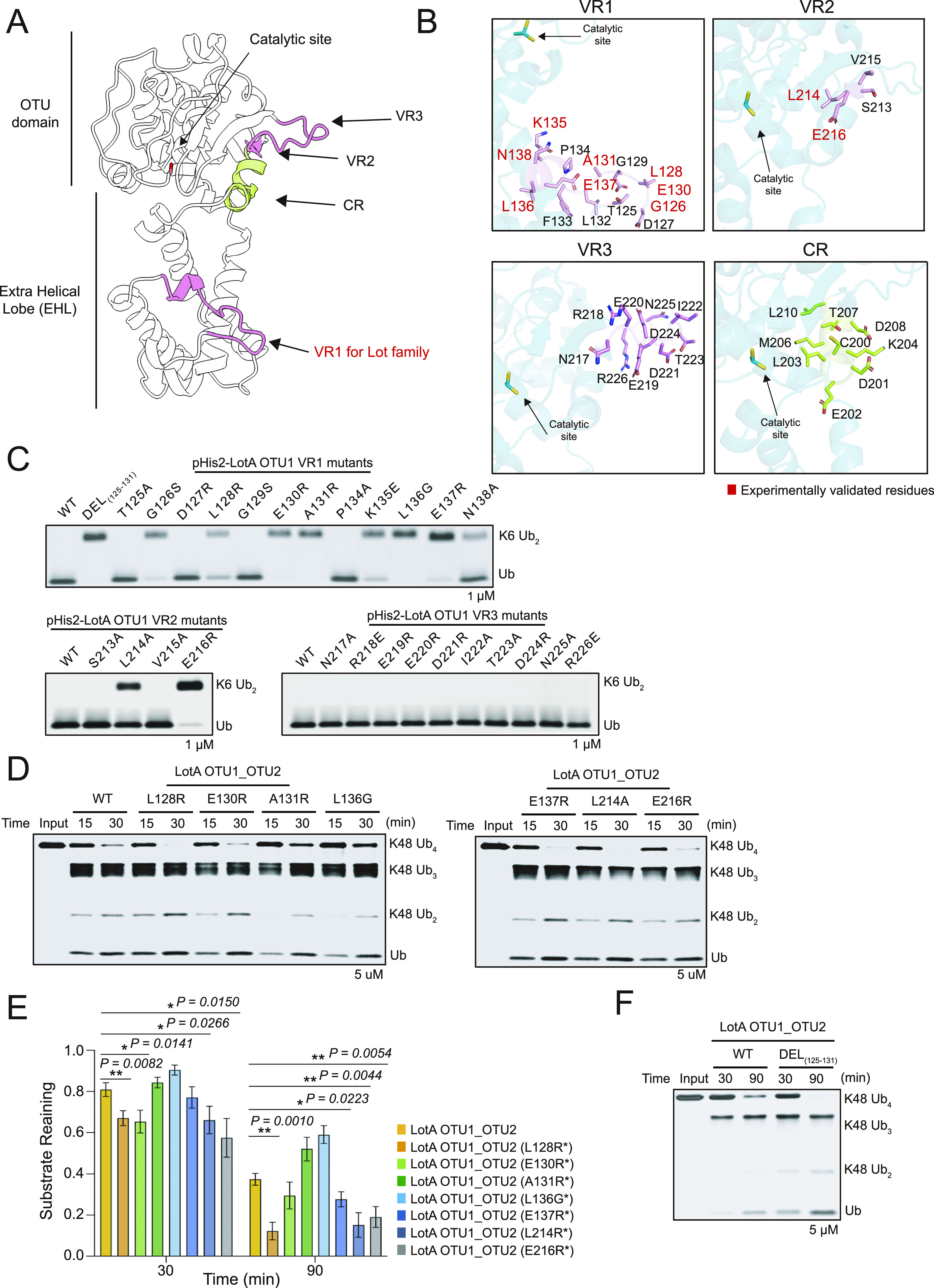
Figure 7. S1 ubiquitin-binding site of LotA OTU1. (A) Cartoon representation of LotA OTU1. The constant region (CR, green) and three variable regions (VRs, pink) are the parts of the S1 ubiquitin-binding site. The EHL domain contains the VR1 region of the LOT DUB family. (B) Close-up views of the VRs and CR of LotA OTU1. Three VRs (pink) and CR (green) residues are represented as a stick model, and experimentally identified residues are highlighted in red color. (C) K6-linked diubiquitin cleavage assay by LotA OTU1 WT and its mutants or deletion of VRs (VR1, VR2, and VR3) in LotA OTU1. Reactions were quenched at the indicated time-points and resolved by SDS–PAGE with silver staining. (D) Polyubiquitin cleavage assay by LotA OTU1_OTU2 WT and VR1 mutants of LotA OTU1 as labeled. (D, E) Quantification of remaining K48-linked tetraubiquitin chains shown in (D). Data are presented as the mean ± S.D (n = 3, * 0.01 < P < 0.05 and ** 0.001 < P < 0.01). (F) Gel-based K48-linked polyubiquitin cleavage assay by LotA OTU1_OTU2 WT and VR1 deletion of LotA (residues 125–131) mutant of LotA OTU1_OTU2. Source data are available for this figure.
Figure 7. S1 ubiquitin-binding site of LotA OTU1. (A) Cartoon representation of LotA OTU1. The constant region (CR, green) and three variable regions (VRs, pink) are the parts of the S1 ubiquitin-binding site. The EHL domain contains the VR1 region of the LOT DUB family. (B) Close-up views of the VRs and CR of LotA OTU1. Three VRs (pink) and CR (green) residues are represented as a stick model, and experimentally identified residues are highlighted in red color. (C) K6-linked diubiquitin cleavage assay by LotA OTU1 WT and its mutants or deletion of VRs (VR1, VR2, and VR3) in LotA OTU1. Reactions were quenched at the indicated time-points and resolved by SDS–PAGE with silver staining. (D) Polyubiquitin cleavage assay by LotA OTU1_OTU2 WT and VR1 mutants of LotA OTU1 as labeled. (D, E) Quantification of remaining K48-linked tetraubiquitin chains shown in (D). Data are presented as the mean ± S.D (n = 3, * 0.01 < P < 0.05 and ** 0.001 < P < 0.01). (F) Gel-based K48-linked polyubiquitin cleavage assay by LotA OTU1_OTU2 WT and VR1 deletion of LotA (residues 125–131) mutant of LotA OTU1_OTU2. Source data are available for this figure.CC BY · PubMed Central

Verification

Claims the paper makes

The claims and statistics of this paper haven’t been checked yet.

Run the check — free, no account needed. It reads the open-access text and checks the sources it cites.

A few free checks a day · papers already verified open instantly · results are public

Self-check

Percentages, sums and reported differences re-computed from the paper’s own numbers.

The self-check needs the paper’s full text.

Citations

Cites

Show all 4 references

Research works printed but not linked: 81

  1. 10.1016/j.molcel.2016.05.009 · DOI
  2. 10.1016/j.molcel.2021.08.024 · DOI
  3. 10.1107/S0907444912001308 · DOI · Paper page
  4. 10.1073/pnas.1015287108 · DOI
  5. 10.1038/nrmicro3259 · DOI
  6. 10.1128/JVI.67.10.6056-6063.1993 · DOI
  7. 10.1038/s41467-020-18952-1 · DOI
  8. 10.1016/j.cell.2016.11.019 · DOI
  9. 10.1038/s41586-019-1440-8 · DOI
  10. 10.1126/science.aaw7446 · DOI
  11. 10.1073/pnas.1221050110 · DOI
  12. 10.1038/s41580-019-0099-1 · DOI
  13. 10.1038/nsmb1346 · DOI
  14. 10.1038/s41580-022-00543-1 · DOI
  15. 10.1038/nature09964 · DOI
  16. 10.1042/BJ20081318 · DOI
  17. 10.1021/ja309802n · DOI
  18. 10.1002/anie.201005995 · DOI
  19. 10.1021/acscatal.7b01505 · DOI
  20. 10.1107/S0907444910007493 · DOI · Paper page
  21. 10.1128/IAI.00344-10 · DOI
  22. 10.1107/S0907444907051554 · DOI
  23. 10.1016/j.molcel.2011.06.034 · DOI
  24. 10.3389/fchem.2021.692168 · DOI
  25. 10.1038/ncomms14004 · DOI
  26. 10.1016/j.chembiol.2016.03.009 · DOI
  27. 10.1038/nsmb.3475 · DOI
  28. 10.1016/j.molcel.2018.02.024 · DOI
  29. 10.1038/s41467-018-03148-5 · DOI
  30. 10.1038/s41467-018-03511-6 · DOI
  31. 10.1073/pnas.1013388108 · DOI
  32. 10.1038/s41586-018-0145-8 · DOI
  33. 10.1016/j.cell.2013.05.014 · DOI
  34. 10.3389/fcimb.2020.00448 · DOI
  35. 10.1042/BJ20071399 · DOI
  36. 10.1146/annurev-biochem-060310-170328 · DOI · Paper page
  37. 10.1038/nrm2731 · DOI · Paper page
  38. 10.1111/j.1365-2958.2008.06124.x · DOI
  39. 10.1111/cmi.12840 · DOI
  40. 10.1016/j.molcel.2018.02.023 · DOI
  41. 10.1242/jcs.188482 · DOI
  42. 10.7554/eLife.58114 · DOI
  43. 10.1074/jbc.RA119.011758 · DOI
  44. 10.1038/nsmb.2792 · DOI
  45. 10.1107/S0021889807021206 · DOI · Paper page
  46. 10.1038/35086067 · DOI · Paper page
  47. 10.1371/journal.ppat.1002743 · DOI
  48. 10.1146/annurev-biochem-061516-044916 · DOI
  49. 10.1016/j.cell.2013.05.046 · DOI · Paper page
  50. 10.1038/nature19836 · DOI
  51. 10.1038/s41592-022-01488-1 · DOI · Paper page
  52. 10.1146/annurev-micro-041020-025803 · DOI
  53. 10.1002/cbic.201402012 · DOI
  54. 10.1016/j.chom.2013.11.012 · DOI
  55. 10.1038/nm.3739 · DOI
  56. 10.1016/j.molcel.2016.06.015 · DOI
  57. 10.1038/s41564-018-0271-y · DOI
  58. 10.1038/nmeth.4169 · DOI · Paper page
  59. 10.3389/fcimb.2017.00487 · DOI
  60. 10.1038/nature17657 · DOI
  61. 10.1016/j.str.2015.05.020 · DOI
  62. 10.1021/acscatal.0c03420 · DOI
  63. 10.1016/j.jsb.2015.08.008 · DOI
  64. 10.1107/S0907444913011426 · DOI
  65. 10.1016/j.jsb.2012.09.006 · DOI · Paper page
  66. 10.1038/s41418-020-00706-7 · DOI
  67. 10.15252/embj.2020105127 · DOI
  68. 10.1016/j.femsre.2004.07.001 · DOI
  69. 10.1006/prep.1998.1003 · DOI
  70. 10.7554/eLife.58277 · DOI
  71. 10.1016/j.molcel.2019.10.019 · DOI
  72. 10.1016/j.bmc.2013.02.039 · DOI
  73. 10.1038/cr.2016.39 · DOI
  74. 10.1128/JB.00376-21 · DOI
  75. 10.1016/j.semcdb.2022.02.008 · DOI
  76. 10.3389/fphar.2016.00107 · DOI
  77. 10.1038/s41564-019-0454-1 · DOI
  78. 10.1101/2022.05.11.491541 · DOI
  79. 10.3390/v7092849 · DOI
  80. 10.1155/2009/315423 · DOI
  81. 10.7554/eLife.42166 · DOI

Metadata

Authors
Kang, S., Kim, G., Choi, M., Jeong, M., van der Heden van Noort, G. J., Roh, S.-H., & Shin, D.
Affiliations
  • Yonsei University (KR)
  • Seoul National University (KR)
  • Department of Cell and Chemical Biology, Leiden University Medical Centre, Leiden, The Netherlands
Corresponding author
Shin, Donghyuk
Venue
Life Science Alliance
Published
2023-04-26
Volume
6
Issue
7
Pages
e202201876
ISSN
2575-1077 (ISSN-L)
OpenAlex
W4367059319
Cited by
9
References
4
Citations by year
2026: 2 · 2025: 2 · 2024: 2 · 2023: 3
FWCI
0.80
Type
journal article
Language
en
Open access
Locations
  • Life Science Alliance (publishedVersion, cc-by)
  • bioRxiv (Cold Spring Harbor Laboratory) (acceptedVersion, cc-by-nc-nd)
  • PubMed Central (publishedVersion, cc-by)
  • PubMed (publishedVersion)
  • Leiden Repository (Leiden University) (submittedVersion, cc-by)
  • DOAJ (DOAJ: Directory of Open Access Journals) (submittedVersion)
  • Seoul National University Open Repository (Seoul National University) (submittedVersion)
  • Leiden Repository (Leiden University) (submittedVersion)
Funders
  • National Research Foundation of Korea (2021R1C1C1003961, 2018R1A6A1A03025607, 2021M3A9I4021220, 2021R1I1A1A01049284, 2021R1A6C103B381)
  • ZonMw (451001026)
  • Nederlandse Organisatie voor Wetenschappelijk Onderzoek (VI.VIDI.192.011)
  • Yonsei University (2021-22-0050)
Topics
  • Life Sciences › Biochemistry, Genetics and Molecular Biology › Molecular Biology › Ubiquitin and proteasome pathways
  • Life Sciences › Biochemistry, Genetics and Molecular Biology › Structural Biology › Enzyme Structure and Function
  • Life Sciences › Biochemistry, Genetics and Molecular Biology › Molecular Biology › Protein Degradation and Inhibitors
Keywords
Legionella, OTU domain, Lota, catalytic triad, K48-linked ubiquitin chains, protein structure, bacterial virulence
MeSH terms
Deubiquitinating Enzymes, Female, Humans, Legionella, Ovarian Neoplasms, Ubiquitin, Polyubiquitin

About these checks

Registry, retraction, Cites and statistics results are computed, not judged, with one exception: a printed reference that no index lists is read once by a model before it counts as a citation error. Claims are extracted by a model and kept only when their quote appears verbatim in the text; each cited source is then checked, with a model’s help, for whether it says what the claim relies on. Those verdicts can be wrong — read the claim’s quote and the cited source before relying on one.

Statistics and arithmetic are recomputed, not judged.

Confidence bands are shown only for fields calibrated on our annotated test set. None is calibrated yet, so no band is shown.

What counts as a claim ›How we measure confidence ›How we check this ›

Are you an author, or do you see a problem with this page? Request a review

How to cite

Cite CoChat for the checks on this page, and the paper for its own findings.

This page
CoChat. (2026). Citation check: Structural insights into ubiquitin chain cleavage by Legionella ovarian tumor deubiquitinases. https://app.cochat.ai/papers/10.26508/lsa.202201876
The paper
Kang, S., Kim, G., Choi, M., Jeong, M., van der Heden van Noort, G. J., Roh, S.-H., & Shin, D. (2023). Structural insights into ubiquitin chain cleavage by Legionella ovarian tumor deubiquitinases. Life Science Alliance. https://doi.org/10.26508/lsa.202201876