

The DOI matches its Crossref record; 318 of the 319 references it cites were found in a registry or an index, and 1 are not checked yet.
Design Challenges in Polymeric Scaffolds for Tissue Engineering
Open accessAbstract
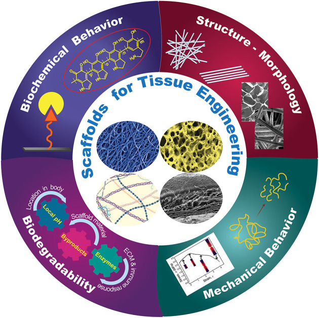
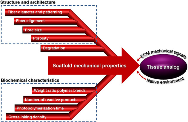
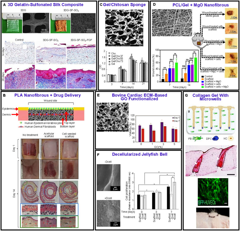
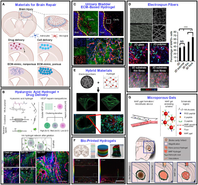
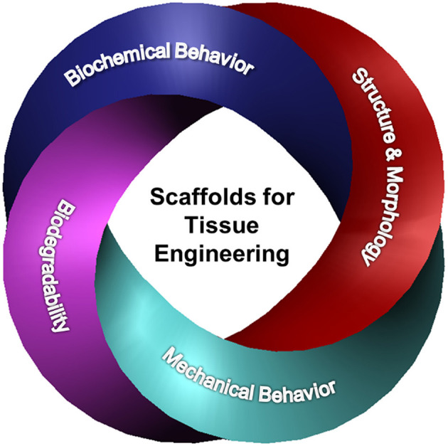
Numerous surgical procedures are daily performed worldwide to replace and repair damaged tissue. Tissue engineering is the field devoted to the regeneration of damaged tissue through the incorporation of cells in biocompatible and biodegradable porous constructs, known as scaffolds. The scaffolds act as host biomaterials of the incubating cells, guiding their attachment, growth, differentiation, proliferation, phenotype, and migration for the development of new tissue. Furthermore, cellular behavior and fate are bound to the biodegradation of the scaffold during tissue generation. This article provides a critical appraisal of how key biomaterial scaffold parameters, such as structure architecture, biochemistry, mechanical behavior, and biodegradability, impart the needed morphological, structural, and biochemical cues for eliciting cell behavior in various tissue engineering applications. Particular emphasis is given on specific scaffold attributes pertaining to skin and brain tissue generation, where further progress is needed (skin) or the research is at a relatively primitive stage (brain), and the enumeration of some of the most important challenges regarding scaffold constructs for tissue engineering.
Overview
The claims and statistics of this paper haven’t been checked yet.
Run the check — free, no account needed. It reads the open-access text and checks the sources it cites.
Integrity signals
Registry
Cites
Statistics
Figures
Verification
Claims the paper makes
The claims and statistics of this paper haven’t been checked yet.
Run the check — free, no account needed. It reads the open-access text and checks the sources it cites.
Claims other papers make about this one
Each is checked against this paper’s own text: does it say what the citing paper says it does?
The source backs part of the claim, not all of it — or backs it for a different population, condition or comparison.
In the citing paper
“This can be related to the fact that fabricated materials were mainly developed as biologically inert, so they would not have provoked any reaction from the recipient’s side [199].”
From Resorbable Biomaterials Used for 3D Scaffolds in Tissue Engineering: A Review (2023) · cites this paper as [199]
Experimental — accuracy not yet measuredThe source backs part of the claim, not all of it — or backs it for a different population, condition or comparison.
In the citing paper
“Although many scaffolds have been successfully applied in vivo or utilized in clinical medicine to restore damaged tissues, adverse effects such as extensive inflammatory response leading to transplant rejection were often observed.”
From Resorbable Biomaterials Used for 3D Scaffolds in Tissue Engineering: A Review (2023) · cites this paper as [199]
Experimental — accuracy not yet measuredThe source backs part of the claim, not all of it — or backs it for a different population, condition or comparison.
In the citing paper
“Furthermore, the synthetic material’s prolonged presence causes an inflammatory response mediated by phagocytes and foreign body giant cells.”
From Resorbable Biomaterials Used for 3D Scaffolds in Tissue Engineering: A Review (2023) · cites this paper as [199]
Experimental — accuracy not yet measuredThe cited source says what the claim attributes to it.
In the citing paper
“By-effects of this phenomenon include fibrous encapsulation of the implanted scaffold [199].”
From Resorbable Biomaterials Used for 3D Scaffolds in Tissue Engineering: A Review (2023) · cites this paper as [199]
The check wasn’t confident either way, so it gives no verdict — never disputed.
In the citing paper
“The implantation site, as well as the procedure of scaffold implantation itself, affect the recruitment and composition of adjacent cell populations.”
From Resorbable Biomaterials Used for 3D Scaffolds in Tissue Engineering: A Review (2023) · cites this paper as [199]
Experimental — accuracy not yet measured
Self-check
Percentages, sums and reported differences re-computed from the paper’s own numbers.
The self-check needs the paper’s full text.
Citations
Cites
Mussel-inspired surface chemistry for multifunctional coatings
Lee, H., Dellatore, S. M., Miller, W. M., & Messersmith, P. B. (2007) · Science · doi:10.1126/science.1147241 · Cited by 10,965
Origin and physiological roles of inflammation
Medzhitov, R. (2008) · Nature · doi:10.1038/nature07201 · Cited by 6,618
Factors affecting wound healing
Guo, S., & DiPietro, L. A. (2010) · Journal of Dental Research · doi:10.1177/0022034509359125 · Cited by 5,792
Astrocyte-endothelial interactions at the blood-brain barrier
Abbott, N. J., Rönnbäck, L., & Hansson, E. (2006) · Nature Reviews Neuroscience · doi:10.1038/nrn1824 · Cited by 5,595
Hydrogels for Tissue Engineering
Lee, K. Y., & Mooney, D. J. (2001) · Chemical Reviews · doi:10.1021/cr000108x · Cited by 5,295
Astrocytes: Biology and pathology
Sofroniew, M. V., & Vinters, H. V. (2010) · Acta Neuropathologica · doi:10.1007/s00401-009-0619-8 · Cited by 5,196
Structure and function of the blood–brain barrier
Abbott, N. J., Patabendige, A. A. K., Dolman, D. E. M., Yusof, S. R., & Begley, D. J. (2010) · Neurobiology of Disease · doi:10.1016/j.nbd.2009.07.030 · Cited by 5,185
Foreign body reaction to biomaterials
Anderson, J. M., Rodriguez, A., & Chang, D. T. (2008) · Seminars in Immunology · doi:10.1016/j.smim.2007.11.004 · Cited by 5,138
Hydrogels for tissue engineering: scaffold design variables and applications
Drury, J. L., & Mooney, D. J. (2003) · Biomaterials · doi:10.1016/s0142-9612(03)00340-5 · Cited by 5,136
Cu-Catalyzed Azide−Alkyne Cycloaddition
Meldal, M., & Tornøe, C. W. (2008) · Chemical Reviews · doi:10.1021/cr0783479 · Cited by 4,775
Research works printed but not linked: 2
- 2020, Tissue Engineering Market Size, Share & Trends Analysis Report by Application (Cord Blood & Cell Banking, Cancer, GI & Gynecology, Dental, Orthopedics, Musculoskeletal, & Spine), by Region, and Segment Forecasts, 2020-2027
- Janeway, 2001, Part V - The immune system in health and disease
Cited by
Sorted by citations
Optimizing scaffold pore size for tissue engineering: insights across various tissue types
Mukasheva, F., Adilova, L., Dyussenbinov, A., Yernaimanova, B., Аbilev, М., & Akilbekova, D. (2024) · Frontiers in Bioengineering and Biotechnology · doi:10.3389/fbioe.2024.1444986 · Cited by 492
Salthouse, D., Novakovic, K., Hilkens, C. M. U., & Ferreira, A. M. (2022) · Acta Biomaterialia · doi:10.1016/j.actbio.2022.11.003 · Cited by 196
Drug Delivery Systems in Regenerative Medicine: An Updated Review
Mansour, A., Romani, M., Acharya, A. B., Rahman, B., Verron, É., & Badran, Z. (2023) · Pharmaceutics · doi:10.3390/pharmaceutics15020695 · Cited by 135
Viscoelastic Biomaterials for Tissue Regeneration
Wu, D. T. S., Jeffreys, N. C., Diba, M., & Mooney, D. J. (2022) · Tissue Engineering Part C Methods · doi:10.1089/ten.tec.2022.0040 · Cited by 126
Polycaprolactone Composites/Blends and Their Applications Especially in Water Treatment
Kayan, G. Ö., & Kayan, A. (2023) · ChemEngineering · doi:10.3390/chemengineering7060104 · Cited by 120
Chitosan-based injectable hydrogel with multifunction for wound healing: A critical review
Li, A., Ma, B., Hua, S., Ping, R., Ding, L., Tian, B., & Zhang, X. (2024) · Carbohydrate Polymers · doi:10.1016/j.carbpol.2024.121952 · Cited by 118
Resorbable Biomaterials Used for 3D Scaffolds in Tissue Engineering: A Review
Vach Agocsova, S., Culenova, M., Birova, I., Omanikova, L., Moncmanova, B., Danisovic, L., Ziaran, S., Bakos, D., & Alexy, P. (2023) · Materials · doi:10.3390/ma16124267 · Cited by 98
High-resolution 3D printing of xanthan gum/nanocellulose bio-inks
Baniasadi, H., Kimiaei, E., Polez, R. T., Ajdary, R., Rojas, O. J., Österberg, M., & Seppälä, J. V. (2022) · International Journal of Biological Macromolecules · doi:10.1016/j.ijbiomac.2022.04.183 · Cited by 98
Zheng, X., Chen, T., Jiang, X., Naito, M., & Watanabe, I. (2022) · Science and Technology of Advanced Materials · doi:10.1080/14686996.2022.2157682 · Cited by 94
Advances of naturally derived biomedical polymers in tissue engineering
Hu, T., Fang, J., Shen, Y., Li, M., Wang, B., Xu, Z., & Hu, W. (2024) · Frontiers in Chemistry · doi:10.3389/fchem.2024.1469183 · Cited by 92
Recent Applications of Electrospun Nanofibrous Scaffold in Tissue Engineering
Owida, H. A., Al-Nabulsi, J., Alnaimat, F., Al-Ayyad, M. S., Turab, N. M., Sharah, A. A., & Shakur, M. (2022) · Applied Bionics and Biomechanics · doi:10.1155/2022/1953861 · Cited by 80
A Review of Biomimetic and Biodegradable Magnetic Scaffolds for Bone Tissue Engineering and Oncology
Păltânea, G., Mănescu, V., Antoniac, I., Antoniac, A., Nemoianu, I. V., Robu, A., & Dura, H. (2023) · International Journal of Molecular Sciences · doi:10.3390/ijms24054312 · Cited by 77
Cell-Tissue Interaction: The Biomimetic Approach to Design Tissue Engineered Biomaterials
Nitti, P., Narayanan, A., Pellegrino, R., Villani, S., Madaghiele, M., & Demitri, C. (2023) · Bioengineering · doi:10.3390/bioengineering10101122 · Cited by 76
3D Printing of Polysaccharide-Based Hydrogel Scaffolds for Tissue Engineering Applications: A Review
Tamo, A. K., Djouonkep, L. D. W., & Selabi, N. B. S. (2024) · International Journal of Biological Macromolecules · doi:10.1016/j.ijbiomac.2024.132123 · Cited by 72
Masri, S., Mazlan, Z., Zulkiflee, I., Salleh, A., Fadilah, N. I. M., Maarof, M., Wen, A. P. Y., Duman, F., Tabata, Y., Aziz, I. A., et al. (2022) · International Journal of Molecular Sciences · doi:10.3390/ijms23010476 · Cited by 63
Girard, F., Lajoye, C., Camman, M., Tissot, N., Pedurand, F. B., Tandon, B., Moedder, D., Liashenko, I., Salameh, S., Dalton, P. D., et al. (2024) · Advanced Functional Materials · doi:10.1002/adfm.202314757 · Cited by 62
Clinical challenges in bone tissue engineering - A narrative review
Hoveidaei, A. H., Sadat‐Shojai, M., Nabavizadeh, S. S., Niakan, R., Shirinezhad, A., Mosalamiaghili, S., & Tabaie, S. A. (2024) · Bone · doi:10.1016/j.bone.2024.117363 · Cited by 62
Polymeric Scaffolds Used in Dental Pulp Regeneration by Tissue Engineering Approach
Sugiaman, V. K., Jeffrey, J., Naliani, S., Pranata, N., Djuanda, R., & Saputri, R. I. (2023) · Polymers · doi:10.3390/polym15051082 · Cited by 58
Water-powered, electronics-free dressings that electrically stimulate wounds for rapid wound closure
Rajaram, K., Jakus, M. A., Chen, H., Jain, B., Kennedy, D. G., Caso, E. A., Mishra, N., Sharma, N., Uzunoğlu, B. E., Han, W. B., et al. (2024) · Science Advances · doi:10.1126/sciadv.ado7538 · Cited by 58
Tamo, A. K., Tran, T. A., Doench, I., Jahangir, S., Lall, A., David, L., Peniche-Covas, C., Walther, A., & Osorio‐Madrazo, A. (2022) · Materials · doi:10.3390/ma15176039 · Cited by 57
Toward a New Generation of Bio-Scaffolds for Neural Tissue Engineering: Challenges and Perspectives
Villanueva‐Flores, F., Garcia-Atutxa, I., Santos, A., & Armendáriz‐Borunda, J. (2023) · Pharmaceutics · doi:10.3390/pharmaceutics15061750 · Cited by 57
Manivannan, R., Sharma, N., Kumar, V., Iyyappan, J., Vimal, S., & Umesh, M. (2024) · Carbohydrate Polymer Technologies and Applications · doi:10.1016/j.carpta.2024.100536 · Cited by 57
Sustainable biofabrication: from bioprinting to AI-driven predictive methods
Filippi, M., Mekkattu, M., & Katzschmann, R. K. (2024) · Trends in biotechnology · doi:10.1016/j.tibtech.2024.07.002 · Cited by 56
Enhancing Stem Cell-Based Therapeutic Potential by Combining Various Bioengineering Technologies
Hong, I. (2022) · Frontiers in Cell and Developmental Biology · doi:10.3389/fcell.2022.901661 · Cited by 58
Wang, Q., Ma, J., Chen, S., & Wu, S. (2023) · Nanomaterials · doi:10.3390/nano13071150 · Cited by 54
Conducting polymer scaffolds: a new frontier in bioelectronics and bioengineering
Nasser, R. A., Arya, S. S., Alshehhi, K. H., Teo, J. C. M., & Pitsalidis, C. (2024) · Trends in biotechnology · doi:10.1016/j.tibtech.2023.11.017 · Cited by 52
Electrically conductive coatings in tissue engineering
Kohestani, A. A., Xu, Z., Baştan, F. E., Boccaccini, A. R., & Pishbin, F. (2024) · Acta Biomaterialia · doi:10.1016/j.actbio.2024.08.007 · Cited by 51
Additive Manufacturing of Polymer/Bioactive Glass Scaffolds for Regenerative Medicine: A Review
Martelli, A., Bellucci, D., & Cannillo, V. (2023) · Polymers · doi:10.3390/polym15112473 · Cited by 49
LCST/UCST behavior of polysaccharides for hydrogel fabrication
Moon, S. H., Park, S. J., Lee, Y. W., & Yang, Y. J. (2024) · RSC Advances · doi:10.1039/d4ra06240j · Cited by 48
Vascularized 3D Human Skin Models in the Forefront of Dermatological Research
Rimal, R., Muduli, S., Desai, P., Marquez, A. B., Möller, M., Platzman, I., Spatz, J., & Singh, S. (2024) · Advanced Healthcare Materials · doi:10.1002/adhm.202303351 · Cited by 46
Gelatin-based scaffolds: An intuitive support structure for regenerative therapy
Rashid, A. B., Showva, N.-N., & Hoque, M. E. (2023) · Current Opinion in Biomedical Engineering · doi:10.1016/j.cobme.2023.100452 · Cited by 44
Norouzi, S., Shemshaki, N. S., Norouzi, E., Latifi, M., Azimi, B., Danti, S., Qiao, X., Miao, Y., Yang, S., Gorji, M., et al. (2024) · Materials Today Chemistry · doi:10.1016/j.mtchem.2024.102016 · Cited by 42
Li, L., Xu, J., Ye, C., Zhou, Y., Yan, F., Chen, Z., & Xiao, Y. (2025) · Biomaterials · doi:10.1016/j.biomaterials.2025.123714 · Cited by 42
Milan, P. B., Masoumi, F., Biazar, E., Jalise, S. Z., & Mehrabi, A. (2024) · Macromolecular Bioscience · doi:10.1002/mabi.202400322 · Cited by 42
Cryopreservation of Tissue-Engineered Scaffold-Based Constructs: from Concept to Reality
Arutyunyan, I. V., Elchaninov, A. V., Sukhikh, G. T., & Fatkhudinov, T. (2021) · Stem Cell Reviews and Reports · doi:10.1007/s12015-021-10299-4 · Cited by 34
Yazdanpanah, A., Madjd, Z., Pezeshki‐Modaress, M., Khosrowpour, Z., Farshi, P., Eini, L., Kiani, J., Seifi, M., Kundu, S. C., Ghods, R., et al. (2022) · Artificial Organs · doi:10.1111/aor.14169 · Cited by 33
Öztemur, J., Özdemir, S., Tezcan-Unlu, H., Çeçener, G., Sezgin, H., & Yalçın-Eniş, İ. (2023) · Biopolymers · doi:10.1002/bip.23564 · Cited by 32
Chernonosova, V. S., Kuzmin, I. E., Shundrina, I. K., Korobeynikov, M. V., Golyshev, V. M., Chelobanov, B. P., & Laktionov, P. P. (2023) · Journal of Functional Biomaterials · doi:10.3390/jfb14020070 · Cited by 31
Codrea, C. I., Baykara, D., Mitran, R., Koyuncu, A. C. Ç., Gündüz, O., & Ficai, A. (2024) · Polymers · doi:10.3390/polym16131932 · Cited by 31
A Novel Antimicrobial Hydrogel for the Management of Periodontal Diseases
Wong, P. Y., Soo, S., Wong, E. S.-C., Praveen, P., Clode, P. L., Baker, M. V., & Matsubara, V. H. (2023) · International Dental Journal · doi:10.1016/j.identj.2023.01.002 · Cited by 30
Chitosan/hydroxyapatite hydrogels for localized drug delivery and tissue engineering: A review
Khodaverdi, K., Naghib, S. M., Mozafari, M., & Rahmanian, M. (2024) · Carbohydrate Polymer Technologies and Applications · doi:10.1016/j.carpta.2024.100640 · Cited by 27
Kumar, P., Sharma, J., Kumar, R., Najser, J., Frantík, J., Manuja, A., Sunnam, N., & Praveenkumar, S. (2025) · Biomaterials Science · doi:10.1039/d4bm01606h · Cited by 25
Cabral, . S. D., de Melo‐Diogo, D., Ferreira, P. C. N., Moreira, A. F., & Correia, I. J. (2024) · International Journal of Biological Macromolecules · doi:10.1016/j.ijbiomac.2024.129210 · Cited by 25
Biomaterials for cardiovascular diseases
Huang, Y., & Ding, Z. (2024) · Biomedical Technology · doi:10.1016/j.bmt.2024.05.001 · Cited by 25
3D Engineered scaffolds of conjugated polymers/metal organic frameworks for biomedical applications
Sharifabad, A. H., Ghanbari, R., Saeb, M. R., Zare, E. N., Rossi, F., Salami‐Kalajahi, M., & Makvandi, P. (2025) · International Materials Reviews · doi:10.1177/09506608241299174 · Cited by 24
Negrescu, A. M., & Cîmpean, A. (2025) · Gels · doi:10.3390/gels11070527 · Cited by 24
Talouki, P. Y., Tackallou, S. H., Shojaei, S., Benisi, S. Z., & Goodarzi, V. (2023) · Biological Procedures Online · doi:10.1186/s12575-023-00197-z · Cited by 23
Random cellulose acetate nanofibers: a breakthrough for cultivated meat production
dos Santos, A. E. A., Guadalupe, J. L., Albergaria, J. D. S., Almeida, I. A., Moreira, A. M. S., Copola, A. G. L., de Araújo, I. P., de Paula, A. M., Neves, B. R. A., Santos, J. P. F., et al. (2024) · Frontiers in Nutrition · doi:10.3389/fnut.2023.1297926 · Cited by 23
Shakir, S., Hackett, T., & Mostaço-Guidolin, L. B. (2022) · Frontiers in Bioengineering and Biotechnology · doi:10.3389/fbioe.2022.1011800 · Cited by 22
Therapeutic Agent-Loaded Fibrous Scaffolds for Biomedical Applications
Park, D., Lee, S. J., Choi, D. K., & Park, J.-W. (2023) · Pharmaceutics · doi:10.3390/pharmaceutics15051522 · Cited by 23
The potential of graphene coatings as neural interfaces
Lopes, V., Moreira, G., Bramini, M., & Capasso, A. (2024) · Nanoscale Horizons · doi:10.1039/d3nh00461a · Cited by 22
Keratin nanofibers in tissue engineering: bridging nature and innovation
Aadil, K. R., Bhange, K., Kumar, N., & Mishra, G. (2024) · Biotechnology for Sustainable Materials · doi:10.1186/s44316-024-00019-6 · Cited by 22
Mohiuddin, M., Rahman, M. M., Uddin, M. N., Hasan, R., & Rahman, I. M. M. (2025) · RSC Advances · doi:10.1039/d5ra06280b · Cited by 21
Future Frontiers in Bioinspired Implanted Biomaterials
Gu, Q., Yuan, R., Sun, D., & Wallace, G. G. (2025) · Advanced Materials · doi:10.1002/adma.202506323 · Cited by 20
New Polymeric Composites Based on Two-Dimensional Nanomaterials for Biomedical Applications
Pires, L. S., Magalhães, F. D., & Pinto, A. M. (2022) · Polymers · doi:10.3390/polym14071464 · Cited by 20
Nanofiber Scaffold-Based Tissue Engineering for the Treatment of Acute Liver Failure
Liu, X., Yao, X., Ouyang, Q., Oliveira, A. L., Li, Y., & Zhang, Y. (2024) · Advanced Fiber Materials · doi:10.1007/s42765-024-00395-8 · Cited by 21
Emerging Strategies in Mesenchymal Stem Cell-Based Cardiovascular Therapeutics
Kumar, R., Mishra, N., Tran, T., Kumar, M., Vijayaraghavalu, S., & Gurusamy, N. (2024) · Cells · doi:10.3390/cells13100855 · Cited by 22
Harnessing native blueprints for designing bioinks to bioprint functional cardiac tissue
Akter, M. Z., Tufail, F., Ahmad, A., Oh, Y. W., Kim, J. M., Kim, S., Hasan, M. M., Li, L., Lee, D., Kim, Y. S., et al. (2025) · iScience · doi:10.1016/j.isci.2025.111882 · Cited by 21
Mohammadzadeh, M., Farzin, A., Pazhouhnia, Z., Hoseinpour, M., & Beheshtizadeh, N. (2026) · Journal of Biological Engineering · doi:10.1186/s13036-025-00607-8 · Cited by 19
Paetzold, R., Coulter, F. B., Singh, G., Kelly, D. J., & O’Cearbhaill, E. D. (2022) · Bioprinting · doi:10.1016/j.bprint.2022.e00206 · Cited by 19
Stavarache, C. E., Ghebaur, A., Serafim, A., Vlăsceanu, G. M., Vasile, E., Gȃrea, S. A., & Iovu, H. (2024) · Polymers · doi:10.3390/polym16111592 · Cited by 19
Subramanian, S. A., Oh, S., Mariadoss, A. V. A., Chae, S., Dhandapani, S., sekar, P. P., Song, S. Y., Woo, C., Xue, D. Q., Choi, J., et al. (2022) · International Journal of Biological Macromolecules · doi:10.1016/j.ijbiomac.2022.04.211 · Cited by 19
Krishnan, Y., & Sampath, S. (2025) · Discover Materials · doi:10.1007/s43939-025-00435-z · Cited by 18
Recent progresses in neural tissue engineering using topographic scaffolds
Han, S. (2024) · American Journal of Stem Cells · doi:10.62347/wmdz8890 · Cited by 18
Zhang, G., Zhang, M., Feng, Q., Wang, R., Mei, H., Xing, K., & Li, J. (2024) · ACS Applied Materials & Interfaces · doi:10.1021/acsami.4c07210 · Cited by 18
Krutko, M., Poling, H. M., Sheth, M., Kongsomros, S., Bryan, A. E., Sharma, M., Singh, A., Reza, H. A., Wikenheiser‐Brokamp, K. A., Takebe, T., et al. (2025) · Advanced Materials Technologies · doi:10.1002/admt.202401513 · Cited by 17
Ohno, Y., Nakajima, C., Ajioka, I., Muraoka, T., Yaguchi, A., Fujioka, T., Akimoto, S., Matsuo, M., Lotfy, A., Nakamura, S., et al. (2023) · Biomaterials · doi:10.1016/j.biomaterials.2023.122003 · Cited by 17
Chan, Y. M., Judge, N. G., Hu, Y., Willits, R. K., Li, N. Y., & Becker, M. L. (2025) · Biomacromolecules · doi:10.1021/acs.biomac.5c00558 · Cited by 16
Advancement in Cancer Vasculogenesis Modeling through 3D Bioprinting Technology
Shukla, A. K., Yoon, S., Oh, S., Lee, D., Ahn, M., & Kim, B. S. (2024) · Biomimetics · doi:10.3390/biomimetics9050306 · Cited by 17
Kopf, S., Root, A., Heinmaa, I., de Lima, J. A., Åkesson, D., & Skrifvars, M. (2024) · ACS Omega · doi:10.1021/acsomega.4c02241 · Cited by 16
Noori, F., Bozorgi, A., Farmani, A. R., Abbasi, A., Ai, J., Tavassoli, A., Ghasemian, A., Morovvati, H., Alipanah, H., Ataollahi, M. R., et al. (2025) · Journal of Polymers and the Environment · doi:10.1007/s10924-025-03543-2 · Cited by 16
de Souza, J. R., Rahimnejad, M., Soares, I. P. M., Anselmi, C., Oliveira, P., dos Reis‐Prado, A. H., Maglaras, V., Dal‐Fabbro, R., Trichês, E. S., & Bottino, M. C. (2025) · Bioprinting · doi:10.1016/j.bprint.2025.e00413 · Cited by 16
Shestovskaya, M. V., Божкова, С. А., Sopova, J. V., Георгиевич, Х. М., & Bozhokin, M. S. (2021) · Biomedicines · doi:10.3390/biomedicines9111666 · Cited by 16
Caronna, F., Glimpel, N., Paar, G.-P., Gries, T., Blaeser, A., Do, K., Dolan, E. B., & Ronan, W. (2022) · Biomaterials Science · doi:10.1039/d1bm02027g · Cited by 16
Novel Electrospun Polycaprolactone/Calcium Alginate Scaffolds for Skin Tissue Engineering
Molina, M. I. E., Chen, C., Martinez, J., Tran, P., & Komvopoulos, K. (2022) · Materials · doi:10.3390/ma16010136 · Cited by 16
Self-citationFadilah, N. I. M., Riha, S. M., Mazlan, Z., Wen, A. P. Y., Hao, L. Q., Joseph, B., Maarof, M., Thomas, S., Motta, A., & Fauzi, M. B. (2023) · Frontiers in Bioengineering and Biotechnology · doi:10.3389/fbioe.2023.1160577 · Cited by 16
Control and interplay of scaffold–biomolecule interactions applied to cartilage tissue engineering
Dupuy, S., Salvador, J., Morille, M., Noël, D., & Belamie, E. (2025) · Biomaterials Science · doi:10.1039/d5bm00049a · Cited by 15
Advances in stimuli-responsive polymers for biomedical and environmental applications
Kang, T. W., Park, K., & Kim, M. S. (2025) · Materials Science and Engineering R Reports · doi:10.1016/j.mser.2025.101140 · Cited by 16
Advances in 3D Printed Scaffolds for Periodontal Regeneration
Daghrery, A., Soares, I. P. M., dos Reis‐Prado, A. H., Araújo, I. J. D. S., Dal‐Fabbro, R., & Bottino, M. C. (2026) · Current Oral Health Reports · doi:10.1007/s40496-025-00421-7 · Cited by 15
Bioengineering toolkits for potentiating organoid therapeutics
Park, S., & Cho, S. (2024) · Advanced Drug Delivery Reviews · doi:10.1016/j.addr.2024.115238 · Cited by 16
Electroconductive Gelatin/Alginate/ Graphene Hydrogel Based Scaffold for Neural Tissue Repair
Madaninasab, P., Mohammadi, M., & Labbaf, S. (2024) · Macromolecular Materials and Engineering · doi:10.1002/mame.202400229 · Cited by 15
Jenvoraphot, T., Thapsukhon, B., Daranarong, D., Molloy, R., Supanchart, C., Krisanaprakornkit, S., Topham, P. D., Tighe, B. J., Mahomed, A., & Punyodom, W. (2022) · ACS Applied Polymer Materials · doi:10.1021/acsapm.1c01794 · Cited by 15
Soman, S. S., Govindharaj, M., Hashimi, N. A., Zhou, J., & Vijayavenkataraman, S. (2022) · International Journal of Bioprinting · doi:10.18063/ijb.v8i4.604 · Cited by 15
García‐Briones, G. S., Laga, R., Černochová, Z., Arjona-Ruiz, C., Janoušková, O., Šlouf, M., Pop‐Georgievski, O., & Kubies, D. (2023) · European Polymer Journal · doi:10.1016/j.eurpolymj.2023.111976 · Cited by 15
ON INDIVIDUAL ASPECTS OF CHOTISAN SAMPLES CHARACTERISATION IN BIOMEDICAL RESEARCH
Khalimov, R. I., Omelko, N. A., & Rusin, E. E. (2022) · Научное обозрение Биологические науки (Scientific Review Biological Sciences) · doi:10.17513/srbs.1305 · Cited by 14
Application of terahertz spectroscopy and imaging techniques in biomedicine
Duan, X., Han, S., Yao, Y., Shen, J., Dai, Y., Mi, J., & Wang, Z. (2025) · iScience · doi:10.1016/j.isci.2025.113990 · Cited by 14
Cesquim, M. G., Lopes, L. R., da Silva, L. D., da Silva, M. B., Donato, G. D., Machado, W. F., Noronha, P. B., de Carvalho, L. T., & Medeiros, S. F. (2025) · Carbohydrate Polymers · doi:10.1016/j.carbpol.2025.124658 · Cited by 14
Paknia, S., Izadi, Z., Moosaipour, M., Moradi, S., Khalilzadeh, B., Jaymand, M., & Samadian, H. (2022) · Journal of Molecular Liquids · doi:10.1016/j.molliq.2022.119701 · Cited by 14
Zdraveva, E., Bendelja, K., Bočkor, L., Dolenec, T., & Mijović, B. (2023) · Polymers · doi:10.3390/polym15030777 · Cited by 14
Enhanced degradation and bioactivity in polysiloxane-based shape memory polymer (SMP) scaffolds
Beltran, F. O., Arabiyat, A. S., Culibrk, R. A., Yeisley, D. J., Houk, C. J., Hicks, A. J., Negrón-Hernández, J., Nitschke, B. M., Hahn, M. S., & Grunlan, M. A. (2023) · Polymer · doi:10.1016/j.polymer.2023.126291 · Cited by 14
Bider, F., Gunnella, C., Reh, J. T., Clejanu, C.-E., Kuth, S., Beltrán, A. M., & Boccaccini, A. R. (2024) · Journal of Biomaterials Applications · doi:10.1177/08853282241280768 · Cited by 14
Bioengineering Tooth and Periodontal Organoids from Stem and Progenitor Cells
Torizal, F. G., Noorintan, S. T., & Gania, Z. (2024) · Organoids · doi:10.3390/organoids3040015 · Cited by 14
Mohammadzadeh, M., Goli, M., Shahrebabaki, K. E., Golshirazi, A., & Labbaf, S. (2025) · Bioprinting · doi:10.1016/j.bprint.2025.e00421 · Cited by 14
Microporous/Macroporous Polycaprolactone Scaffolds for Dental Applications
Shabab, T., Bas, O., Dargaville, B. L., Ravichandran, A., Tran, P. A., & Hutmacher, D. W. (2023) · Pharmaceutics · doi:10.3390/pharmaceutics15051340 · Cited by 13
Ferenčík, N., Danko, M., Nadova, Z., Kolembusová, P., & Steingartner, W. (2024) · Bioengineering · doi:10.3390/bioengineering11050442 · Cited by 13
Intervertebral disc degeneration and regenerative medicine
Farag, M., Rezk, R., Hutchinson, H. J., Zankevich, A., & Lucke-Wold, B. P. (2024) · Clinical and Translational Discovery · doi:10.1002/ctd2.289 · Cited by 13
Kaya, B., & Yeşil-Çeliktaş, Ö. (2024) · Bio-Design and Manufacturing · doi:10.1007/s42242-024-00289-z · Cited by 14
Edible electrospun materials for scalable cultivated beef production
Dagès, B. A. S., Fabian, J. A., Poláková, D., Rysová, M., Topham, P. D., Souppez, J.-B. R. G., Hanga, M. P., & Theodosiou, E. (2024) · Food and Bioproducts Processing · doi:10.1016/j.fbp.2024.11.012 · Cited by 13
Effanga, V. E., Akilbekova, D., Mukasheva, F., Zhao, X., Kalyon, D. M., & Erişken, C. (2024) · Gels · doi:10.3390/gels10110745 · Cited by 13
Khanbaba, A., Mozaffari, H., Majidi, R. F., & Derakhshan, M. A. (2024) · BioNanoScience · doi:10.1007/s12668-024-01652-1 · Cited by 13
Metadata
- Authors
- Echeverria Molina, M. I., Malollari, K. G., & Komvopoulos, K.
- Affiliations
- University of California, Berkeley (US)
- Corresponding author
- Komvopoulos, Kyriakos
- Venue
- Frontiers in Bioengineering and Biotechnology
- Publisher
- Frontiers Media SA
- Published
- 2021-06-14
- Volume
- 9
- Pages
- 617141-617141
- ISSN
- 2296-4185 (ISSN-L)
- PMC
- PMC8236583
- OpenAlex
- W3171498185
- Cited by
- 397
- References
- 318
- Citations by year
- 2026: 103 · 2025: 124 · 2024: 89 · 2023: 49 · 2022: 30 · 2021: 2
- FWCI
- 3.59
- Type
- journal article
- Language
- en
- Open access
- gold
- doi.org
- Licence
- Locations
- Frontiers in Bioengineering and Biotechnology (publishedVersion, cc-by)
- PubMed Central (publishedVersion, cc-by)
- PubMed (publishedVersion)
- DOAJ (DOAJ: Directory of Open Access Journals) (submittedVersion, cc-by-sa)
- eScholarship (California Digital Library) (submittedVersion)
- eScholarship (California Digital Library) (submittedVersion, cc-by)
- Topics
- Physical Sciences › Engineering › Biomedical Engineering › Bone Tissue Engineering Materials
- Health Sciences › Medicine › Surgery › Tissue Engineering and Regenerative Medicine
- Physical Sciences › Materials Science › Biomaterials › Hydrogels: synthesis, properties, applications
- Keywords
- polymer scaffolds, tissue engineering, biodegradable biomaterials, scaffold design, biodegradation, cell behavior, scaffold architecture, mechanical properties, skin regeneration, brain tissue regeneration
About these checks
Registry, retraction, Cites and statistics results are computed, not judged, with one exception: a printed reference that no index lists is read once by a model before it counts as a citation error. Claims are extracted by a model and kept only when their quote appears verbatim in the text; each cited source is then checked, with a model’s help, for whether it says what the claim relies on. Those verdicts can be wrong — read the claim’s quote and the cited source before relying on one.
Statistics and arithmetic are recomputed, not judged.
Confidence bands are shown only for fields calibrated on our annotated test set. None is calibrated yet, so no band is shown.
What counts as a claim ›How we measure confidence ›How we check this ›
Are you an author, or do you see a problem with this page? Request a review
