

The DOI matches its Crossref record; 58 of the 58 references it cites were found in a registry or an index.
Mesenchymal Stromal Cells and Their Extracellular Vesicles Enhance the Anti-Inflammatory Phenotype of Regulatory Macrophages by Downregulating the Production of Interleukin (IL)-23 and IL-22
Open accessAbstract
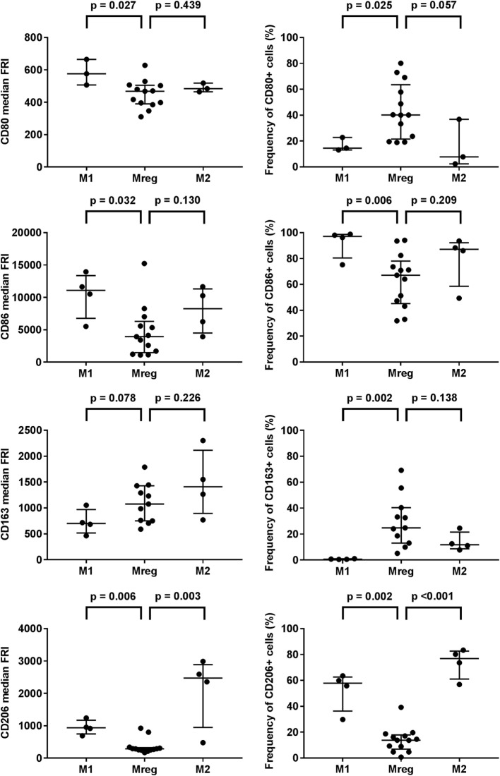
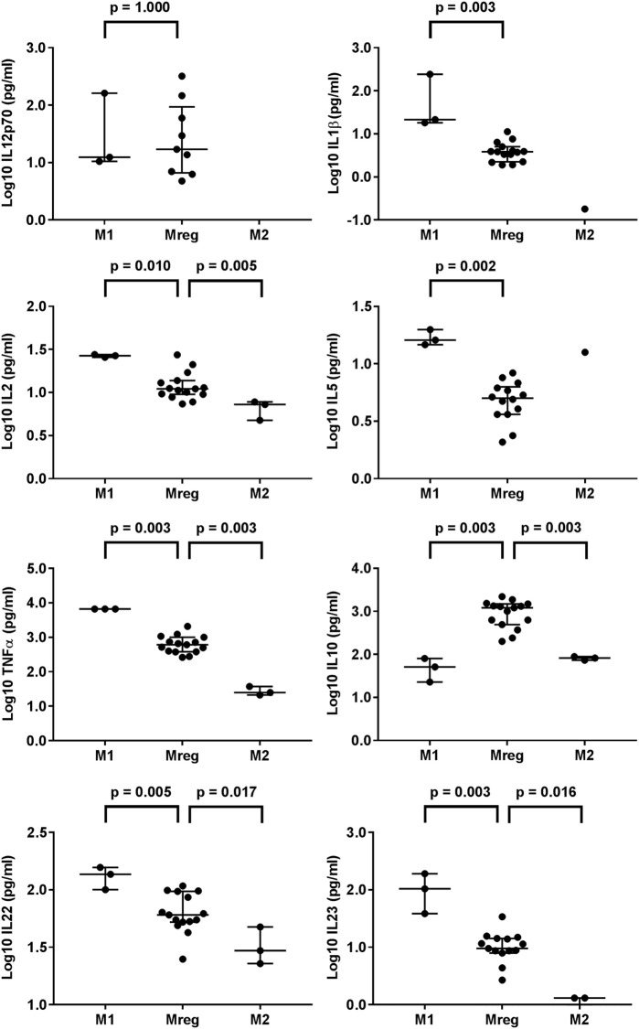
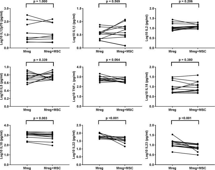
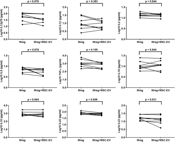
Resolution-phase macrophage population orchestrates active dampening of the inflammation by secreting anti-inflammatory and proresolving products including interleukin (IL)-10 and lipid mediators. We investigated the effects of both human bone marrow-derived mesenchymal stromal cells (MSCs) and MSC-derived extracellular vesicles (MSC-EVs) on mature human regulatory macrophages (Mregs). The cytokines and lipid mediators were determined from cell culture media of Mregs cultivated with MSCs and MSC-EVs. Additionally, the alterations in the expression of cell surface markers and the phagocytic ability of Mregs were investigated. Our novel findings indicate that both MSC co-culture and MSC-EVs down-regulated the production of IL-23 and IL-22 enhancing the anti-inflammatory phenotype of Mregs and amplifying proresolving properties. The levels of prostaglandin E2 (PGE2) were substantially upregulated in MSC co-culture media, which may endorse proresolving lipid mediator class switching. Additionally, our results manifest, for the first time, that MSC-EVs mediate the Mreg phenotype change via PGE2. These data suggest that both human MSC and MSC-EVs may potentiate tolerance-promoting proresolving phenotype of human Mregs.
Overview
The claims and statistics of this paper haven’t been checked yet.
Run the check — free, no account needed. It reads the open-access text and checks the sources it cites.
Integrity signals
Registry
Cites
Statistics
Figures
Verification
Claims the paper makes
The claims and statistics of this paper haven’t been checked yet.
Run the check — free, no account needed. It reads the open-access text and checks the sources it cites.
Claims other papers make about this one
Each is checked against this paper’s own text: does it say what the citing paper says it does?
The source backs part of the claim, not all of it — or backs it for a different population, condition or comparison.
In the citing paper
“The authors also demonstrated that MSC-EVs mediate their action, at least in part, via PGE2 (Table 1) [75].”
From Mesenchymal stem cell secretome for regenerative medicine: Where do we stand? (2025) · cites this paper as [75]
Experimental — accuracy not yet measuredThe cited source says what the claim attributes to it.
In the citing paper
“Hyvarinen and co-workers have shown that BM-MSCs co-culture with regulatory macrophages and MSC-EVs decrease the levels of IL-22 and IL-23 in the macrophages-produced CM [75].”
From Mesenchymal stem cell secretome for regenerative medicine: Where do we stand? (2025) · cites this paper as [75]
Self-check
Percentages, sums and reported differences re-computed from the paper’s own numbers.
The self-check needs the paper’s full text.
Citations
Cites
Exploring the full spectrum of macrophage activation
Mosser, D. M., & Edwards, J. P. (2008) · Nature Reviews Immunology · doi:10.1038/nri2448 · Cited by 9,144
Bettelli, E., Carrier, Y., Gao, W., Korn, T., Strom, T. B., Oukka, M., Weiner, H. L., & Kuchroo, V. K. (2006) · Nature · doi:10.1038/nature04753 · Cited by 6,992
Macrophage activation and polarization: nomenclature and experimental guidelines
Murray, P. J., Allen, J. E., Biswas, S. K., Fisher, E. A., Gilroy, D. W., Goerdt, S., Gordon, S., Hamilton, J. A., Ivashkiv, L. B., Lawrence, T., et al. (2014) · Immunity · doi:10.1016/j.immuni.2014.06.008 · Cited by 6,337
Human mesenchymal stem cells modulate allogeneic immune cell responses
Aggarwal, S., & Pittenger, M. F. (2005) · Blood · doi:10.1182/blood-2004-04-1559 · Cited by 4,511
Macrophages in Tissue Repair, Regeneration, and Fibrosis
Wynn, T. A., & Vannella, K. M. (2016) · Immunity · doi:10.1016/j.immuni.2016.02.015 · Cited by 4,481
Fadok, V. A., Bratton, D. L., Konowal, A., Freed, P. W., Westcott, J. Y., & Henson, P. M. (1998) · Journal of Clinical Investigation · doi:10.1172/jci1112 · Cited by 3,166 · cited 2×
“Kalim KW, Groettrup M. Prostaglandin E2 inhibits IL-23 and IL-12 production by human monocytes through down-regulation of their common p40 subunit.”
BackgroundMurray, P. J. (2017) · Annual Review of Physiology · doi:10.1146/annurev-physiol-022516-034339 · Cited by 2,981
Németh, K., Leelahavanichkul, A., Yuen, P. S. T., Mayer, B., Parmelee, A., Doi, K., Robey, P. G., Leelahavanichkul, K., Koller, B. H., Brown, J. M., et al. (2009) · Nature Medicine · doi:10.1038/nm.1905 · Cited by 2,364
Exosome secreted by MSC reduces myocardial ischemia/reperfusion injury
Lai, R. C., Arslan, F., Lee, M. M., Sze, N. S. K., Choo, A., Chen, T. S., Salto-Tellez, M., Timmers, L., Lee, C. N., El Oakley, R. M., et al. (2010) · Stem Cell Research · doi:10.1016/j.scr.2009.12.003 · Cited by 2,215
Interleukin-22, a TH17 cytokine, mediates IL-23-induced dermal inflammation and acanthosis
Zheng, Y., Danilenko, D. M., Valdez, P. A., Kasman, I. M., Eastham‐Anderson, J., Wu, J., & Ouyang, W. (2006) · Nature · doi:10.1038/nature05505 · Cited by 1,888
Meisel, R., Zibert, A., Laryea, M. D., Göbel, U., Däubener, W., & Dilloo, D. (2004) · Blood · doi:10.1182/blood-2003-11-3909 · Cited by 1,638
Lipid mediator class switching during acute inflammation: signals in resolution
Levy, B. D., Clish, C. B., Schmidt, B., Gronert, K., & Serhan, C. N. (2001) · Nature Immunology · doi:10.1038/89759 · Cited by 1,453
Induction and molecular signature of pathogenic TH17 cells
Lee, Y., Awasthi, A., Yosef, N., Quintana, F. J., Xiao, S., Peters, A., Wu, C., Kleinewietfeld, M., Kunder, S., Hafler, D. A., et al. (2012) · Nature Immunology · doi:10.1038/ni.2416 · Cited by 1,174
Resolvins and protectins in inflammation resolution
Serhan, C. N., & Petasis, N. A. (2011) · Chemical Reviews · doi:10.1021/cr100396c · Cited by 996
Th17 cells transdifferentiate into regulatory T cells during resolution of inflammation
Gagliani, N., Vesely, M. C. A., Iseppon, A., Brockmann, L., Xu, H. Y., Palm, N. W., de Zoete, M. R., Licona-Limón, P., Paiva, R., Ching, T. H., et al. (2015) · Nature · doi:10.1038/nature14452 · Cited by 830
Mesenchymal stem cell–educated macrophages: A novel type of alternatively activated macrophages
Kim, J., & Hematti, P. (2009) · Experimental Hematology · doi:10.1016/j.exphem.2009.09.004 · Cited by 785
Resolution of inflammation: an integrated view
Ortega‐Gómez, A., Perretti, M., & Soehnlein, O. (2013) · EMBO Molecular Medicine · doi:10.1002/emmm.201202382 · Cited by 776
Current and emerging therapeutic targets for IBD
Neurath, M. F. (2017) · Nature Reviews Gastroenterology & Hepatology · doi:10.1038/nrgastro.2016.208 · Cited by 717
Mesenchymal Stem Cells Secrete Immunologically Active Exosomes
Zhang, B., Yin, Y., Lai, R. C., Tan, S. S., Choo, A. B. H., & Lim, S. K. (2013) · Stem Cells and Development · doi:10.1089/scd.2013.0479 · Cited by 710
Resolvin D2 is a potent regulator of leukocytes and controls microbial sepsis
Spite, M., Norling, L. V., Summers, L., Yang, R., Cooper, D., Petasis, N. A., Flower, R. J., Perretti, M., & Serhan, C. N. (2009) · Nature · doi:10.1038/nature08541 · Cited by 695
Jackson, M. V., Morrison, T. J., Doherty, D. F., McAuley, D. F., Matthay, M. A., Kissenpfennig, A., O'Kane, C. M., & Krasnodembskaya, A. D. (2016) · Stem Cells · doi:10.1002/stem.2372 · Cited by 582
Sicco, C. L., Reverberi, D., Balbi, C., Ulivi, V., Principi, E., Pascucci, L., Becherini, P., Bosco, M. C., Varesio, L., Franzin, C., et al. (2017) · Stem Cells Translational Medicine · doi:10.1002/sctm.16-0363 · Cited by 579
Dalli, J., & Serhan, C. N. (2012) · Blood · doi:10.1182/blood-2012-04-423525 · Cited by 572
Mechanisms of mesenchymal stromal cell immunomodulation
English, K. (2012) · Immunology and Cell Biology · doi:10.1038/icb.2012.56 · Cited by 533
A new view of prostaglandin E regulation of the immune response
Phipps, R. P., Stein, S. H., & Roper, R. L. (1991) · Immunology Today · doi:10.1016/0167-5699(91)90064-z · Cited by 528
Therapeutic Effects of Human Mesenchymal Stem Cell–derived Microvesicles in Severe Pneumonia in Mice
Monsel, A., Zhu, Y., Gennai, S., Qi, H., Hu, S., Rouby, J., Rosenzwajg, M., Matthay, M. A., & Lee, J. W. (2015) · American Journal of Respiratory and Critical Care Medicine · doi:10.1164/rccm.201410-1765oc · Cited by 468
Haworth, O., Cernadas, M., Yang, R., Serhan, C. N., & Levy, B. D. (2008) · Nature Immunology · doi:10.1038/ni.1627 · Cited by 436
YlÖstalo, J. H., Bartosh, T. J., Coble, K., & Prockop, D. J. (2012) · STEM CELLS · doi:10.1002/stem.1191 · Cited by 427
Vasandan, A. B., Jahnavi, S., Chandanala, S., Prasad, P., Kumar, A., & Prasanna, S. J. (2016) · Scientific Reports · doi:10.1038/srep38308 · Cited by 415
Melief, S. M., Schrama, E., Brugman, M. H., Tiemessen, M. M., Hoogduijn, M. J., Fibbe, W. E., & Roelofs, H. (2013) · Stem Cells · doi:10.1002/stem.1432 · Cited by 408
A novel phenotype for an activated macrophage: the type 2 activated macrophage
Anderson, C. F., & Mosser, D. M. (2002) · Journal of Leukocyte Biology · doi:10.1189/jlb.72.1.101 · Cited by 347
Cyclooxygenases, prostanoids, and tumor progression
Wang, M., Honn, K. V., & Nie, D. (2007) · Cancer and Metastasis Reviews · doi:10.1007/s10555-007-9096-5 · Cited by 344
Resolution phase lipid mediators of inflammation: agonists of resolution
Serhan, C. N., & Chiang, N. (2013) · Current Opinion in Pharmacology · doi:10.1016/j.coph.2013.05.012 · Cited by 343
Trapani, M. D., Bassi, G., Midolo, M., Gatti, A., Kamga, P. T., Cassaro, A., Carusone, R., Adamo, A., & Krampera, M. (2016) · Scientific Reports · doi:10.1038/srep24120 · Cited by 299
Transcriptomic analyses of murine resolution-phase macrophages
Stables, M. J., Shah, S. H., Camon, E. B., Lovering, R. C., Newson, J., Byström, J., Farrow, S. N., & Gilroy, D. W. (2011) · Blood · doi:10.1182/blood-2011-04-345330 · Cited by 291
Reversing Lipopolysaccharide Toxicity by Ligating the Macrophage Fcγ Receptors
Gerber, J. S., & Mosser, D. M. (2001) · The Journal of Immunology · doi:10.4049/jimmunol.166.11.6861 · Cited by 263
Hutchinson, J. A., Riquelme, P., Sawitzki, B., Tomiuk, S., Miqueu, P., Zuhayra, M., Oberg, H., Pascher, A., Lützen, U., Janßen, U., et al. (2011) · The Journal of Immunology · doi:10.4049/jimmunol.1100762 · Cited by 239
Resolvin D1 and Aspirin-Triggered Resolvin D1 Promote Resolution of Allergic Airways Responses
Rogério, A. D. P., Haworth, O., Croze, R. H., Oh, S. F., Uddin, M., Carlo, T., Pfeffer, M. A., Priluck, R., Serhan, C. N., & Levy, B. D. (2012) · The Journal of Immunology · doi:10.4049/jimmunol.1101665 · Cited by 237
Schif‐Zuck, S., Gross, N., Assi, S., Rostoker, R., Serhan, C. N., & Ariel, A. (2010) · European Journal of Immunology · doi:10.1002/eji.201040801 · Cited by 236
Faouder, P. L., Baillif, V., Spreadbury, I., Motta, J., Rousset, P., Chêne, G., Guigné, C., Tercé, F., Vanner, S. J., Vergnolle, N., et al. (2013) · Journal of Chromatography B · doi:10.1016/j.jchromb.2013.06.014 · Cited by 226
Chiossone, L., Conte, R., Spaggiari, G. M., Serra, M., Romei, C., Bellora, F., Becchetti, F., Andaloro, A., Moretta, L., & Bottino, C. (2016) · Stem Cells · doi:10.1002/stem.2369 · Cited by 181
Kilpinen, L., Impola, U., Sankkila, L., Ritamo, I., Aatonen, M., Kilpinen, S. K., Tuimala, J., Valmu, L., Levijoki, J. M., Finckenberg, P., et al. (2013) · Journal of Extracellular Vesicles · doi:10.3402/jev.v2i0.21927 · Cited by 169
Self-citationFang, X., Abbott, J. A., Cheng, L., Colby, J. K., Lee, J. W., Levy, B. D., & Matthay, M. A. (2015) · The Journal of Immunology · doi:10.4049/jimmunol.1500244 · Cited by 157
Saldanha‐Araújo, F., Ferreira, F. I. S., Palma, P. V. B., Araújo, A. G., Queiróz, R. H. C., Covas, D. T., Zago, M. A., & Panepucci, R. A. (2011) · Stem Cell Research · doi:10.1016/j.scr.2011.04.001 · Cited by 137
Osteoblast recruitment from stem cells does not decrease by age at late adulthood
Leskelä, H., Risteli, J., Niskanen, S., Koivunen, J. P., Ivaska, K. K., & Lehenkari, P. P. (2003) · Biochemical and Biophysical Research Communications · doi:10.1016/j.bbrc.2003.10.095 · Cited by 128
Self-citationAmarnath, S., Foley, J., Farthing, D. E., Gress, R. E., Laurence, A. D. J., Eckhaus, M., Métais, J., Rose, J. J., Hakim, F. T., Felizardo, T. C., et al. (2014) · Stem Cells · doi:10.1002/stem.1934 · Cited by 117
Kerkelä, E., Laitinen, A., Räbinä, J., Valkonen, S., Takatalo, M. S., Larjo, A., Veijola, J., Lampinen, M., Siljander, P. R. M., Lehenkari, P. P., et al. (2016) · Stem Cells · doi:10.1002/stem.2280 · Cited by 114
Self-citationLiu, X., Qu, X., Chen, Y., Liao, L., Cheng, K., Shao, C., Zenke, M., Keating, A., & Zhao, R. C. (2012) · The Journal of Immunology · doi:10.4049/jimmunol.1102996 · Cited by 87
Newson, J., Motwani, M. P., Kendall, A. C., Nicolaou, A., Muccioli, G. G., Alhouayek, M., Bennett, M., van de Merwe, R. C., Naomi-James, S., Maeyer, R. P. H. D., et al. (2017) · Cell Reports · doi:10.1016/j.celrep.2017.08.098 · Cited by 78
IL-23 and Th17 Disease in Inflammatory Arthritis
Yago, T., Nanke, Y., Kawamoto, M., Kobashigawa, T., Yamanaka, H., & Kotake, S. (2017) · Journal of Clinical Medicine · doi:10.3390/jcm6090081 · Cited by 68
Hutchinson, J. A., Riquelme, P., Geissler, E. K., & Fändrich, F. (2010) · Methods in molecular biology · doi:10.1007/978-1-60761-869-0_13 · Cited by 67
Poloso, N. J., Urquhart, P., Nicolaou, A., Wang, J., & Woodward, D. F. (2013) · Molecular Immunology · doi:10.1016/j.molimm.2012.12.010 · Cited by 65
Tsoyi, K., Hall, S. R. R., Dalli, J., Colas, R. A., Ghanta, S., Ith, B., Coronata, A. A., Fredenburgh, L. E., Baron, R. M., Choi, A. M. K., et al. (2016) · Critical Care Medicine · doi:10.1097/ccm.0000000000001999 · Cited by 61
Regulatory macrophages as therapeutic targets and therapeutic agents in solid organ transplantation
Broichhausen, C., Riquelme, P., Geissler, E. K., & Hutchinson, J. A. (2012) · Current Opinion in Organ Transplantation · doi:10.1097/mot.0b013e328355a979 · Cited by 59
The generation of macrophages with anti-inflammatory activity in the absence of STAT6 signaling
Fleming, B. D., Chandrasekaran, P., Dillon, L. A. L., Dalby, E., Suresh, R., Sarkar, A., El-Sayed, N. M., & Mosser, D. M. (2015) · Journal of Leukocyte Biology · doi:10.1189/jlb.2a1114-560r · Cited by 49
Kalim, K. W., & Groettrup, M. (2012) · Molecular Immunology · doi:10.1016/j.molimm.2012.08.014 · Cited by 42
Peura, M., Bízik, J., Salmenperä, P., Noro, A., Korhonen, M. O., Pätilä, T., Vento, A. E., Vaheri, A. I., Alitalo, R., Vuola, J., et al. (2009) · Wound Repair and Regeneration · doi:10.1111/j.1524-475x.2009.00507.x · Cited by 36
Self-citationReview of phase III trial data on IL ‐23 inhibitors tildrakizumab and guselkumab for psoriasis
Amin, M., Darji, K., No, D. J., & Wu, J. J. (2017) · Journal of the European Academy of Dermatology and Venereology · doi:10.1111/jdv.14451 · Cited by 31
Cited by
Sorted by citations
Mesenchymal Stem Cell Immunomodulation: Mechanisms and Therapeutic Potential
Song, N., Scholtemeijer, M., & Shah, K. (2020) · Trends in Pharmacological Sciences · doi:10.1016/j.tips.2020.06.009 · Cited by 948
The Immunomodulatory Functions of Mesenchymal Stromal/Stem Cells Mediated via Paracrine Activity
Zhou, Y., Yamamoto, Y., Xiao, Z., & Ochiya, T. (2019) · Journal of Clinical Medicine · doi:10.3390/jcm8071025 · Cited by 303
Reis, M., Mavin, E., Nicholson, L., Green, K., Dickinson, A. M., & Wang, X.-N. (2018) · Frontiers in Immunology · doi:10.3389/fimmu.2018.02538 · Cited by 286
Effects of Mesenchymal Stem Cell-Derived Exosomes on Autoimmune Diseases
Shen, Z., Huang, W., Liu, J., Tian, J., Wang, S., & Rui, K. (2021) · Frontiers in Immunology · doi:10.3389/fimmu.2021.749192 · Cited by 285
Biology and therapeutic potential of mesenchymal stem cell‐derived exosomes
Zhou, X., & Kalluri, R. (2020) · Cancer Science · doi:10.1111/cas.14563 · Cited by 266
Metadata
- Authors
- Hyvärinen, K., Holopainen, M., Skirdenko, V., Ruhanen, H., Lehenkari, P., Korhonen, M., Käkelä, R., Laitinen, S., & Kerkelä, E.
- Affiliations
- Finnish Red Cross (FI)
- University of Helsinki (FI)
- Oulu University Hospital (FI)
- Corresponding author
- Hyvärinen, Kati
- ORCID
- Venue
- Frontiers in Immunology
- Publisher
- Frontiers Media SA
- Published
- 2018-04-12
- Volume
- 9
- Pages
- 771-771
- ISSN
- 1664-3224 (ISSN-L)
- PMC
- PMC5906545
- OpenAlex
- W2797721539
- Cited by
- 111
- References
- 58
- Citations by year
- 2026: 4 · 2025: 7 · 2024: 17 · 2023: 19 · 2022: 17 · 2021: 17 · 2020: 20 · 2019: 4 · 2018: 5
- FWCI
- 6.52
- Type
- journal article
- Language
- en
- Open access
- gold
- doi.org
- Locations
- Frontiers in Immunology (publishedVersion, cc-by)
- Europe PMC (PubMed Central) (publishedVersion, cc-by)
- PubMed Central (publishedVersion, cc-by)
- PubMed (publishedVersion)
- Työväentutkimus Vuosikirja (submittedVersion)
- DOAJ (DOAJ: Directory of Open Access Journals) (submittedVersion)
- Funders
- Tekes (3986/31/2013)
- Topics
- Life Sciences › Biochemistry, Genetics and Molecular Biology › Cell Biology › Mesenchymal stem cell research
- Life Sciences › Biochemistry, Genetics and Molecular Biology › Molecular Biology › Extracellular vesicles in disease
- Life Sciences › Immunology and Microbiology › Immunology › Immune cells in cancer
- Keywords
- mesenchymal stem cells, extracellular vesicles, regulatory macrophages, IL-23, IL-22, prostaglandin E2, inflammation resolution, specialized pro-resolving mediators, phagocytosis, cell surface markers, bone marrow-derived cells
- MeSH terms
- Extracellular Vesicles, Interleukin-22, Humans, Immune Tolerance, Inflammation, Interleukins, Macrophages, Phenotype, Down-Regulation, Interleukin-23, Mesenchymal Stem Cells
About these checks
Registry, retraction, Cites and statistics results are computed, not judged, with one exception: a printed reference that no index lists is read once by a model before it counts as a citation error. Claims are extracted by a model and kept only when their quote appears verbatim in the text; each cited source is then checked, with a model’s help, for whether it says what the claim relies on. Those verdicts can be wrong — read the claim’s quote and the cited source before relying on one.
Statistics and arithmetic are recomputed, not judged.
Confidence bands are shown only for fields calibrated on our annotated test set. None is calibrated yet, so no band is shown.
What counts as a claim ›How we measure confidence ›How we check this ›
Are you an author, or do you see a problem with this page? Request a review
