

The DOI matches its Crossref record; 3 of the 3 references it cites were found in a registry or an index.
miR-128 Regulates Tumor Cell CD47 Expression and Promotes Anti-tumor Immunity in Pancreatic Cancer
Overview
The claims and statistics of this paper haven’t been checked yet.
Run the check — free, no account needed. It reads the open-access text and checks the sources it cites.
Integrity signals
Registry
Cites
Statistics
Figures
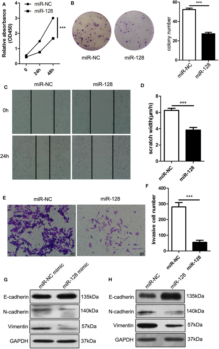
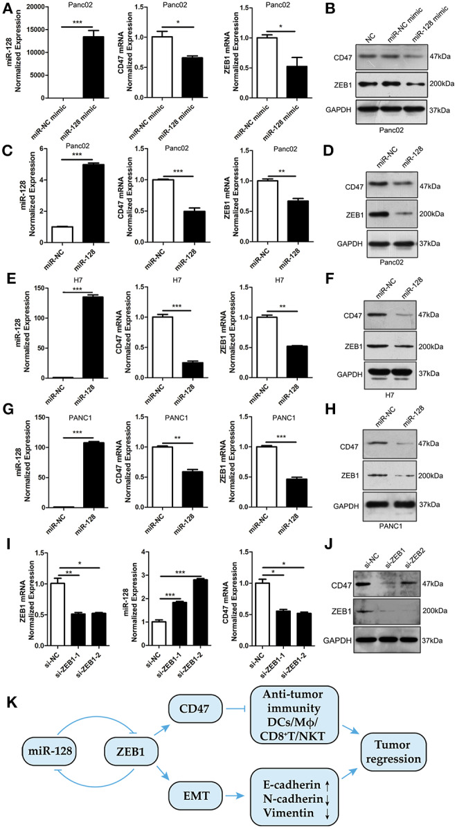
Verification
Claims the paper makes
The claims and statistics of this paper haven’t been checked yet.
Run the check — free, no account needed. It reads the open-access text and checks the sources it cites.
Claims other papers make about this one
Each is checked against this paper’s own text: does it say what the citing paper says it does?
The source backs part of the claim, not all of it — or backs it for a different population, condition or comparison.
In the citing paper
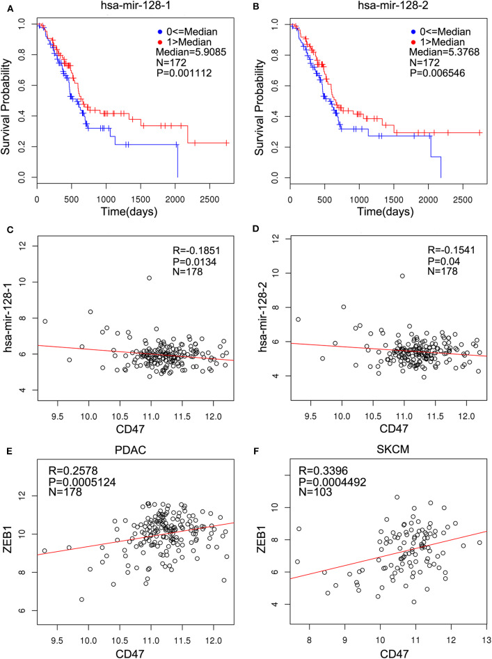
“Overexpression miR-128-3p in pancreatic adenocarcinoma was inversely correlated with CD47 (cluster of differentiation 47),177”
From MiR-128-3p – a gray eminence of the human central nervous system (2024) · cites this paper as [177]
Experimental — accuracy not yet measuredThe cited source says what the claim attributes to it.
In the citing paper
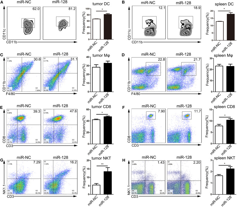
“Overexpression of miR-128-3p considerably increased the percentages of DCs, CD8+ T cells, and natural killer T cells (NKT) within the tumor and spleen.”
From MiR-128-3p – a gray eminence of the human central nervous system (2024) · cites this paper as [177]
In the citing paper
“Several reports demonstrated the high level of miR-128-3p as a pro-inflammatory and anti-immunosuppressive factor in immune cells, potentially significantly enhancing antitumor immunity.”
From MiR-128-3p – a gray eminence of the human central nervous system (2024) · cites this paper as [177]
Self-check
Percentages, sums and reported differences re-computed from the paper’s own numbers.
The self-check needs the paper’s full text.
Citations
Cites
MicroRNA therapeutics: towards a new era for the management of cancer and other diseases
Rupaimoole, R., & Slack, F. J. (2017) · Nature Reviews Drug Discovery · doi:10.1038/nrd.2016.246 · Cited by 4,741
Mechanism-driven biomarkers to guide immune checkpoint blockade in cancer therapy
Topalian, S. L., Taube, J. M., Anders, R. A., & Pardoll, D. M. (2016) · Nature Reviews Cancer · doi:10.1038/nrc.2016.36 · Cited by 2,733
Regulatory networks defining EMT during cancer initiation and progression
Craene, B. D., & Berx, G. (2013) · Nature Reviews Cancer · doi:10.1038/nrc3447 · Cited by 2,487
Research works printed but not linked: 44
- Kleeff, 2016, Pancreatic cancer, Nat Rev Dis Primers., 2, 16022, 10.1038/nrdp.2016.22 · DOI · Paper page
- Siegel, 2017, Cancer statistics, 2017, CA Cancer J Clin., 67, 7, 10.3322/caac.21387 · DOI · Paper page
- Rahib, 2014, Projecting cancer incidence and deaths to 2030: the unexpected burden of thyroid, liver, and pancreas cancers in the United States, Cancer Res., 74, 2913, 10.1158/0008-5472.CAN-14-0155 · DOI · Paper page
- Sharma, 2015, The future of immune checkpoint therapy, Science., 348, 56, 10.1126/science.aaa8172 · DOI · Paper page
- Brown, 2001, Integrin-associated protein (CD47) and its ligands, Trends Cell Biol., 11, 130, 10.1016/S0962-8924(00)01906-1 · DOI
- Jaiswal, 2009, CD47 is upregulated on circulating hematopoietic stem cells and leukemia cells to avoid phagocytosis, Cell., 138, 271, 10.1016/j.cell.2009.05.046 · DOI
- Willingham, 2012, The CD47-signal regulatory protein alpha (SIRPa) interaction is a therapeutic target for human solid tumors, Proc Natl Acad Sci U S A., 109, 6662, 10.1073/pnas.1121623109 · DOI
- Michaels, 2018, CD47 blockade as an adjuvant immunotherapy for resectable pancreatic cancer, Clin Cancer Res., 24, 1415, 10.1158/1078-0432.CCR-17-2283 · DOI
- Murata, 2018, CD47-signal regulatory protein α signaling system and its application to cancer immunotherapy, Cancer Sci., 109, 2349, 10.1111/cas.13663 · DOI
- Matlung, 2017, The CD47-SIRPα signaling axis as an innate immune checkpoint in cancer, Immunol Rev., 276, 145, 10.1111/imr.12527 · DOI
- Weiskopf, 2017, Cancer immunotherapy targeting the CD47/SIRPα axis, Eur J Cancer., 76, 100, 10.1016/j.ejca.2017.02.013 · DOI
- Liu, 2015, CD47 blockade triggers T cell-mediated destruction of immunogenic tumors, Nat Med., 21, 1209, 10.1038/nm.3931 · DOI
- Nieto, 2016, EMT: 2016, Cell., 166, 21, 10.1016/j.cell.2016.06.028 · DOI · Paper page
- Puisieux, 2014, Oncogenic roles of EMT-inducing transcription factors, Nat Cell Biol., 16, 488, 10.1038/ncb2976 · DOI
- Caramel, 2018, Pleiotropic roles for ZEB1 in cancer, Cancer Res., 78, 30, 10.1158/0008-5472.CAN-17-2476 · DOI
- Krebs, 2017, The EMT-activator Zeb1 is a key factor for cell plasticity and promotes metastasis in pancreatic cancer, Nat Cell Biol., 19, 518, 10.1038/ncb3513 · DOI
- Noman, 2018, CD47 is a direct target of SNAI1 and ZEB1 and its blockade activates the phagocytosis of breast cancer cells undergoing EMT, Oncoimmunology, 7, e1345415, 10.1080/2162402X.2017.1345415 · DOI
- Yang, 2015, miR-192 suppresses leptomeningeal dissemination of medulloblastoma by modulating cell proliferation and anchoring through the regulation of DHFR. integrins, and CD47, Oncotarget., 6, 43712, 10.18632/oncotarget.6227 · DOI
- Huang, 2018, MIR-708 promotes phagocytosis to eradicate T-ALL cells by targeting CD47, Mol Cancer., 17, 12, 10.1186/s12943-018-0768-2 · DOI
- Bruno, 2011, Identification of a microRNA that activates gene expression by repressing nonsense-mediated RNA decay, Mol Cell., 42, 500, 10.1016/j.molcel.2011.04.018 · DOI
- Hsu, 2006, miRNAMap: genomic maps of microRNA genes and their target genes in mammalian genomes, Nucleic Acids Res., 34, D135, 10.1093/nar/gkj135 · DOI
- Woo, 2012, Regulation of colony stimulating factor-1 expression and ovarian cancer cell behavior in vitro by miR-128 and miR-152, Mol Cancer., 11, 58, 10.1186/1476-4598-11-58 · DOI
- Lu, 2017, Posttranscriptional regulation of Galectin-3 by miR-128 contributes to colorectal cancer progression, Oncotarget, 8, 15242, 10.18632/oncotarget.14839 · DOI
- Xiao, 2018, MiR-128 regulation of glucose metabolism and cell proliferation in triple-negative breast cancer, BJS., 105, 75, 10.1002/bjs.10646 · DOI
- Corbett, 1984, Induction and chemotherapeutic response of two transplantable ductal adenocarcinomas of the pancreas in C57BL/6 mice, Cancer Res., 44, 717
- Vasaikar, 2018, LinkedOmics: analyzing multi-omics data within and across 32 cancer types, Nucleic Acids Res, 46, D956, 10.1093/nar/gkx1090 · DOI
- Subramani, 2015, Emerging roles of microRNAs in pancreatic cancer diagnosis, therapy and prognosis (Review), Int J Oncol., 47, 1203, 10.3892/ijo.2015.3129 · DOI
- Li, 2016, MicroRNA targeted therapeutic approach for pancreatic cancer, Int J Biol Sci., 12, 326, 10.7150/ijbs.15017 · DOI
- Frampton, 2015, Integrated molecular analysis to investigate the role of microRNAs in pancreatic tumour growth and progression, Lancet., 385, S37, 10.1016/S0140-6736(15)60352-X · DOI
- Sharpe, 2017, Introduction to checkpoint inhibitors and cancer immunotherapy, Immunol Rev., 276, 5, 10.1111/imr.12531 · DOI
- Cioffi, 2017, The miR-25-93-106b cluster regulates tumor metastasis and immune evasion via modulation of CXCL12 and PD-L1, Oncotarget., 8, 21609, 10.18632/oncotarget.15450 · DOI
- Mari, 2018, microRNA 125a regulates MHC-I expression on esophageal adenocarcinoma cells, associated with suppression of antitumor immune response and poor outcomes of patients, Gastroenterology., 155, 784, 10.1053/j.gastro.2018.06.030 · DOI
- Zhao, 2011, CD47-signal regulatory protein-α (SIRPα) interactions form a barrier for antibody-mediated tumor cell destruction, Proc Natl Acad Sci U S A., 108, 18342, 10.1073/pnas.1106550108 · DOI
- Liu, 2015, Pre-clinical development of a humanized anti-CD47 antibody with anti-cancer therapeutic potential, PLoS One., 10, e0137345, 10.1371/journal.pone.0137345 · DOI
- Brabletz, 2010, The ZEB/miR-200 feedback loop–a motor of cellular plasticity in development and cancer?, EMBO Rep., 11, 670, 10.1038/embor.2010.117 · DOI
- Vandewalle, 2009, The role of the ZEB family of transcription factors in development and disease, Cell Mol Life Sci., 66, 773, 10.1007/s00018-008-8465-8 · DOI
- Burk, 2008, A reciprocal repression between ZEB1 and members of the miR-200 family promotes EMT and invasion in cancer cells, EMBO Rep., 9, 582, 10.1038/embor.2008.74 · DOI
- Sun, 2015, miR-128 modulates chemosensitivity and invasion of prostate cancer cells through targeting ZEB1, Jpn J Clin Oncol., 45, 474, 10.1093/jjco/hyv027 · DOI
- Zhao, 2018, Tumor suppressor miR-128-3p inhibits metastasis and epithelial-mesenchymal transition by targeting ZEB1 in esophageal squamous-cell cancer, Acta Biochim Biophys Sin., 50, 171, 10.1093/abbs/gmx132 · DOI
- Xu, 2017, Dendritic cells but not macrophages sense tumor mitochondrial DNA for cross-priming through signal regulatory protein α signaling, Immunity., 47, 363, 10.1016/j.immuni.2017.07.016 · DOI
- Terabe, 2008, The role of NKT cells in tumor immunity, Adv Cancer Res., 101, 277, 10.1016/S0065-230X(08)00408-9 · DOI
- Bendelac, 2007, The biology of NKT cells, Annu Rev Immunol., 25, 297, 10.1146/annurev.immunol.25.022106.141711 · DOI
- Brennan, 2013, Invariant natural killer T cells: an innate activation scheme linked to diverse effector functions, Nat Rev Immunol., 13, 101, 10.1038/nri3369 · DOI
- Brigl, 2003, Mechanism of CD1d-restricted natural killer T cell activation during microbial infection, Nat Immunol., 4, 1230, 10.1038/ni1002 · DOI
Cited by
Sorted by citations
Poh, A. R., & Ernst, M. (2021) · Cancers · doi:10.3390/cancers13122860 · Cited by 106
Xie, Y., Wang, X., Wang, W., Pu, N., & Liu, L. (2025) · Journal of Translational Medicine · doi:10.1186/s12967-025-06422-5 · Cited by 80
McCubrey, J. A., Yang, L. V., Abrams, S. L., Steelman, L. S., Follo, M. Y., Cocco, L., Ratti, S., Martelli, A. M., Augello, G., & Cervello, M. (2022) · Cells · doi:10.3390/cells11142155 · Cited by 49
MicroRNAs as Regulators of Phagocytosis
Gierlikowski, W., & Gierlikowska, B. (2022) · Cells · doi:10.3390/cells11091380 · Cited by 25
MiR-128-3p – a gray eminence of the human central nervous system
Kiel, K., Król, S. K., Bronisz, A., & Godlewski, J. (2024) · Molecular Therapy Nucleic Acids · doi:10.1016/j.omtn.2024.102141 · Cited by 15
Metadata
- Authors
- Xi, Q., Chen, Y., Yang, G.-Z., Zhang, J.-Y., Zhang, L.-J., Guo, X.-D., Zhao, J.-Y., Xue, Z.-Y., Li, Y., & Zhang, R.
- Venue
- Frontiers in Immunology
- Publisher
- Frontiers Media SA
- Published
- 2020-05-27
- Volume
- 11
- ISSN
- 1664-3224
- PMC
- PMC7267029
- Cited by
- 47
- References
- 3
- Funders
- National Natural Science Foundation of China
About these checks
Registry, retraction, Cites and statistics results are computed, not judged, with one exception: a printed reference that no index lists is read once by a model before it counts as a citation error. Claims are extracted by a model and kept only when their quote appears verbatim in the text; each cited source is then checked, with a model’s help, for whether it says what the claim relies on. Those verdicts can be wrong — read the claim’s quote and the cited source before relying on one.
Statistics and arithmetic are recomputed, not judged.
Confidence bands are shown only for fields calibrated on our annotated test set. None is calibrated yet, so no band is shown.
What counts as a claim ›How we measure confidence ›How we check this ›
Are you an author, or do you see a problem with this page? Request a review
