

The DOI matches its Crossref record; 21 of the 21 references it cites were found in a registry or an index.
Honing the Double-Edged Sword: Improving Human iPSC-Microglia Models
Abstract
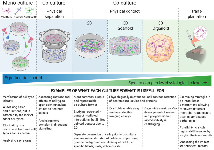
Human induced Pluripotent Stem Cell (hiPSC) models are a valuable new tool for research into neurodegenerative diseases. Neuroinflammation is now recognized as a key process in neurodegenerative disease and aging, and microglia are central players in this. A plethora of hiPSC-derived microglial models have been published recently to explore neuroinflammation, ranging from monoculture through to xenotransplantation. However, combining physiological relevance, reproducibility, and scalability into one model is still a challenge. We examine key features of the in vitro microglial environment, especially media composition, extracellular matrix, and co-culture, to identify areas for improvement in current hiPSC-microglia models.
Overview
The claims and statistics of this paper haven’t been checked yet.
Run the check — free, no account needed. It reads the open-access text and checks the sources it cites.
Integrity signals
Registry
Cites
Statistics
Figures
Verification
Claims the paper makes
The claims and statistics of this paper haven’t been checked yet.
Run the check — free, no account needed. It reads the open-access text and checks the sources it cites.
Claims other papers make about this one
Each is checked against this paper’s own text: does it say what the citing paper says it does?
In the citing paper
“However, our sample size of two donors per group may have been insufficient to detect subtle differences, while more physiological conditions, such as co-cultures with other cell types [124], may be necessary to reveal such changes in future studies.”
From microRNA-132 attenuates inflammation in induced pluripotent stem cell-derived microglia from Alzheimer’s disease patients (2026) · cites this paper as [124]
Self-check
Percentages, sums and reported differences re-computed from the paper’s own numbers.
The self-check needs the paper’s full text.
Citations
Cites
Neurotoxic reactive astrocytes are induced by activated microglia
Liddelow, S. A., Guttenplan, K. A., Clarke, L. E., Bennett, F. C., Bohlen, C. J., Schirmer, L., Bennett, M. L., Münch, A. E., Chung, W.-S., Peterson, T. C., et al. (2017) · Nature · doi:10.1038/nature21029 · Cited by 7,971
Identification of a unique TGF-β–dependent molecular and functional signature in microglia
Butovsky, O., Jedrychowski, M. P., Moore, C. S., Cialic, R., Lanser, A. J., Gabriely, G., Koeglsperger, T., Dake, B., Wu, P. M., Doykan, C. E., et al. (2014) · Nature Neuroscience · doi:10.1038/nn.3599 · Cited by 2,543
Elmore, M. R. P., Najafi, A. R., Koike, M. A., Dagher, N. N., Spangenberg, E. E., Rice, R. A., Kitazawa, M., Matusow, B., Nguyen, H., West, B. L., et al. (2014) · Neuron · doi:10.1016/j.neuron.2014.02.040 · Cited by 1,968
Marschallinger, J., Iram, T., Zardeneta, M., Lee, S. E., Lehallier, B., Haney, M. S., Pluvinage, J. V., Mathur, V., Hahn, O., Morgens, D. W., et al. (2020) · Nature Neuroscience · doi:10.1038/s41593-019-0566-1 · Cited by 1,384 · cited 1×
“Both scenarios are likely occurring in tandem in various neurodegenerative diseases, as well as in normal ageing, where defective phagocytosis as well as pro-inflammatory profiles have recently been reported (94, 95).”
BackgroundAn environment-dependent transcriptional network specifies human microglia identity
Gosselin, D., Skola, D., Coufal, N. G., Holtman, I. R., Schlachetzki, J. C. M., Sajti, E., Jaeger, B. N., O’Connor, C., Fitzpatrick, C., Pasillas, M. P., et al. (2017) · Science · doi:10.1126/science.aal3222 · Cited by 1,220
Microglia development follows a stepwise program to regulate brain homeostasis
Matcovitch-Natan, O., Winter, D. R., Giladi, A., Vargas Aguilar, S., Spinrad, A., Sarrazin, S., Ben-Yehuda, H., David, E., Zelada González, F., Perrin, P., et al. (2016) · Science · doi:10.1126/science.aad8670 · Cited by 1,180 · cited 1×
“The cytokine response can exacerbate neuronal damage if prolonged, contributing to chronic neurodegenerative disease [(6), reviewed by (7)].”
BackgroundLin, Y.-T., Seo, J., Gao, F., Feldman, H. M., Wen, H.-L., Penney, J., Cam, H. P., Gjoneska, E., Raja, W. K., Cheng, J., et al. (2018) · Neuron · doi:10.1016/j.neuron.2018.05.008 · Cited by 1,125
Bohlen, C. J., Bennett, F. C., Tucker, A. F., Collins, H. Y., Mulinyawe, S. B., & Barres, B. A. (2017) · Neuron · doi:10.1016/j.neuron.2017.04.043 · Cited by 658
A 3D human triculture system modeling neurodegeneration and neuroinflammation in Alzheimer’s disease
Park, J., Wetzel, I., Marriott, I., Dréau, D., D’Avanzo, C., Kim, D. Y., Tanzi, R. E., & Cho, H. (2018) · Nature Neuroscience · doi:10.1038/s41593-018-0175-4 · Cited by 644
Haenseler, W., Sansom, S. N., Buchrieser, J., Newey, S. E., Moore, C. S., Nicholls, F. J., Chintawar, S., Schnell, C., Antel, J. P., Allen, N. D., et al. (2017) · Stem Cell Reports · doi:10.1016/j.stemcr.2017.05.017 · Cited by 593
Self-citation
Research works printed but not linked: 75
- Ginhoux, 2010, Fate mapping analysis reveals that adult microglia derive from primitive macrophages, Science (80), 330, 10.1126/science.1194637 · DOI · Paper page
- Swinnen, 2013, Complex invasion pattern of the cerebral cortex bymicroglial cells during development of the mouse embryo, Glia, 61, 10.1002/glia.22421 · DOI
- Paolicelli, 2011, Synaptic pruning by microglia is necessary for normal brain development, Science (80), 333, 10.1126/science.1202529 · DOI · Paper page
- Sakai, 2020, Core Concept: How synaptic pruning shapes neural wiring during development and, possibly, in disease, Proc Natl Acad Sci USA, 117, 10.1073/pnas.2010281117 · DOI
- Li, 2018, Microglia and macrophages in brain homeostasis and disease, Nat Rev Immunol, 18, 10.1038/nri.2017.125 · DOI · Paper page
- Sabogal-Guáqueta, 2020, Microglia alterations in neurodegenerative diseases and their modeling with human induced pluripotent stem cell and other platforms, Prog Neurobiol, 190, 10.1016/j.pneurobio.2020.101805 · DOI
- Mancuso, 2019, Stem-cell-derived human microglia transplanted in mouse brain to study human disease, Nat Neurosci, 22, 10.1038/s41593-019-0525-x · DOI · Paper page
- Dubbelaar, 2018, The Kaleidoscope of Microglial Phenotypes, Front Immunol, 9, 10.3389/fimmu.2018.01753 · DOI · Paper page
- Sayed, 2016, Translation of Human-Induced Pluripotent Stem Cells from Clinical Trial in a Dish to Precision Medicine, J Am Coll Cardiol, 67, 10.1016/j.jacc.2016.01.083 · DOI
- Rehbach, 2020, Publicly available hiPSC lines with extreme polygenic risk scores for modeling schizophrenia, bioRxiv, 10.1101/2020.07.04.185348 · DOI
- Bennett, 2020, The influence of environment and origin on brain resident macrophages and implications for therapy, Nat Neurosci, 23, 10.1038/s41593-019-0545-6 · DOI
- Bian, 2020, Deciphering human macrophage development at single-cell resolution, Nature, 582, 10.1038/s41586-020-2316-7 · DOI
- Askew, 2017, Coupled Proliferation and Apoptosis Maintain the Rapid Turnover of Microglia in the Adult Brain, Cell Rep, 18, 391, 10.1016/j.celrep.2016.12.041 · DOI · Paper page
- Abud, 2017, iPSC-derived human microglia-like cells to study neurological diseases, Neuron, 94, 10.1016/j.neuron.2017.03.042.iPSC-derived · DOI
- Brownjohn, 2018, Functional Studies of Missense TREM2 Mutations in Human Stem Cell-Derived Microglia, Stem Cell Rep, 10, 10.1016/j.stemcr.2018.03.003 · DOI · Paper page
- Claes, 2019, Human stem cell–derived monocytes and microglia-like cells reveal impaired amyloid plaque clearance upon heterozygous or homozygous loss of TREM2, Alzheimer’s Dement, 15, 10.1016/j.jalz.2018.09.006 · DOI
- Douvaras, 2017, Directed Differentiation of Human Pluripotent Stem Cells to Microglia, Stem Cell Rep, 8, 10.1016/j.stemcr.2017.04.023 · DOI · Paper page
- Pandya, 2017, Differentiation of human and murine induced pluripotent stem cells to microglia-like cells, Nat Neurosci, 20, 10.1038/nn.4534 · DOI · Paper page
- Takata, 2017, Induced-Pluripotent-Stem-Cell-Derived Primitive Macrophages Provide a Platform for Modeling Tissue-Resident Macrophage Differentiation and Function, Immunity, 47, 183, 10.1016/j.immuni.2017.06.017 · DOI
- Muffat, 2016, Efficient derivation of microglia-like cells from human pluripotent stem cells, Nat Med, 22, 10.1038/nm.4189 · DOI · Paper page
- Grubman, 2020, A CX3CR1 Reporter hESC Line Facilitates Integrative Analysis of In-Vitro-Derived Microglia and Improved Microglia Identity upon Neuron-Glia Co-culture, Stem Cell Rep, 14, 10.1016/j.stemcr.2020.04.007 · DOI
- Gutbier, 2020, Large-scale production of human IPSC-derived macrophages for drug screening, Int J Mol Sci, 21, 1, 10.3390/ijms21134808 · DOI
- Hasselmann, 2019, Development of a Chimeric Model to Study and Manipulate Human Microglia In Vivo, Neuron, 103, 1016, 10.1016/j.neuron.2019.07.002 · DOI · Paper page
- Svoboda, 2019, Human iPSC-derived microglia assume a primary microglia-like state after transplantation into the neonatal mouse brain, Proc Natl Acad Sci USA, 116, 10.1073/pnas.1913541116 · DOI
- Xu, 2020, Human iPSC-derived mature microglia retain their identity and functionally integrate in the chimeric mouse brain, Nat Commun, 11, 1, 10.1038/s41467-020-15411-9 · DOI
- Haenseler, 2019, Concise Review: Modeling Neurodegenerative Diseases with Human Pluripotent Stem Cell-Derived Microglia, Stem Cells, 37, 10.1002/stem.2995 · DOI
- Miron, 2020, Investigating Microglia in Health and Disease : Challenges and Opportunities, Trends Immunol, 41, 10.1016/j.it.2020.07.002 · DOI
- Speicher, 2019, Generating microglia from human pluripotent stem cells: Novel in vitro models for the study of neurodegeneration, Mol Neurodegener, 14, 1, 10.1186/s13024-019-0347-z · DOI
- Hanger, 2020, Emerging Developments in Human Induced Pluripotent Stem Cell-Derived Microglia: Implications for Modelling Psychiatric Disorders With a Neurodevelopmental Origin, Front Psychiatry, 11, 10.3389/fpsyt.2020.00789 · DOI
- Kleman, 2008, Physiological glucose is critical for optimized neuronal viability and AMPK responsiveness in vitro, J Neurosci Methods, 167, 292, 10.1016/j.jneumeth.2007.08.028 · DOI
- Rocktäschel, 2019, High glucose concentrations mask cellular phenotypes in a stem cell model of tuberous sclerosis complex, Epilepsy Behav, 101, 10.1016/j.yebeh.2019.106581 · DOI
- Giulian, 1986, Characterization of ameboid microglia isolated from developing mammalian brain, J Neurosci, 6, 10.1523/jneurosci.06-08-02163.1986 · DOI
- Witting, 2011, Microglia Cell Culture: A Primer for the Novice, 49, 10.1007/978-1-61779-170-3_4 · DOI
- Schumann, 1990, Structure and Function of Lipopolysaccharide Binding Protein, Science (80), 249, 10.1126/science.2402637 · DOI
- Liu, 2018, Corticosterone preexposure increases NF-κB translocation and sensitizes IL-1β responses in BV2 microglia-like cells, Front Immunol, 9, 10.3389/fimmu.2018.00003 · DOI
- Bardy, 2015, Neuronal medium that supports basic synaptic functions and activity of human neurons in vitro, Proc Natl Acad Sci USA, 112, 10.1073/pnas.1504393112 · DOI · Paper page
- Abutbul, 2012, TGF-β signaling through SMAD2/3 induces the quiescent microglial phenotype within the CNS environment, Glia, 60, 10.1002/glia.22343 · DOI · Paper page
- Elmore, 2014, CSF1 receptor signaling is necessary for microglia viability, which unmasks a cell that rapidly repopulates the microglia- depleted adult brain, Neuron, 82, 10.1016/j.neuron.2014.02.040.CSF1 · DOI
- Nieweg, 2009, Marked differences in cholesterol synthesis between neurons and glial cells from postnatal rats, J Neurochem, 109, 10.1111/j.1471-4159.2009.05917.x · DOI · Paper page
- McIlvain, 2018, Mechanical properties of the in vivo adolescent human brain, Dev Cognit Neurosci, 34, 27, 10.1016/j.dcn.2018.06.001 · DOI
- Lam, 2019, Tissue-specific extracellular matrix accelerates the formation of neural networks and communities in a neuron-glia co-culture on a multi-electrode array, Sci Rep, 9, 1, 10.1038/s41598-019-40128-1 · DOI
- Chaudhuri, 2020, Effects of extracellular matrix viscoelasticity on cellular behaviour, Nature, 584, 10.1038/s41586-020-2612-2 · DOI · Paper page
- Jensen, 2020, Is It Time to Start Transitioning From 2D to 3D Cell Culture, Front Mol Biosci, 7, 10.3389/fmolb.2020.00033 · DOI · Paper page
- Frampton, 2011, Fabrication and optimization of alginate hydrogel constructs for use in 3D neural cell culture, Biomed Mater, 6, 015002, 10.1088/1748-6041/6/1/015002 · DOI
- Jeffery, 2014, Hyaluronic acid-based 3D culture model for in vitro testing of electrode biocompatibility, Biomacromolecules, 15, 10.1021/bm500318d · DOI
- Koss, 2017, Improved 3D hydrogel cultures of primary glial cells for in vitro modelling of neuroinflammation, J Vis Exp, 2017, 10.3791/56615 · DOI
- Leite, 2020, A human co-culture cell model incorporating microglia supports glioblastoma growth and migration, and confers resistance to cytotoxics, FASEB J, 34, 10.1096/fj.201901858RR · DOI
- Cho, 2017, Development of an in vitro 3D brain tissue model mimicking in vivo-like pro-inflammatory and pro-oxidative responses, Ann Biomed Eng, 46, 10.1007/s10439-018-2004-z · DOI
- Song, 2014, Anti-inflammatory effects of three-dimensional graphene foams cultured with microglial cells, Biomaterials, 35, 10.1016/j.biomaterials.2014.05.002 · DOI
- Schwartz, 2015, Human pluripotent stem cell-derived neural constructs for predicting neural toxicity, Proc Natl Acad Sci USA, 112, 10.1073/pnas.1516645112 · DOI · Paper page
- Jiang, 2016, Enhanced migration of neural stem cells by microglia grown on a three- dimensional graphene scaffold ACS, Appl. Mater. Interfaces, 8, 10.1021/acsami.6b06780 · DOI
- Chronopoulou, 2012, Self-assembling peptide hydrogels promote microglial cells proliferation and NGF production, Soft Matter, 8, 10.1039/c2sm25528f · DOI
- Pires, 2015, The role of the surface on microglia function: Implications for central nervous system tissue engineering, J R Soc Interface, 12, 1, 10.1098/rsif.2014.1224 · DOI
- Liu, 2019, Three-dimensional (3D) brain microphysiological system for organophosphates and neurochemical agent toxicity screening, PLoS One, 14, 10.1371/journal.pone.0224657 · DOI
- Blazquez, 2017, 3D coculture model of the brain parenchyma–metastasis interface of brain metastasis
- Hermida, 2020, Three dimensional in vitro models of cancer: Bioprinting multilineage glioblastoma models, Adv Biol Regul, 75, 10.1016/j.jbior.2019.100658 · DOI
- Sridar, 2017, Peptide modification of polyimide-insulated microwires: Towards improved biocompatibility through reduced glial scarring, Acta Biomater, 60, 10.1016/j.actbio.2017.07.026 · DOI
- Qin, 2005, LPS induces CD40 gene expression through the activation of NF-κB and STAT-1α in macrophages and microglia, Blood, 106, 10.1182/blood-2005-02-0759 · DOI · Paper page
- Blasi, 1990, Immortalization of murine microglial cells by a v-raf/v-myc carrying retrovirus, J Neuroimmunol, 27, 10.1016/0165-5728(90)90073-V · DOI
- Amadio, 2013, Plasticity of primary microglia on micropatterned geometries and spontaneous long-distance migration in microfluidic channels, BMC Neurosci, 14, 10.1186/1471-2202-14-121 · DOI
- Guttenplan, 2019, Astrocytes and microglia: Models and tools, J Exp Med, 216, 71, 10.1084/jem.20180200 · DOI
- Wright, 2000, Lymphoid/neuronal cell surface OX2 glycoprotein recognizes a novel receptor on macrophages implicated in the control of their function, Immunity, 13, 10.1016/S1074-7613(00)00023-6 · DOI
- Biber, 2007, Neuronal “On” and “Off” signals control microglia, Trends Neurosci, 30, 596, 10.1016/j.tins.2007.08.007 · DOI · Paper page
- Mott, 2004, Neuronal expression of CD22: Novel mechanism for inhibiting microglial proinflammatory cytokine production, Glia, 46, 10.1002/glia.20009 · DOI
- Maciejewski-Lenoir, 1999, Characterization of fractalkine in rat brain cells: migratory and activation signals for CX3CR-1-expressing microglia, J Immunol, 163, 10.4049/jimmunol.163.3.1628 · DOI
- Szepesi, 2018, Bidirectional Microglia–Neuron Communication in Health and Disease, Front Cell Neurosci, 12, 10.3389/fncel.2018.00323 · DOI · Paper page
- Neumann, 1998, Neuronal Control of the Immune Response in the Central Nervous System: Linking Brain Immunity to Neurodegeneration, J Neuropathol Exp Neurol, 57, 1, 10.1097/00005072-199801000-00001 · DOI
- Nimmerjahn, 2005, Neuroscience: Resting microglial cells are highly dynamic surveillants of brain parenchyma in vivo, Science (80), 308, 10.1126/science.1110647 · DOI · Paper page
- Liddelow, 2020, Microglia and astrocyte interactions in health and disease: Dynamic Duo or Partners in Crime, Trends Immunol, 41, 1, 10.1016/j.it.2020.07.006 · DOI · Paper page
- Liddelow, 2017, Reactive Astrocytes: Production, Function, and Therapeutic Potential, Immunity, 46, 10.1016/J.IMMUNI.2017.06.006 · DOI · Paper page
- Goshi, 2020, A primary neural cell culture model to study neuron, astrocyte, and microglia interactions in neuroinflammation, J Neuroinflammation, 17, 155, 10.1186/s12974-020-01819-z · DOI
- Nimmervoll, 2013, LPS-induced microglial secretion of TNFα increases activity-dependent neuronal apoptosis in the neonatal cerebral cortex, Cereb Cortex, 23, 10.1093/cercor/bhs156 · DOI
- Wang, 2005, Involvement of proinflammatory factors, apoptosis, caspase-3 activation and Ca2+ disturbance in microglia activation-mediated dopaminergic cell degeneration, Mech Ageing Dev, 126, 10.1016/j.mad.2005.06.012 · DOI
- Ormel, 2018, Microglia innately develop within cerebral organoids, Nat Commun, 9, 1, 10.1038/s41467-018-06684-2 · DOI
- Mathews, 2019, Human Interleukin-34 facilitates microglia-like cell differentiation and persistent HIV-1 infection in humanized mice, Mol Neurodegener, 14, 12, 10.1186/s13024-019-0311-y · DOI
Cited by
Sorted by citations
Penning, A., Snoeck, S., Ormaechea, O. R., Ayyildiz, D., Polzer, O., Buitrago-Arango, M., Capobianco, R., de Winter, F., Balusu, S., Verhaagen, J., et al. (2026) · Acta Neuropathologica Communications · doi:10.1186/s40478-026-02228-8 · Cited by 6
Metadata
- Authors
- Hedegaard, A., Stodolak, S., James, W. S., & Cowley, S. A.
- Venue
- Frontiers in Immunology
- Publisher
- Frontiers Media SA
- Published
- 2020-12-08
- Volume
- 11
- ISSN
- 1664-3224
- PMC
- PMC7753623
- Cited by
- 29
- References
- 21
- Funders
- Oxford Martin School, University of Oxford
- Biotechnology and Biological Sciences Research Council
- Eli Lilly and Company
About these checks
Registry, retraction, Cites and statistics results are computed, not judged, with one exception: a printed reference that no index lists is read once by a model before it counts as a citation error. Claims are extracted by a model and kept only when their quote appears verbatim in the text; each cited source is then checked, with a model’s help, for whether it says what the claim relies on. Those verdicts can be wrong — read the claim’s quote and the cited source before relying on one.
Statistics and arithmetic are recomputed, not judged.
Confidence bands are shown only for fields calibrated on our annotated test set. None is calibrated yet, so no band is shown.
What counts as a claim ›How we measure confidence ›How we check this ›
Are you an author, or do you see a problem with this page? Request a review
